Iran Red Crescent Med J. 2016 July; 18(7):e22932. Published online 2016 February 20.
doi:10.5812/ircmj.22932. Case Report
Solitary Plasmacytoma of the Mandible: An Uncommon Entity
Ezher Hamza Dayisoylu,
1,*Ozcan Ceneli,
1and Esra Zeypep Coskunoglu
11Department of Oral and Maxillofacial Surgery, Faculty of Dentistry, Baskent University, Bahcelievler, Ankara, Turkey
*Corresponding author: Ezher Hamza Dayisoylu, Department of Oral and Maxillofacial Surgery, Faculty of Dentistry, Baskent University, Bahcelievler, Ankara, Turkey. Tel: +90-5326624778, Fax: +90-3322570637, E-mail: [email protected]
Received2014 September 21; Revised 2015 March 23; Accepted 2015 April 25.
Abstract
Introduction:Plasma cell dyscrasias are characterized by a monoclonal neoplastic proliferation of plasma cells. Solitary bone plasmacytoma (SBP) is a local form of the disease with the vertebrae and long bones being the most frequently encountered sites. Its prevalence in the maxillofacial area is extremely rare.
Case Presentation:A 70-year-old Caucasian male patient was referred for the extraction of his mobile premolar tooth with a poorly-defined radiolucent lesion. Histopathological analysis revealed an SBP and no distant lesion or serum M protein was noted on radi-ological and hematradi-ological examinations. The patient was under follow-up care with no recurrence at 2 years of follow up. Conclusions:Diagnosis of an SBP is based on local radiological and neurological symptoms and similar systemic manifestations of multiple myeloma that are also distinctive for SBP. Skeletal radiological analysis including CT and PET-CT, bone marrow biopsy, and serum protein electrophoresis are essential for confirmation of the diagnosis. Although surgery, chemotherapy, and radiation, or a combination of these modalities, have been successfully used in the treatment of SBP, it should be managed in relation to its possible long-term evolution.
Keywords:Multiple Myeloma, Solitary Plasmacytoma, Mandible
1. Introduction
Plasma cell neoplasms are characterized by uncon-trolled proliferation of plasma cells. Solitary bone plasma-cytomas (SBPs) and extramedullary plasmaplasma-cytomas (EPs) are the localized forms of the disease, whereas multiple myeloma (MM) is a systemic clonal proliferation of plasma cells based in the bone marrow (1). Fewer than 5% of pa-tients with plasma cell dyscrasias present as SBP or EP with-out distinct evidence of systemic myeloma (2). The diag-nosis of SBP is based on a biopsy of the specimen, skeletal radiological evaluation with a lack of any other distant le-sions, and a negative result for monoclonal plasma cells on bone marrow aspirate to rule out MM. The majority of SBP lesions either progress to MM over a period of 2 - 3 years af-ter diagnosis or local asymptomatic lesions tend to occur. Therefore, early diagnosis, proper treatment, and close fol-low up are crucial for survival (3). Clinical symptoms of SBP consist of jaw pain, paresthesia, and mobility of the sur-rounding teeth, or pathologic bone fractures. Radiological signs of SBP include ill defined unilocular or multilocular radiolucent lesions or a mass with cortical expansion (4). Although osteolytic jaw lesions commonly appear in pa-tients with MM, the ileum, femur, humerus, and thoracic vertebrae are the most frequent locations for SBP and its incidence in the maxillofacial area is extremely rare. SBP
treatment consists of surgery, chemotherapy, or a combi-nation of these modalities. There is also some controversy concerning management solely with surgery or radiother-apy alone (5). In this case report, the treatment of a 70-year-old Caucasian male patient with SBP of the mandible is pre-sented.
2. Case Presentation
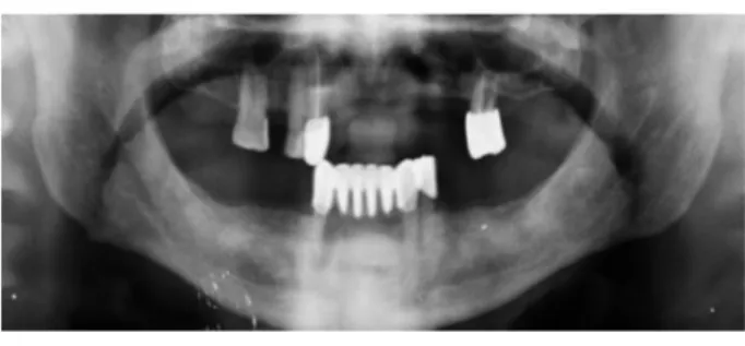
A 70-year-old Caucasian male patient was referred to Baskent University Department of Oral and Maxillofacial Surgery for the extraction of mobile tooth # 44. The pa-tient’s medical history was noncontributory, and the phys-ical examination revealed no other abnormalities. An in-traoral examination showed a mobile tooth with poor oral hygiene and pain on palpation. A radiological examina-tion revealed a poorly-defined destructed radiolucent area around the mandibular premolar region (Figure 1). Numb-ness of the chin, expansion of the bone cortices, and other complaints were not observed. The mobile tooth was ex-tracted and the lesion was enucleated. Following the curet-tage, an alveoloplasty was performed under local anesthe-sia. The histopathological examination of the specimen showed diffuse stromal infiltration by plasma cells with eosinophilic cytoplasm and eccentric nuclei. The immuno-histochemical examination showed positivity with CD138
Copyright © 2016, Iranian Red Crescent Medical Journal. This is an open-access article distributed under the terms of the Creative Commons Attribution-NonCommercial 4.0 International License (http://creativecommons.org/licenses/by-nc/4.0/) which permits copy and redistribute the material just in noncommercial usages, provided the original work is properly cited.
Dayisoylu EH et al.
and a monoclonal restriction for the Kappa chain. The pa-tient was consulted to the hematology department and no monoclonal protein was observed with serum and urine immunofixation electrophoresis. The sedimentation rate was 5 mm/h and the beta 2 microglobulin level was 2.2 mg/L (reference range: 0.97 - 2.64 mg/L). Additionally, no re-nal dysfunction, hypercalcemia, or anemia were observed in age-related reference ranges. PET CT scans and cone beam CT (CBCT) evaluations revealed a lack of any local or distant lytic lesions except for an increased FDG uptake in the right hemimandible during PET-CT scan analysis. Therefore, we concluded that adequate surgical debride-ment had been achieved. The patient was followed up on a monthly basis for 6 months and no further lesions were observed at the 2-year follow-up examination.
Figure 1.Note the Irregular Radiolucent Area Around Tooth # 44
3. Discussion
Plasma cell neoplasms are classified into three sub-groups as multiple myeloma (MM), solitary bone plasma-cytoma (SBP), and extramedullary plasmaplasma-cytoma (EP). A plasmacytoma is a result of uncontrolled proliferation of a single clone of B cells with no distant spread that are able to evolve up to the stage of plasma cells (6). Whereas MM is the disseminated form of the plasma cell neoplasms, SBP and EP are separate entities that are observed as local-ized lesions in the bone or extramedullary soft tissue, re-spectively (4). Systemic alterations such as hypercalcemia, impaired renal function, anemia, bone lesions, leukope-nia, thrombocytopeleukope-nia, and proliferation of monoclonal plasma cells in the bone marrow are frequently observed in MM patients (7). Daghighi et al. noted that bone damage may also be responsible for alteration of blood calcium lev-els, especially in MM cases compared with SBP. Therefore, complete blood cell count (CBC), calcium, phosphorous, C-reactive protein (CRP), beta 2-microglobulin analyses, and skeletal bone surveys are highly recommended (8). On the other hand, SBP is a rare disease that constitutes approxi-mately 3% - 10% of all plasma cell neoplasms and clinical
signs and symptoms of SBP may be really poor that differs from MM. Therefore, an early diagnosis of SBP may be chal-lenging to perform (3,9). Moreover, SBP commonly occurs in long bones and vertebrae, however, its involvement in the mandible is only 4.4% of the cases, which may also com-plicate its diagnosis (3). Pisano et al. noted SBP most fre-quently occurs at the posterior mandible, which is consis-tent with our case (10). SBP is frequently misdiagnosed as benign lesions, inflammatory diseases, and less frequently as malignant tumors. Clinical signs and symptoms indi-cated by patients include localized pain and paresthesia, but bone-impaired functions are also frequently reported (4). The most frequent clinical symptoms of SBP are re-ferred pain in the jaws and teeth that may also be related to other jaw lesions, and less commonly seen are swelling, soft tissue masses, and pathologic fractures. The tumors are found as unilocular or multilocular radiolucent de-structive lesions on radiological analysis that are also con-sistent with our case as a unilocular ill-defined lesion. Al-though no spontaneous jaw pain was observed in our pa-tient except tooth mobility the OPG and CBCT revealed a lytic radiolucent area for SBP. Canger et al. (4) noted that patients are generally male with a 2:1 male to female ratio, and they are typically in their sixth or seventh decades of life; thus, our patient’s profile was also consistent with the literature (11,12).
Elevated serum or urine calcium levels, renal failure, anemia, bone lesions, and consequently pathological frac-tures are typically seen in MM patients. Alternatively, a lack of these findings increases the median survival rate in SBP patients. Furthermore, SBP may be an isolated dis-ease or the first manifestation of a subsequent MM. Lesmes et al. claimed there is some controversy about SBP man-agement solely with surgery or radiotherapy. On the other hand, there is often a need for a second surgical approach for biopsy for the eradication of the disease and the possi-ble need for further radiotherapy (5). Daghighi et al. also noted CT and MRI findings are not specific for plasmacy-toma and histopathological examinations are crucial for the diagnosis (8). Therefore, a surgery-first approach may be advantageous over solely using radiotherapy depend-ing on the size and location of the lesion. In our case, fol-lowing surgical debridement, no sign of local or distant lesions were noted and no radiotherapy was planned. Al-though radiation, chemotherapy, surgery, or a combina-tion of these modalities provides good results with low re-currence rates, close follow up is essential (13). The progno-sis of SBP could be worse if recurrence is present as in cases of evolution toward systemic diseases such as MM (13).
Dayisoylu EH et al.
Footnote
Authors’ Contribution: Ezher Hamza Dayisoylu: first visit of the patient, operation, and follow up of the patient and preparation of the manuscript; Ozcan Ceneli: diagno-sis and follow up of the patient concerning a hematolog-ical viewpoint; Esra Zeypep Coskunoglu: pathologhematolog-ical ex-amination, diagnosis, and preparation of the paper.
References
1. Knowling MA, Harwood AR, Bergsagel DE. Comparison of ex-tramedullary plasmacytomas with solitary and multiple plasma cell tumors of bone. J Clin Oncol. 1983;1(4):255–62. [PubMed:6668499]. 2. Munshi NC, Anderson KC. New strategies in the treatment of
mul-tiple myeloma. Clin Cancer Res. 2013;19(13):3337–44. doi:
10.1158/1078-0432.CCR-12-1881. [PubMed:23515406].
3. Kaur H, Parhar S, Kaura S, Bansal S, Jawanda M, Madhushankari GS, et al. A large painless swelling of the posterior mandible. Oral Surg Oral Med Oral Pathol Oral Radiol. 2013;115(2):152–6. doi:
10.1016/j.oooo.2012.01.007. [PubMed:23312915].
4. Canger EM, Celenk P, Alkan A, Gunhan O. Mandibular involvement of solitary plasmocytoma: a case report. Med Oral Patol Oral Cir Bucal. 2007;12(1):E7–9. [PubMed:17195833].
5. Lesmes D, Laster Z. Plasmacytoma in the temporomandibular joint: a case report. Br J Oral Maxillofac Surg. 2008;46(4):322–4. doi:
10.1016/j.bjoms.2007.06.009. [PubMed:17698264].
6. Ariyarathenam A, Galvin N, Akoh JA. Secondary extramedullary plas-macytoma causing small bowel intussusception in a patient with multiple myeloma - A case report. Int J Surg Case Rep. 2013;4(5):486– 8. doi:10.1016/j.ijscr.2013.02.018. [PubMed:23562898].
7. Talamo G, Farooq U, Zangari M, Liao J, Dolloff NG, Loughran TJ, et al. Beyond the CRAB symptoms: a study of presenting clinical manifestations of multiple myeloma. Clin Lymphoma Myeloma Leuk. 2010;10(6):464–8. doi:10.3816/CLML.2010.n.080. [PubMed:21156463]. 8. Daghighi MH, Poureisa M, Shimia M, Mazaheri-Khamene R, Daghighi
S. Extramedullary plasmacytoma presenting as a solitary mass in the intracranial posterior fossa. Iran J Radiol. 2012;9(4):223–6. doi:
10.5812/iranjradiol.8759. [PubMed:23408237].
9. Seoane J, Aguirre-Urizar JM, Esparza-Gomez G, Suarez-Cunqueiro M, Campos-Trapero J, Pomareda M. The spectrum of plasma cell neopla-sia in oral pathology. Medicina oral. 2003;8(4):269–80.
10. Pisano JJ, Coupland R, Chen SY, Miller AS. Plasmacytoma of the oral cavity and jaws: a clinicopathologic study of 13 cases. Oral Surg Oral Med Oral Pathol Oral Radiol Endod. 1997;83(2):265–71. [PubMed:
9117760].
11. Rodriguez-Caballero B, Sanchez-Santolino S, Garcia-Montesinos-Perea B, Garcia-Reija MF, Gomez-Roman J, Saiz-Bustillo R. Mandibular solitary plasmocytoma of the jaw: a case report. Med Oral Patol Oral Cir Bucal.2011;16(5):e647–50. [PubMed:20711133].
12. Baad R, Kapse SC, Rathod N, Sonawane K, Thete SG, Kumar MN. Soli-tary plasmacytoma of the mandible - a rare entity. J Int Oral Health. 2013;5(3):97–101. [PubMed:24155610].
13. Marotta S, Di Micco P. Solitary plasmacytoma of the jaw. J Blood Med. 2010;1:33–6. doi:10.2147/JBM.S8385. [PubMed:22282681].