Gökhan At›fl*, Serdar Ar›san and
Ayhan Dalk›l›nç
fiiflli Etfal Research and Training Hospital, 1st Urology Clinics, fiiflli-Istanbul - Turkey
Abstract
HER2/neu 185 kDa is a transmembrane receptor tyrosine kinase being a part of epidermal growth factor receptor family. Especially overexpression was shown in breast cancer and treatment procedures targeting this protein are in clinical use. The aim of this study is to show the overexpression of HER2/neu in bladder TCC patients by using immunohistochemical methods. 46 TCC and 15 healthy bladder tissues as a control group were used in the study. Deparafinized tumor specimens were marked by using monoclonal antihuman HER2 protein antibody and the protein was shown by using peroxidase secondary antibody. Non-staining or membrane staining less than 10% were classified as 0, while membrane staining partly more than 10% were 1, poor complete membrane staining or moderate staining more than 10% were 2 and complete membrane strongly staining were classified as 3. Classification 2 and 3 were evaluated as over staining of HER2/neu. Overexpression of HER2/Neu protein were shown in 12/46 (24.5%) of TCC specimens. Overexpression of HER2/neu were detected in 2 (12.5%) of Grade 1, 1 (16.6%) of Grade 2 and 12 (50%) of Grade 3 patients. Overexpression of HER2/Neu protein were positive in 5 (17.8%) of 28 superficial (Ta-T1) tumor specimens and 7 (38.8%) of 18 invasive (T2-T3) tumor specimens. Overexpression of HER2/neu in
Grade III tumors were statistically significant when compared with Grade I and Grade II (p=0.012). Overexpression of HER2/neu detected in bladder TCC will be used in clinical setting as a treatment option targeting this protein as in the breast cancer in the future.
Keywords: Bladder carcinoma, HER2/neu, protein overexpression, trastuzumab, immunohistochemistry
Introduction
HER2/neu, a 185 kDa transmembrane receptor tyrosine kinase, is a member of the epidermal growth factor receptor (EGFR) family. The Her2 gene is localized to chromosome 17q and encodes a glycoprotein with intrinsic tyrosine kinase activity. The HER2/neu-encoded protein molecule occupies a critical position in the biochemical pathways responsible for the transduction of mitogenic signals from a variety of growth factor receptors. In addition to its role in regulating normal cellular proliferation, overexpression of the HER2/neu gene appears to play a role in neoplastic cell growth.
In breast carcinoma, HER2/neu gene amplification and receptor protein overexpression are tightly correlated and have prognostic and therapeutic implications. Approximately 15% to 25% of all breast cancers are HER2/neu positive for protein overexpression or gene amplification, and is associated with a poor clinical outcome (Slamon et al., 1989; Slamon et al., 2001). HER2/neu positive patients benefit from treatment with a monoclonal antibody to the HER2/neu protein (trastuzumab). This treatment modality has been shown to improve survival significantly in patients with metastatic breast cancer (Slamon et al., 2001)
The incidence of overexpression of HER2/neu in bladder cancer is one of the highest among all human malignancies, ranging from 9% to 34% of cancers tested (Sato et al., 1992; Coombs et al., 1991). Its prognostic value and correlation with tumor stage and grade has been variably reported. In transitional cell carcinoma of the bladder, it was found that HER2 is
*Correspondence Author:
fiiflli Etfal Research and Training Hospital, 1st Urology Clinics, fiiflli-Istanbul
E-mail: [email protected]
Received: July 5, 2007; Accepted: August 21, 2007.
Determination of HER2/NEU gene amplification and protein
overexpression in bladder transitional cell carcinoma
overexpressed with a greater frequency in higher grades (40%) and stages (38%) than lower grades (0%) and stages (8%) (Kruger et al., 2002). With respect to the potential application of HER2/neu to the treatment of TCC, protein overexpression or gene amplification may predict the clinical response to treatment with trastuzumab. Furthermore, trastuzumab acts synergistically, in breast cancer, with
agents more commonly used in treatment of TCCs such as cisplatin, with minimal additional toxic side-effects, suggesting it may be of value in the treatment of TCC (Pegram et al., 1999; Burris et al., 2000).
In this study, we examined bladder specimens with grade 1 to 3 and superficial (Stage Ta ,T1) and invasive (Stage T2, T3) TCC for the overexpression of the Her-2/neu protein.
Control (n) 15
Man
Age (Range, years)
12 28‐81 Woman
Age (Range, years)
3 35‐76
Patients 46
Man
Age (Range, years) Grade I Grade II Grade III Ta‐T1 T2‐T3 31 41‐81 10 4 17 16 12 Woman
Age (Range, years) Grade I Grade II Grade III Ta‐T1 T2‐T3 15 44‐67 6 2 7 8 6
Materials and methods
The tissue samples used in this study were obtained from 15 patients with benign and 46 patients with malignant bladder tumors. Tumor tissues were analyzed for HER2/neu expression by immunohistochemistry (IHC) using a commercial monoclonal antibody on 4-µ-thick, formalin fixed, parafin embedded tissue sections from archival blocks using standardized avidin-biotin techniques. Briefly, sections were deparaffinized and cleared. Endogenous peroxidase activity was blocked by a 3% solution of hydrogen peroxide and methanol. Sections were then hydrated in graded alcohol to distilled water. Microwave heat-induced (100 ºC) antigen retrieval was performed in citrate-buffered solution for a total of 4 minutes (twice times 2-minute cycles). Slides were then cooled to room temperature and placed in a humidified chamber. A solution of 10% normal horse serum was applied for 20 minutes. After blotting, the primary antibody (c-erb B2; Biogenex; 1:50 dilution) was applied and incubated overnight at 4 ºC. After rinsing in phosphate-buffered saline (PBS), a horse-antimouse biotinylated antibody applied at a dilution of 1:800 and incubated for 60 minutes at room temperature. Two PBS rinses were followed by 30-minute incubation with avidin-biotin complex. Color development was achieved by application of the chromagen 3,3´diaminobenzidine tetrahydrocloride and careful monitoring of staining by direct light microscopy visualization (generally for 2-3 minutes per section). The slides were then rinsed thoroughly in running tap water, counterstained in mayers hematoxylin, dehydrated, cleared, and coverslipped. Duplicate sections that were subjected to all phases of
staining except the primary antibody served as negative controls. Positive controls were paraffin embedded sections of breast carcinoma that had been identified as positive for HER2/neu by IHC.
Sections were analyzed with light microscopy by a single pathologist, and the presence of malignant cells was confirmed. Sections were scored as positive only if membrane staining was evident and ranked as 0 (no staining or <10% staining of tumor cells), 1+ (faint staining in >10% of cells; however, only partial staining of the membrane is seen), 2+ (weak-to-moderate, complete membrane staining in >10% of tumor cells), or 3+ (strong, complete membrane staining in >10% of tumor cells) (Figure 1).
Results
Tissue samples were collected from 15 patients with benign bladder lesions and 46 patients with malignant bladder tumors. There were 43 males and 18 females, with a mean age of 65 years (range 28-81 years). Overall, 12 (24.5%) of the 46 TCC specimens were positive for HER2/Neu protein overexpression using IHC (Table 1).
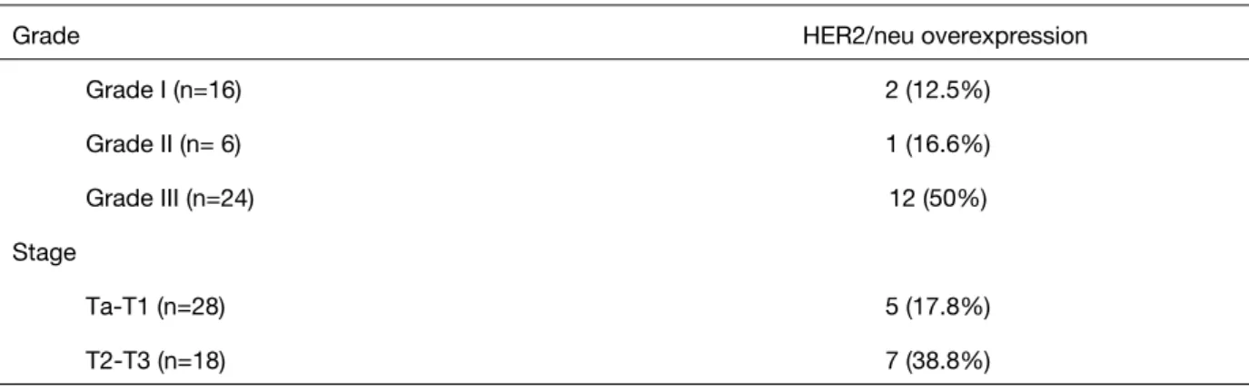
Overexpression of HER2/neu were detected in 2 (12.5%) of the 16 patients (Grade 1), 1 (16.6%) of the 6 patients (Grade 2) and 12 (50%) of the 24 patients (Grade 3). Overexpression of HER2/Neu protein were positive in 5 (17.8%) of 28 superficial (Ta-T1) tumor specimens and 7 (38.8%) of 18 invasive (T2-T3) tumor specimens (Table 2). Overexpression of HER2/neu in Grade 3 tumors was statistically significant when compared with Grade 1 and Grade 2 (p=0.012).
Figure 1. HER2/neu immunoreactivity staining scores. Left figure shows negative staining, middle is +1 score staining and right
Discussion
HER-2 gene received attention in the early 1990s, with several studies aimed at defining its role in bladder TCC and its capacity as a prognostic indicator. The prognostic value of HER2 in bladder carcinoma has not been established; however, the success of trastuzumab therapy in patients with breast carcinoma has stimulated interest in exploring the potential of this therapy for patients with urothelial carcinoma.
Early studies of the expression of HER2 protein in bladder carcinoma found a correlation between increased HER2 expression and both higher tumor stage and grade (Christopher et al., 2004; Gorgoulis et
al., 1995; Moch et al., 1993; Moriyama et al., 1991).
Others suggested that the overexpression of HER2 was an independent variable in determining patient survival (Sato et al., 1991; Korkolopoulou et al., 1997). A confounding variable in these studies was that several different antibodies were used for IHC and that the criteria for IHC positivity were based on cytoplasmic and/or membrane staining patterns.
HER2/neu overexpression could be evaluated by IHC, fluorescence in situ hybridization (FISH), and serum analysis. The best method to detect HER2/neu overexpression in urothelial cancer is not known. Because the greatest clinical benefit is observed in patients with the highest levels of Her-2/neu expression, trastuzumab trials in breast cancer often require eligible patients to have either 3+ overexpression by IHC or 2+ overexpression by IHC with positive FISH results. However, initial studies permitted patients with 2+ or 3+ overexpression by IHC, as we did in this trial (Slamon et al., 2001; Cobleigh et al., 1999).
HER2/neu protein overexpression is believed to arise from a combination of two possible mechanisms. The first is gene amplification which as several reports suggests that it is not a common mechanism in bladder cancer (Coombs et al., 1991; Kruger et al., 2002). The other mechanism is upregulated transcription which is possibly reflective of the nature of the HER2/neu protein (as a growth factor). Higher levels of transcription factors, even in the absence of gene amplification result in increased HER2/neu protein overexpression (Latif et al., 2004).
Using IHC, we found a statistically significant difference in HER2/neu protein overexpression in grade 3 TCC compared with grade 1 and 2 and in invasive TCC (T2 or T3) versus superficial TCC (Ta or T1), with overexpression more prevalent in the high grade and invasive specimens. This is consistent with the findings of several recent reports (Christopher et
al., 2004; Ravery et al., 1997; Jimenez et al., 2001;
Kruger et al., 2002; Gandour-Edwards et al., 2002). Despite the inconclusive data on the prognostic value of HER2 as an independent marker of tumor progression, there may be a therapeutic role for trastuzumab, the humanized monoclonal antibody directed against the HER2 surface receptor, in combination with a taxane in patients with HER2 positive disease. It has been shown previously in pivotal trials of trastuzumab plus paclitaxel that this combination is efficacious in patients with metastatic, HER2 positive breast carcinoma (Slamon et al., 2001). A multicenter National Cancer Institute-sponsored study conducted a trial to investigate the safety and efficacy of trastuzumab, carboplatin, gemcitabine, and paclitaxel in patients with advanced urothelial carcinoma. In this study the overall response rate was 70%; myelosuppression and cardiac toxicity were
Grade HER2/neu overexpression
Grade I (n=16) 2 (12.5%) Grade II (n= 6) 1 (16.6%) Grade III (n=24) 12 (50%) Stage Ta‐T1 (n=28) 5 (17.8%) T2‐T3 (n=18) 7 (38.8%)
high, and in the context of the setting is felt to be clinically acceptable and consistent with that observed in breast cancer trials. Determining the true contribution of trastuzumab requires randomized trials (Hussain et al., 2007).
Conclusions
Using IHC, the results of our study demonstrated that overexpression of the HER2/neu protein is present in human bladder TCC. Moreover, a statistically significant trend of an increased prevalence of overexpression in grade 3, invasive bladder TCC specimens compared with grade 1 and 2, superficial TCC was observed. Larger studies that include metastatic TCC specimens for HER2/neu protein overexpression and gene amplification are necessary to provide more complete information on HER-2/neu in bladder TCC. Once these data are obtained, the appropriateness of clinical trials with trastuzumab can be ascertained.
References
Slamon DJ, Godolphin W, Jones LA, Holt JA, Wong SG, Keith DE, Levin WJ, Stuart SG, Udove J, Ullrich A: Studies of the HER-2/neu proto-oncogene in human breast and ovarian cancer.
Science. 12: 244: 707-712, 1989.
Slamon DJ, Leyland-Jones B, Shak S, Fuchs H, Paton V, Bajamonde A, Fleming T, Eiermann W, Wolter J, Pegram M, Baselga J, Norton L: Use of chemotherapy plus a monoclonal antibody against HER2 for metastatic breast cancer that overexpresses HER2. N Engl J Med. 15:783-792, 2001.
Sato K, Moriyama M, Mori S, Saito M, Watanuki T, Terada K, Okuhara E, Akiyama T, Toyoshima K, Yamamoto T: An immunohistologic evaluation of c-erbB-2 gene product in patients with urinary bladder carcinoma. Cancer 15:2493-2498, 1992. Coombs LM, Pigott DA, Sweeney E, Proctor AJ,
Eydmann ME, Parkinson C, Knowles MA: Amplification and over-expression of c-erbB-2 in transitional cell carcinoma of the urinary bladder.
Br J Cancer. 63: 601-608, 1991.
Kruger S, Weitsch G, Buttner H, Matthiensen A, Bohmer T, Marquardt T, Sayk F, Feller AC, Bohle A. Overexpression of c-erbB-2 oncoprotein in muscle-invasive bladder carcinoma: relationship with gene amplification, clinicopathological
parameters and prognostic outcome. Int J Oncol. 21: 981-987, 2002.
Pegram MD, Slamon DJ: Combination therapy with trastuzumab (Herceptin) and cisplatin for chemoresistant metastatic breast cancer: evidence for receptor-enhanced chemosensitivity. Semin
Oncol.26 Suppl 12:89-95, 1999.
Burris HA 3rd: Docetaxel (Taxotere) in HER-2-positive patients and in combination with trastuzumab (Herceptin). Semin Oncol. 27: (2 Suppl 3):19-23, 2002.
Coogan CL, Estrada CR, Kapur S, Bloom KJ: HER-2/neu protein overexpression and gene amplification in human transitional cell carcinoma of the bladder. Urology. 63:786-790, 2004. Gorgoulis VG, Barbatis C, Poulias I, Karameris AM:
Molecular and immunohistochemical evaluation of epidermal growth factor receptor and c-erb-B-2 gene product in transitional cell carcinomas of the urinary bladder: a study in Greek patients. Pathol. 8:758-764, 1995.
Moch H, Sauter G, Moore D, Mihatsch MJ, Gudat F, Waldman F: p53 and erbB-2 protein overexpression are associated with early invasion and metastasis in bladder cancer. Virchows Arch A
Pathol Anat Histopathol. 423(5):329-334, 1993.
Moriyama M, Akiyama T, Yamamoto T, Kawamoto T, Kato T, Sato K, Watanuki T, Hikage T, Katsuta N, Mori S: Expression of c-erbB-2 gene product in urinary bladder cancer. J Urol.145:423-427, 1991. Korkolopoulou P, Christodoulou P, Kapralos P,
Exarchakos M, Bisbiroula A, Hadjiyannakis M, Georgountzos C, Thomas-Tsagli E: The role of p53, MDM2 and c-erb B-2 oncoproteins, epidermal growth factor receptor and proliferation markers in the prognosis of urinary bladder cancer. Pathol Res
Pract. 193:767-775, 1997.
Ravery V, Grignon D, Angulo J, Pontes E, Montie J, Crissman J, Chopin D: Evaluation of epidermal growth factor receptor, transforming growth factor alpha, epidermal growth factor and c-erbB2 in the progression of invasive bladder cancer. Urol Res. 25:9-17, 1997.
Jimenez RE, Hussain M, Bianco FJ Jr, Vaishampayan U, Tabazcka P, Sakr WA, Pontes JE, Wood DP Jr, Grignon DJ.: Her-2/neu overexpression in muscle-invasive urothelial carcinoma of the bladder:
prognostic significance and comparative analysis in primary and metastatic tumors. Clin Cancer Res. 7:2440-2447, 2001.
Kruger S, Weitsch G, Buttner H, Matthiensen A, Bohmer T, Marquardt T, Sayk F, Feller AC, Bohle A: HER2 overexpression in muscle-invasive urothelial carcinoma of the bladder: prognostic implications.
Int J Cancer. 102:514-518, 2002.
Gandour-Edwards R, Lara PN Jr, Folkins AK, LaSalle JM, Beckett L, Li Y, Meyers FJ, DeVere-White R: Does HER2/neu expression provide prognostic information in patients with advanced urothelial carcinoma? Cancer. 95:1009-1015, 2002.
Cobleigh MA, Vogel CL, Tripathy D, Robert NJ, Scholl S, Fehrenbacher L, Wolter JM, Paton V, Shak S, Lieberman G, Slamon DJ: Multinational study of the efficacy and safety of humanized anti-HER2 monoclonal antibody in women who have HER2-overexpressing metastatic breast cancer that has progressed after chemotherapy for metastatic disease. J Clin Oncol. 17:2639-2648, 1999. Latif Z, Watters AD, Dunn I, Grigor K, Underwood MA,
Bartlett JM: HER2/neu gene amplification and protein overexpression in G3 pT2 transitional cell carcinoma of the bladder: a role for anti-HER2 therapy? Eur J Cancer. 40:56-63, 2004.
Hussain MH, MacVicar GR, Petrylak DP, Dunn RL, Vaishampayan U, Lara PN Jr, Ch Institute.atta GS, Nanus DM, Glode LM, Trump DL, Chen H, Smith DC: Trastuzumab, paclitaxel, carboplatin, and gemcitabine in advanced human epidermal growth factor receptor-2/neu-positive urothelial carcinoma: results of a multicenter phase II National Cancer Institute trial. J Clin Oncol. 25:2218-2224, 2007.