Van Tıp Derg 24(4): 369-371, 2017 DOI: 10.5505/vtd.2017.63935
*Sorumlu Yazar: Op. Dr. Engin Akgul, Department of Cardiovascular Surgery, Dumlupinar University Evliya Celebi Training and Research
Hospital, Kutahya/Turkey, E-mail: [email protected] GSM: 0 (544) 659 32 22
Geliş Tarihi: 27.03.2017, Kabul Tarihi: 23.06.2017
CASE REPORT / OLGU SUNUMU
8
th
Decades Presentation of Subaortic Fibrous
Membrane with Left Ventricular Outflow Tract
Obstruction
8. Dekatta Tanı Konan Sol Ventrikül Çıkım Yolu Darlığı Oluşturan Subaortik
Fibröz Memban
Engin Akgül*, Asiye Aslı Gözüaçık, Ahmet Hakan Vural
Dumlupinar University Evliya Celebi Training and Research Hospital, Department of Cardiovascular Surgery, Kutahya, Turkey
Introduction
Discrete subaortic stenosis (DSS) is
characterized by a fibromembraneus tissue
which surrounds the aortic region. This is a
rare cause of left ventricular outflow tract
obstruction (LVOT) in the adult age. LVOT
can cause progressive left ventricular
hypertrophy, secondary aortic regurgitation
(AR) and heart failure at the end (1,2,3).
Clinical symptoms are variable for example
chest pain and fatigue, progressive dyspnea,
chief complaints can be seen (4). In this case
we want to describe a subaortic fibrous
membrane in an adult female undergoing
surgical intervention.
Case Report
A 70-year-old female was brought to emergency service with history of 3 syncopes for last one month. Also she had got dyspnea, tachycardia and palpitation too. The symptoms were progressive and she had mild exertional dyspnea New York Heart Association functional (NYHA) class II and edema. An electrocardiogram was at sinus rhythm of 68 beats/min and left ventricular hypertrophy. There was mild cardiomegaly in chest radiography. She had systolic murmur: grade 3-4 /6, harsh midsystolic murmur loudest at the left mid-sternal border. Transthoracic echocardiography (TTE) performed with the GE Vivid S5; revealed the presence of a fixed subaortic membrane coexisting with systolic anterior motion (SAM) of the mitral valve. The interventricular basal septum wall
ABSTRACT
Subaortic membrane is the reason of the left ventricular outflow tract obstruction in the adults and it may causes severe clinical outcomes. A 70-year-old old female patient presented with syncope, tachycardia, anginal pain. Subaortic fibromembraneus ridge was shown by echocardiography. She was treated with surgical resection of fibrous membrane with no any complication. She was discharged from hospital on the 10th postoperative day. At six-month postoperative follow-up there were not any recurrence on echocardiogram. Recurrence is possible and close follow-up is mandatory.
Key Words: Subaortic fibrous membrane, left ventricular
outflow tract obstruction, left ventricular hypertrophy
ÖZET
Subaortik fibröz membran, sol ventrikül çıkım yolunda darlık oluşturabilecek bir durumdur ve farklı klinik tablolarla karşımıza çıkabilir. Senkop, taşikardi ve göğüs ağrısı yakınmalarıyla başvuran 70 yaşındaki bayan hastaya yapılan ekokardiografide subaortik membran tespit edildi. Hastaya komplikasyon olmadan başarılı bir şekilde cerrahi tedavi uygulandı. Operasyon sonrası 10. günde taburcu edilen hastanın 6. ay kontrolünde ise rekürrens tespit edilmedi. Fibromembranöz dokunun tekrar gelişebileceği göz önünde bulundurularak yakın takip önem arzetmektedir.
Anahtar Kelimeler: Subaortik fibröz membran, sol
Akgül et al. / Late Onset Subaortic Fibrous Membrane
Van Tıp Derg Cilt:24, Sayı:4, Ekim/2017 370
thickness was 16 mm at diastolic phase, a resting LVOT peak gradient Doppler-estimated 83 mm Hg, / 38 mm Hg mean gradient with valsalva (Figure 1), left atrium was dilated, measured 44 mm. Ejection fraction was %60-65. To define the anatomy of the subaortic membrane at the patient, a transesophageal echocardiography (TEE) was
our next step. TEE demonstrated the subaortic membrane 15 mm under tri-cuspid aortic valve with minimal aortic regurgitation, peak gradient (Figure 2, Figure 3 and Figure 4) Coronary arteries checked by normal coronary angiogram and she had not any additional cardiac pathology. The patient was operated with midsternal approach.
Fig. 3. Aortic koaptation and subvalvular membrane.
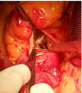
Cardiopulmonary bypass was established by cannulation of the ascending aorta and the right atrium with two-stage cannula. Aortotomy was done with oblique incision and fibromembraneus tissue resected surgically with preservation of her aortic valve without any complications (Figure 4). After resection of membrane, suturing over and over aortotomy and heart started to bit, TEE performed to check the gradient and any residual membrane and there was not any pathological finding. Her complaints reduced at less than one week after surgery. Before discharge TTE showed no significant findings. She was discharged at the 8th day after operation and she had no significant symptoms at her 6 -week follow-up visit.
Fig. 4. Surgical view of fibrous membrane.
Discussion
The etiologic characteristics, prevalence, therapeutic options for DSS in adults have not been established well (3,5). DSS is more common in children and accounts nearly 20% of all cases of LVOT undergo to surgery (5). DSS is a rare pathology and its etiology is unknown. It is one
Akgül et al. / Late Onset Subaortic Fibrous Membrane
Van Tıp Derg Cilt:24, Sayı:4, Ekim/2017 371
of the reasons for LVOT obstruction and rapidly hemodynamic progression. Also it causes secondary aortic regurgitation. Although it can occur as a isolated lesion it may accompany with any other congenital diseases (1,6,7) such as accessory mitral valve tissue, abnormal left ventricular papillary muscle, anomalous muscular band, bicuspid aortic valve, atrioventricular canal defects, ventricular septal defect, interrupted aortic arch, patent ductus arteriosus, coarctation of the aorta, persistent superior left vena cava (1,6,8,9). DSS causes morphologic abnormalities at LVOT. This status includes increased aorto-mitral fibrous distance, arised aorto-septal angle, malalignment ventricular septal defect and increased turbulence (10). Then septal shear stress generate proliferative reaction at endocardium and development of subaortic membrane (3,6,7) At the final producing of left ventricular hypertrophy and aortic regurgitation are inevitable (6,8).
In adults DSS may occur with many different symptoms. For example syncope, palpitation, dyspnea, chest pain but it may be asymptomatic too. Diagnosis can be made by echocardiography (6). A cohort study at 4 centers with 149 patients showed that DSS progressed slowly in adult. The baseline gradient measured 32.3±17 mmHg and <1mmHg rising per year. But if the gradient was more than 50mmHg and continue to increase, had severe AR were acceptable predictors for surgery (3). Timing for surgical treatment is controversial (6,8). But if left ventricular hypertrophy and AR started surgery must be the choice (10). At rapid progression on LVOT obstruction early resection of subaortic membrane with surgery may be recommended (4). Surgical treatment may range from excision of membrane to myectomy or aortic valve replacement (9). In our case subaortic membrane resected and aortic valve preservated. DSS causes aortic stenosis in adults and it must be kept in mind. Treatment with surgery has low morbidity and mortality risk. But the main determinative point is the anatomic findings of the lesion. Long term surgery result is not presumable
for recurrent. So follow-up after surgery is compulsory.
References
1. Marasini M, Zannini L, Ussia GP, Pinto R, Moretti R, Lerzo F, et al. Discrete subaortic stenosis: incidence, morphology and surgical impact of associated subaortic anomalies. Ann Thorac Surg 2003; 75(6): 1763-1768.
2. Erdal AC, Silistreli E, Catalyurek H, Albayrak G, Aykut K, Acıkel U. Timing of surgical treatment in subaortic stenosis. Turk Gogus Kalp Dama 2004; 12(3): 169-171.
3. Van der Linde D, Takkenberg JJ, Rizopoulos D, Heuvelman HJ, Budts W, van Dijk AP, et al. Natural history of discrete subaortic stenosisin adults: a multicentre study. Eur Heart J 2013; 34(21): 1548-1556.
4. Ozsin Kadir K, Toktas F, Sanri Umut S, Yavuz S. Discrete subaortic stenosis in an adult patient eurj.2016.2.1.66.
5. Butany J, Vaideeswar P, David TE. Discrete subaortic membranes in adults--a clinicopathological analysis. Cardiovasc Pathol 2009; 18(4): 236-242.
6. Qureshi A, Awuor S, Martinez M. Adult
presentation of subaortic stenosis: another great hypertrophic cardiomyopathy mimic. Heart Lung Circ 2015; 24(1): 7-10.
7. Silbiger JJ. The role of shear stress in the pathogenesis of discrete subaortic stenosis: implications for surgical treatment. J Heart Valve Dis 2011; 20(2): 123-128.
8. Huang JJ, Azakie A, Russell IA. Echo rounds: discrete subvalvular aortic stenosis. Anesth Analg 2010; 110(4): 1003-1005.
9. Hraska V, Photiadis J, Arenz C. Surgery for subvalvar aortic stenosis resection of discrete subvalvar aortic membrane. Multimed Man Cardiothorac Surg 2007; 007(723): mmcts.2006.002303. 10. Oliver JM, Gonzalez A, Gallego P,
Sanchez-Recalde A, Benito F, Mesa JM. Discrete subaortic stenosis in adults: Increased prevalence and slow rate of progression of the obstruction and aortic regurgitation. J Am Coll Cardiol 2001; 38(3): 835-342.