1 Dumlupınar University Evliya Celebi Hospital, Obstetrics and Gynecology Department Kütahya, Turkey 2 Şişli Etfal Training and Research Hospital, Obstetrics and Gynecology Department İstanbul, Turkey
3 Nenehatun Obstetrics and Gynecology Hospital, Erzurum, Turkey Yazışma Adresi /Correspondence: Suna Kabil Kucur,
Dumlupinar University Evliya Celebi Training and Research Hospital, Obstetrics and Gynecology Department, Kutahya, Turkey Email: dr.suna@hotmail.com
Geliş Tarihi / Received: 27.01.2013, Kabul Tarihi / Accepted: 16.05.2013 Copyright © Dicle Tıp Dergisi 2013, Her hakkı saklıdır / All rights reserved
ORIGINAL ARTICLE / ÖZGÜN ARAŞTIRMA
Contribution of spiral artery blood flow changes assessed by transvaginal color
Doppler sonography for predicting endometrial pathologies
Endometrial patolojileri öngörmede transvajinal renkli Doppler ultrasonografi ile belirlenen
spiral arter akım değişikliklerinin katkısı
Suna Kabil Kucur1, Alev Atış Aydın2, Osman Temizkan2, İlay Gözükara3, Eda Ülkü Uludağ3,
Canan Acar2, İnci Davas2 ÖZET
Amaç: Endometrial patolojilerin değerlendirilmesinde
transvajinal renkli Doppler ultrasonografi (RDU) ile spiral arter akım parametrelerinin katkısını araştırmak.
Yöntemler: Anormal uterin kanama ile başvurmuş ve
endometrial değerlendirme ihtiyacı olan 97 hastanın ka-tıldığı prospektif gözlemsel bir çalışmadır. Endometrial kalınlık, yapı ve ekojenite kaydedildi. Transvajinal RDU ile spiral arter pulsatilite indeksi (Pİ) ve rezistif indeksi (Rİ) ölçüldü. Tüm olgulara endometrial örnekleme yapıldı. Ult-rasonografik ve histopatolojik bulgular karşılaştırıldı.
Bulgular: Histopatolojik tanılar; 39 olgu (%40,2)
endo-metrial polip, 9 olgu (%9,3) endoendo-metrial hiperplazi, 10 olgu (%10,3) submuköz myom, 7 olgu (%7,2) endomet-rium kanseri, 32 olgu (%33) nonspesifik bulgular. En-dometrial kanser olgularında spiral arter Pİ istatistiksel olarak yüksek anlamlılıkta düşük bulundu (p<0,01). spiral arter Rİ da malign histopatoloji olan olgularda anlamlı ola-rak düşük bulundu (p<0,05).
Sonuç: Endometrial patolojiler endometrial spiral arter
Doppler değişiklikleriyle ilişkilidir.
Anahtar kelimeler: Spiral arter, Doppler ultrasonografi,
endometrium.
ABSTRACT
Objective: To investigate the diagnostic value of blood
flow measurements in spiral artery by transvaginal color Doppler sonography (CDS) in predicting endometrial pa-thologies.
Methods: Ninety-seven patients presenting with
abnor-mal uterine bleeding and requiring endometrial assess-ment were included in this prospective observational study. Endometrial thickness, structure and echogenicity were recorded. Pulsatility index (PI) and resistive index (RI) of the spiral artery were measured by transvaginal CDS. Endometrial sampling was performed for all sub-jects. Sonographic and hystopathologic findings were compared.
Results: The histopathological diagnoses were as
fol-lows; 39 cases (40.2%) endometrial polyp, 9 cases (9.3%) endometrial hyperplasia, 10 cases (10.3) submu-cous myoma, 7 cases (7.2%) endometrium cancer, and 32 cases (33%) nonspecific findings. The spiral artery PI in endometrium cancer group was highly significantly lower than other groups (p<0.01). The spiral artery RI was also significantly lower in the patients with malignant his-tology (p<0.05).
Conclusion: Endometrial pathologies are associated
sig-nificantly with endometrial spiral artery Doppler changes.
Key words: Spiral artery, Doppler ultrasonography,
INTRODUCTION
Abnormal uterine bleeding (AUB) is one of the most commonly encountered problems in gyneco-logic practice. Transvaginal sonography (TVS) has become first-line diagnostic tool for patients with abnormal uterine bleeding. It has significantly im-proved our ability to diagnose uterine pathologies accurately. However, we still need second stage invasive tests that cause patient discomfort and in-crease the cost compared to TVS for accurate diag-nosis. Over recent years color Doppler sonography (CDS) have been started to be used to predict endo-metrial pathologies [1,2]. Color Doppler sonogra-phy, a noninvasive and simple tool, is useful in dis-tinguishing endometrial lesions, helps us to decide what is necessary for invasive tests and plans the invasive method to be chosen.
Endometrial cancer is the most common malig-nancy of the female genital tract [3]. Endometrial thickness is a nonspecific finding of endometrial cancer [4]. Hence, CDS of the genital vessels can improve the sensitivity and specificity of TVS for the prediction of endometrial malignancies [5,6].
In this prospective study, we investigated the diagnostic value of the sub endometrial spiral artery blood flow parameters for the prediction of underly-ing endometrial pathology.
METHODS
The study was carried out at Şişli Etfal Training and Research Hospital. Ninety-seven patients present-ing with abnormal uterine bleedpresent-ing and requirpresent-ing endometrial assessment were included in this pro-spective observational study.
Exclusion criteria were pregnancy, pelvic in-flammatory disease, cervicitis, genital tumor, sys-temic diseases causing abnormal uterine bleeding, intrauterine device use and use of drugs affecting uterine vasculature such as hormonal therapy, oral contraceptives or tamoxifen during the previous 3 months,
The study was conducted according to the guidelines for clinical studies described in the Dec-laration of Helsinki (as revised by the World Medi-cal Association, http://www.wma.net). Regional Ethical Committee approved the study. All patients
gave oral and written informed consent prior to the examination.
The patients were examined prospectively by standard B-mode TVS and CDS in the midfollicular phase. All patients were scheduled for an invasive diagnostic procedure like dilatation-curettage or hysteroscopy after sonographic imaging. Histopath-ologic examination was performed in the pathology laboratory of our hospital. All ultrasound scans were performed by the same examiner to avoid interob-server variability. All women were examined firstly using conventional gray-scale TVS with a 5.0-MHz transvaginal probe in the lithotomy position with an empty bladder. The uterus was thoroughly assessed in coronal and sagittal planes using a Siemens Acu-son Antares 4D machine. Endometrial double layer thickness, structure and echogenicity were noted. Endometrial double layer width is measured at the thickest portion of the longitudinal section. Then, vascularization of the uterus is visualized with color Doppler technique. Blood flow velocity waveforms were evaluated in the spiral arteries at the sub endo-metrial region that is within 1 mm of the originally defined myometrial-endometrial contour [7].
The results of the examinations were compared with the histologic diagnosis of the endometrial specimen. The primary outcome measures were spiral artery Pulsatility index (PI) and spiral artery resistive index (RI).
Statistical analysis
Statistical calculations were undertaken using the Number Cruncher Statistical System (NCSS) 2007& PASS 2008 Statistical Software (Utah, USA). Cat-egorical data were compared using the chi-square test. Kruskal Wallis test was used to compare the parameters with abnormal dissociation between the groups for descriptive statistical methods as well as quantitative data. A result was assumed to be statis-tically significant if the P value of each respective test was ≤ 0.05.
RESULTS
Ninety-seven patients who had admitted with AUB were enrolled in this prospective study. Mean age, parity, and endometrial thickness of the participants were 45.11±2.64 years, 3.22±2.16, and 12.56±7.43 mm respectively (Table 1).
Table 1 summarizes the histopathological diag-noses of the subjects. The histopathological diagno-ses were as follows; 39 cadiagno-ses (40.2%) endometrial polyp, 9 cases (9.3%) endometrial hyperplasia, 10 cases (10.3) submucous myoma, 7 cases (7.2%) en-dometrium cancer, and 32 cases (33%) nonspecific findings ( Table 2). Secretory endometrium, prolif-erative endometrium, and atrophic endometrium on histology were considered as nonspecific findings.
On gray scale ultrasonography 35 (36.1%) had uniform endometrium whereas 62 (63.9%) had no uniform endometrium. No uniform endometrium showed a strong correlation with organic endome-trial lesions. All cases with endomeendome-trial cancer had no uniform endometrium. Endometrial double layer thickness was significantly greater in patients with endometrial hyperplasia and cancer than in those with nonspecific pathologies (p<0.05).
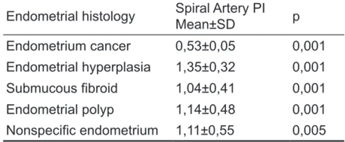
There were significant correlations between spiral artery PI and RI and different endometrial histologies. Table 3 and Table 4 summarize the comparison of the spiral artery Doppler indices between endometrial cancer and different endome-trial histopathologies. In patients with endomeendome-trial cancer spiral artery PI was found to be significantly lower than other groups (p<0.01). Spiral artery RI was also lower in endometrial cancer group than in endometrial polyp, hyperplasia and fibroid group (p<0.05).
Table 1. Demographic and clinical variables of the study
group Mean ± SD Age (years) 45,11±2.64 Gravity 4.4 ± 2.64 Parity 3.2 ± 2.16 Hemoglobin concentration (g/dl) 11.75 ± 1.83 Endometrial thickness (mm) 12,56±7,43 SD: Standard deviation
Table 2. Histological diagnoses of all patients
Endometrial histology n % Endometrial polyp 39 40,2 Endometrial hyperplasia 9 9,3 Submucous fibroid 10 10,3 Endometrium cancer 7 7,2 Nonspecific findings 32 33
Table 3. Comparison of the pulsatility indices (PI) of spiral
artery between endometrial cancer and other pathologies Endometrial histology Spiral Artery PIMean±SD p
Endometrium cancer 0,53±0,05 0,001 Endometrial hyperplasia 1,35±0,32 0,001 Submucous fibroid 1,04±0,41 0,001 Endometrial polyp 1,14±0,48 0,001 Nonspecific endometrium 1,11±0,55 0,005 SD: Standard deviation
Table 4. Comparison of the spiral artery resistive indices
between endometrial cancer and other pathologies Endometrial histology Spiral Artery RIMean±SD p
Endometrium cancer 0,44±0,06
Endometrial hyperplasia 0,70±0,11 0,001
Submucous fibroid 0,56±0,15 0,033
Endometrial polyp 0,59±0,17 0,005
Nonspecific endometrium 0,55±0,18 0,052
RI: Resistive index, SD: Standard deviation
DISCUSSION
Histological examination of the endometrium is gold standard in the diagnosis of endometrial pa-thologies. However, with the advance of high reso-lution ultrasound, studies on noninvasive evaluation of the uterine cavity have dramatically increased. Many studies showed thicker endometrium on gray-scale sonography in neoplastic endometrial lesions [8-11]. However endometrial thickening on TVS is a nonspecific finding, therefore secondary tests are usually required to reduce false positive results. Besides, a cut-off value of 4-5 mm to distinguish benign from malignant endometrial lesions did not have high sensitivity and specificity to replace in-vasive methods in postmenopausal women [12-14]. Color Doppler ultrasonography of uterine and en-dometrial vessels can be used to increase the diag-nostic value of gray- scale TVS. In this study we compared the Doppler indices of uterine and spiral arteries with the final histologic diagnoses.
There are conflicting reports on the Doppler as-sessment of genitalia in differentiating uterine pa-thologies in the literature. The studies in the litera-ture reveal controversial results in utilizing Doppler ultrasound to predict uterine malignancies.
In recent years, many studies have been report-ed in gynecologic Doppler ultrasound assessment of the uterine cavity. Ernest et al studied the relation-ship between uterine blood flow and endometrial and sub endometrial blood flows during stimulated and natural cycles [15]. Many authors have also searched the effect of different medications on uter-ine artery and endometrial blood flow [16-18].
Lower impedance to blood flow in tumoral tis-sues has leaded authors to use Doppler analysis of genital vessels to differentiate malignant lesions from benign lesions. A few authors have studied the role Doppler indices of genital vessels in differenti-ation of premalignant and malignant endometrial le-sions. Some reported Doppler ultrasound to be use-ful but others limited to the values [6,8,11,14,19,20]. The characteristics of endometrial and myometrial vascularization on Doppler sonography were then searched. A few discriminatory vascular patterns have been attributed to endometrial polyps, fibroids and endometrial carcinoma [21-23].
Samulak et al., evaluated uterine artery maxi-mum end-diastolic velocity of blood flow, time-av-eraged maximum velocity (TAMXV) of blood flow, and peak systolic velocity of blood flow in women with postmenopausal bleeding. Although statisti-cally insignificant, these values were found to be highest in the carcinoma group and lowest in the control group [24]. That reflects lower impedance to blood flow in cancerous lesions. Englert-Golon et al., reported significantly lower PI and RI in the en-dometrial vessels and uterine arteries, significantly higher TAMXV in the endometrial vessels and uter-ine arteries in cases with endometrial cancer than in patients with endometrial hyperplasia [25].
The present study showed a correlation be-tween the spiral artery PI and RI and endometrial malignancy. In patients with endometrial carcino-ma, spiral artery PI and RI were both significantly lower than those without malignant histology. En-dometrial thickness was significantly higher in pa-tients with endometrial carcinoma and hyperplasia.
Weiner et al reported that the sensitivity of the uterine artery RI was 100% when a cut-off value of 0.83 was considered in patients with endometri-al cancer [26]. Kurjak et endometri-al., reported significantly lower RI near or <0.40 in cases with endometrial carcinoma. They also said that transvaginal color
myometrial invasion and tumor staging [27]. Bezir-cioglu et al., defined the endometrial thickness of 5 mm, uterine artery PI of 1,450, uterine artery RI of 0.715, radial artery PI of 1.060, radial artery RI of 0.645 as the cut-off points for malignant endome-trium in postmenopausal women [14].
Arslan et al., investigated the role of transvagi-nal color Doppler ultrasonography for the predic-tion of precancerous endometrial lesions. They con-cluded that transvaginal ultrasonography and Dop-pler ultrasonography cannot replace the invasive procedures [8].
A possible limitation of our study is small study population, but strength of this study is that all cases underwent histopathological diagnosis.
In conclusion, endometrial pathologies are as-sociated significantly with endometrial spiral artery changes. Although in patients with malignant en-dometrial lesions blood flow of the spiral arteries displayed lower impedance, the Doppler ultrasound use as a diagnostic test is not accepted now. How-ever, with advancing technology, color Doppler so-nography can replace the invasive diagnostic meth-ods for endometrial pathologies.
REFERENCES
1. Chan FY, Chau MT, Pun TC, et al. Limitations of transvagi-nal sonography and color Doppler imaging in the differ-entiation of endometrial carcinoma from benign lesions. J Ultrasound Med 1994;13:623-628.
2. Kanat-Pektas M, Gungor T, Mollamahmutoglu L. The evalu-ation of endometrial tumors by transvaginal and Doppler ultrasonography. Arch Gynecol Obstet 2008;277:495-499. 3. Epstein E, Skoog L, Isberg PE, et al. An algorithm
includ-ing results of gray-scale and power Doppler ultrasound examination to predict endometrial malignancy in women with postmenopausal bleeding. Ultrasound Obstet Gynecol 2002;20:370–376.
4. Develioglu OH, Bilgin T, Yalcin OT, Ozalp S. Transvaginal ultrasonography and uterine artery Doppler in diagnosis endometrial pathologies and carcinoma in postmenopausal bleeding. Arch Gynecol Obstet 2003;268:175-180. 5. Wilailak S, Jirapinyo M, Theppisai U. Transvaginal Doppler
sonography: is there a role for this modality in the evalu-ation of women with postmenopausal bleeding? Maturita 2005;50:111-116.
6. Wilczak M, Samulak D, Englert-Golon M, Pieta B. Clinical usefulness of evaluation of quality parameters of blood flow: pulsation index and resistance index in the uterine arteries in the initial differential diagnostics of pathology within the endometrium. Eur J Gyneacol Oncol 2010;31:437-439. 7. Ng EH, Chan CC, Tang OS, et al. Endometrial and
subendo-three-dimensional Power Doppler ultrasound in excessive ovarian responders. Hum Reprod 2004;19:924-931. 8. Arslan M, Erdem A, Erdem M, et al. Transvaginal color
Dop-pler ultrasonography for the prediction of precancerous en-dometrial lesions. Int J Gynecol Obstet 2003;80:299-306. 9. Gull B, Carlsson S, Karlsson B, et al. Transvaginal
ultraso-nography of the endometrium in women with postmeno-pausal bleeding: is it always necessary to perform an endo-metrial biopsy? Am J Obstet Gynecol 2000;182:509-515. 10. Briley M, Lindsell DR. The role of transvaginal ultrasound
in the investigation of women with post-menopausal bleed-ing. Clin Radiol 1998;53:502–505.
11. Sladkevicius P, Valentin L, Marsal K. Endometrial thick-ness and Doppler velocimetry of the uterine arteries as dis-criminators of endometrial status in women with postmeno-pausal bleeding: a comparative study. Am J Obstet Gynecol 1994;171:722-728.
12. Conoscenti G, Meir YJ, Fischer-Tamaro L, et al. Endome-trial assessment by transvaginal sonography and histologi-cal findings after D & C in women with postmenopausal bleeding. Ultrasound Obstet Gynecol 1995;6:108-115. 13. Amit A, Weiner Z, Ganem N, et al. The diagnostic value
of power Doppler measurements in the endometrium of women with postmenopausal bleeding. Gynecol Oncol 2000;77:243-247.
14. Bezircioglu I, Baloglu A, Cetinkaya B, et al. The diagnostic value of the Doppler ultrasonography in distinguishing the endometrial malignancies in women with postmenopausal bleeding. Arch Gynecol Obstet 2012;285:1369-1374. 15. Ng EH, Chan CC, Tang OS, et al. Relationship between
uterine blood flow and endometrial and subendometrial blood flows during stimulated and natural cycles. Fertil Steril 2006;85:721-727.
16. Haliloglu B, Celik A, Ilter E, et al. Comparison of uterine artery blood flow with levonorgestrel intrauterine system and copper intrauterine device. Contraception 2011;83:578-581.
17. Dane B, Akca A, Dane C, et al. Comparison of the effects of the levonorgestrel-releasing intrauterine system (Mi-rena) and depot-medroxyprogesterone acetate (Depo-Pro-vera) on subendometrial microvascularisation and uterine artery blood flow. Eur J Contracept Reprod Health Care 2009;14:240-244.
18. Jiménez MF, Arbo E, Vetori D, et al. The effect of the le-vonorgestrel-releasing intrauterine system and the copper intrauterine device on subendometrial microvascularization
and uterine artery blood flow. Fertil Steril 2008;90:1574-1578.
19. Dragojevic S, Mitrovic A, Dikic S, Canovic F. The role of transvaginal colour Doppler sonography in evalua-tion of abnormal uterine bleeding. Arch Gynecol Obstet 2005;271:332–335.
20. Epstein E, Valentin L. Gray-scale ultrasound morphology in the presence or absence of intrauterine fluid and vas-cularity as assessed by color Doppler for discrimination between benign and malignant endometrium in women with postmenopausal bleeding. Ultrasound Obstet Gynecol 2006;28:89-95.
21. Alcazar JL, Castillo G, Minguez JA , Galan MJ. Endome-trial blood flow mapping using transvaginal power Dop-pler sonography in women with postmenopausal bleeding and thickened endometrium. Ultrasound Obstet Gynecol 2003;21:583-588.
22. Opolskiene G, Sladkevicius P and Valentin L. Ultrasound assessment of endometrial morphology and vascularity to predict endometrial malignancy in women with postmeno-pausal bleeding and sonographic endometrial thickness ≥4.5 mm. Ultrasound Obstet Gynecol 2007;30:332-340. 23. Cil A.P, Tulunay G, Kose MF, Haberal A. Power
Dop-pler properties of endometrial polyps and submucosal fi-broids: a preliminary observational study in women with known intracavitary lesions. Ultrasound Obstet Gynecol 2010;35:233-237.
24. Samulak D, Wilczak M, Englert-Golon M, Michalska MM. The diagnostic value of evaluating the maximum velocity of blood flow in the uterine arteries of women with post-menopausal bleeding. Arch Gynecol Obstet 2011;284:1175-1178.
25. Englert-Golon M, Szpurek D, Moszyński R, et al. Clinical value of the measurement of blood flow in uterine arter-ies and endometrial vessels in women with postmenopausal bleeding using “power” angio Doppler technique. Ginekol Pol 2006;77:759-763.
26. Weiner Z, Beck D, Rottem S, et al. Uterine artery flow ve-locity waveforms and color flow imaging in women with perimenopausal and postmenopausal bleeding. Correlation to endometrial histopathology. Acta Obstet Gynecol Scand 1993;72:162-166.
27. Kurjak A, Shalan H, Sosic A, et al. Endometrial carcino-ma in postmenopausal women: evaluation by transvagi-nal color Doppler ultrasonography. Am J Obstet Gyncol 1993;169:1597-1603.
