KADIR HAS UNIVERSITY
GRADUATE SCHOOL OF SCIENCE AND ENGINEERING
IN SILICO SCREENING OF NEURONAL NITRIC OXIDE
SYNTHASE ENZYME INHIBITORS
GRADUATE THESIS
BAHANUR ÖRTMEN
Bahanur Ö rtmen M .S . T hesis 20 14
IN SILICO SCREENING OF NEURONAL NITRIC OXIDE
SYNTHASE ENZYME INHIBITORS
Bahanur ÖRTMEN
Submitted to the Graduate School of Science and Engineering
in partial fulfillment of the requirements for the degree of
Master of Science
in
COMPUTATIONAL BIOLOGY AND BIOINFORMATICS
KADIR HAS UNIVERSITY
IN SILICO SCREENING OF NEURONAL NITRIC OXIDE SYNTHASE
ENZYME INHIBITORS
ABSTRACT
BAHANUR ÖRTMEN
Master of Science in Computational Biology and Bioinformatics May, 2014
Three closely related isoforms of nitric oxide synthases (NOS) catalyze an
important secondary messenger nitric oxide (NO) synthesis through oxidation of
L-arginine to L-citrulline. These three NOS isoforms takes parts in different tissues for
various physiological and pathological processes. Neuronal NOS (nNOS) produce
NO in central and peripheral nervous system, endothelial NOS (eNOS) plays role in
endothelial cells and NO in macrophage cells is produced by inducible NOS (iNOS).
Excessive NO production in nervous cells following pathological conditions is
observed. Dysregulation of NO, therefore, may force NO to act as a neurotoxin that
causes several neurodegenerative diseases including Parkinson’s, Alzheimer’s,
Huntington’s diseases. Considering all these facts, developing a selective and good
potential inhibitor for nNOS is a compulsory task to achieve. However, among all
AP PE
isoforms there is high active site conservation so that no drug that shows these
desired properties has yet been designed and developed.
In this present work, virtual screening techniques were applied to design
selective nNOS inhibitors. Molecular modeling studies were done using already
known crystal structures of all three isoforms. First of all, to find primary lead
candidates, several hundred compounds were screened via ZINCv12 lead library.
Then, modifications were done on the selected scaffolds via de novo design method
to derive our inhibitor candidates. AutoDock 4.02 docking virtual tool was employed
for docking and scoring of inhibitor candidates. Inhibition constants and best pose
predictions of docked ligands within the active sites of three isoforms were
considered for further examinations and comparison analysis. Already bound ligands
in downloaded experimentally determined X-ray structures of all isoforms were
re-docked to crosscheck our studies. In this thesis two lead scaffolds among all and 22
inhibitor candidates derived from these two scaffolds were selected to discuss for
optimization for further development of best potential and selective inhibitor for
nNOS.
SEÇİCİ NİTRİK OKSİT SENTAZ ENZİM İNHİBİTÖRLERİNİN BİLGİSAYAR
ORTAMINDA TARANMASI
ÖZET
BAHANUR ÖRTMEN
Hesaplamalı Biyoloji ve Biyoinformatik, Yüksek Lisans May, 2014
Nitrik oksit sentaz enziminin (NOS) üç izoformu, L-arjininin L-sitruline
oksidasyonu ile önemli bir ikincil haberci olan nitric oksit (NO) sentezler. NOS’un
bu üç izoformu çeşitli fizyolojik ve patolojik süreçte farklı dokularda yer almaktadır.
Nöronal NOS (nNOS) merkezi ve periferik sinir sisteminde, endotelyal NOS (eNOS)
endotel hücrelerinde ve indüklenebilen NOS (iNOS) ise makrofaj hücrelerinde NO
sentezini katalizlemektedir. Parkinson, Alzheimer, Huntington hastalığı gibi çeşitli
nörodejeneratif hastalıklarda aşırı NO üretimi görülebilir ve uygunsuz NO
regülasyonu, NO’nun nörotoksin gibi davranmasına neden olabilir. Tüm bu bulgular
değerlendirildiğinde, nNOS’a seçici ve nNOS üzerinde güçlü inhibitör etki gösteren
ilaçların geliştirilmesinin gerekli olduğu görülmektedir. Ancak NOS izoformlarının
aktif bölgesinde yüksek derecede benzerlik bulunmasından dolayı bu istenilen
Bu yapılan çalışmada, selektif nNOS inhibitörü tasarlamak amacıyla
bilgisayar ortamında sanal tarama teknikleri uygulanmıştır ve NOS’un üç
izoformunun bilinen kristal yapıları kullanılarak moleküler modelleme çalışmaları
yapılmıştır. İlk olarak, öne çıkan küçük molekül iskeletlerini bulmak için ZINCv12
parçaçık kütüphanesi aracılığı ile yüzlerce bileşik taranmıştır. Daha sonra, inhibitör
adaylarının türetilmesi için de novo tasarım yöntemleriyle seçilen modellerde
modifikasyonlar yapılmıştır. Adayların inhibitör derecesi ve inhibisyon bölgesine
hedeflendirilmesi için Autodock 4.02 hedeflendirme aracı kullanılmıştır. İnhibisyon
katsayıları ve hedeflendirilen ligandın üç izoformun aktif bölgelerindeki en iyi
konumu tetkik ve karşılaştırma analizleriyle değerlendirilmiştir. Çalışmalarımızı
doğrulamak için izoformların deneysel sonuçlardan elde edilen X-ışını yapılarına
bağlı ligandlar AutoDock ile yeniden hedeflendirilmiştir. Bu çalışmada tasarlanan
öncü iskeletlerden 2 tanesi ve bu 2 iskeletten türetilen inhibitor adaylarının 22 tanesi,
en güçlü selektif nNOS inhibitörü optimizasyonunun tartışılması için belirlenmiştir.
Anahtar Kelimeler:Nitrik oksit sentaz, eNOS, iNOS, nNOS, de novo dizayn, ilaç
Acknowledgements
First and foremost, I would like to express my sincere and deepest appreciate
to my advisor Prof. Kemal Yelekçi. Thanks for giving me the opportunity to be part
of this group, for his immense knowledge, understanding, encouragement and
continuous support of my master’s study and my research. It was an honor to work
with him. I could not have imagined having a better advisor and mentor for my
scientific study.
Besides my advisor, I would also like to express my gratitude to Assoc. Prof.
Ebru Demet Akdoğan, Dr. Tuğba Arzu Özal and Assist. Prof. Şebnem Eşsiz Gökhan
for their continuous support and encouragement.
I also would like to thank to Mr. Serkan Altuntaş for supporting and
motivating me during entire period of my study.
Last but not least, I owe more than thanks to my precious family for their
unconditional love, endless support and believing in me throughout my life. And I
Table of Contents
Abstract………..vi
Özet………...viii
Acknowledgement………..x
1 Introduction 1
1.1 Nitric Oxide Synthase and Nitric Oxide………...1
1.2 Structure of Nitric Oxide Synthases……… 3
1.3 Regulation and Function of Nitric Oxide Synthases………6
1.4 Neurodegenerative Diseases and nNOS inhibition………..7
1.5 Drug Design and In Silico Approach……….11
1.6 Selective Inhibition of Neuronal Nitric Oxide Synthase………16
2 Materials and Methods 18
2.1 Ligands and Enzyme Preparations……….………18
2.2 Generation of Potential Inhibitors………..20
2.2.1 Test Inhibitors……….20
2.2.2 Inhibitor Candidates………...21
2.3 Docking………..25
3 Results and Discussion 26
Conclusion……….46
List of Tables
Table 1 : List of Some Commercially Available VS Tools and Their Vendors...15
Table 2 : Some Residues in Three Isoforms’ Active Sites……….18
Table 3 : Lead IDs of First Group of Potential Inhibitors and Corresponding
Functional Groups………22
Table 4 : Lead IDs of Second Group of Potential Inhibitors and Corresponding
Functional Groups………24
Table 5 : Chemical Structures of 22 Designed Potential NOS Inhibitors and Their
List of Figures
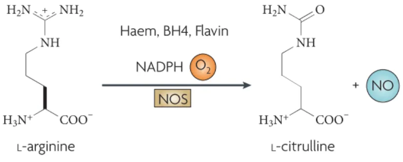
Figure 1 : Chemical Reaction of NO Synthesis carried by NOS……….1
Figure 2 : Roles of Nitric Oxide in Central and Peripheral Nervous System………..2
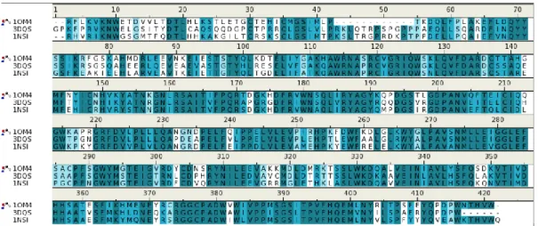
Figure 3 : Alignment of Amino Acid Residues of Three NOS Isoforms……….4
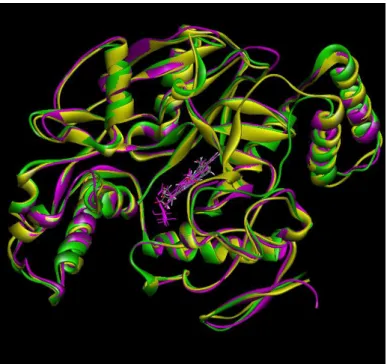
Figure 4 : Superposition of Chain A Backbone of 1OM4 (nNOS), 3DQS (eNOS) and 1NSI (iNOS)………...…5
Figure 5 : General Structure of NOS enzymes……….5
Figure 6 : Mechanism of Nitric Oxide Synthases………7
Figure 7 : Neurotoxic Effects of Nitric Oxide………10
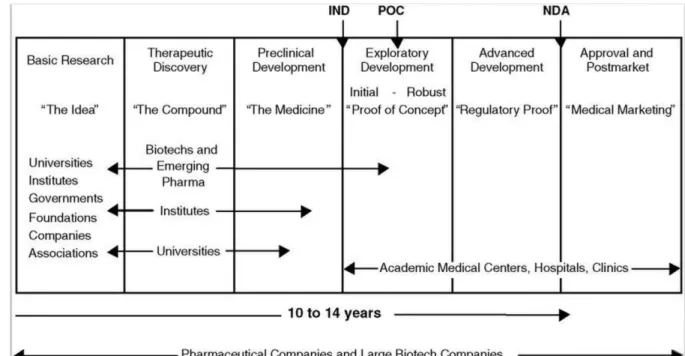
Figure 8 : Biomedical Research from Idea to Market………12
Figure 9 : In silico Approach during Drug Discovery Process………..13
Figure 10 : Designed and Synthesized Ligands by Richard B. Silverman…………21
Figure 11 : First Scaffold Used in This Study………22
Figure 12 : Second Scaffold Used in This Study………...23
Figure 13 : Plots of Experimentally and Computationally Obtained Inhibition Constant Values (Ki)………29
Best-1 INTRODUCTION
1.1 Nitric Oxide Synthase and Nitric Oxide
Over three decades, structure of nitric oxide synthase, function of nitric oxide
synthases (NOS) and inhibition of NOS enzyme are important subjects for many
researches since it synthesizes an important signaling molecule in various tissues,
nitric oxide (NO). 1 NOS catalyzes a NADPH- dependent formation of NO and
citrulline from L-arginine. 2, 3 ( Figure 1)
Figure 1 | Chemical Reaction of NO synthesis carried by NOS. 3
This free radical gas, nitric oxide is produced essentially in endothelials,
macrophages and neuronal cells with the reaction in Figure 1 carried by three
isoforms of NOS enzyme; endothelial NOS (eNOS), neuronal NOS (nNOS) and
and Ca2+ - dependent isoforms. Whereas there is another isoform Ca2+ independent
that depends on mechanism of action, inducible NOS (iNOS). 4
All three isoforms take part in many important physiological and
pathophysiological processes in mammalian cells. iNOS- derived NO in macrophage
cells has important role as cytotoxic agent to destroy pathogens and microorganisms
during immune and inflammatory response. eNOS-derived vascular NO plays
significant role in controlling vascular protection such as blood pressure, protection
from platelet aggregation. 5 Main function of nNOS-derived NO is releasing
neurotransmitters and nNOS-derived NO has been indicated in Figure 2 showing
central effects and peripheral effects. The nNOS-derived NO has important roles in
various synaptic signaling, synaptogenesis events and in modulation of actions such
1.2 Structure of Nitric Oxide Synthases
Common three isoforms of NOS are inducible NOS, endothelial NOS and
neuronal NOS. In different chromosomes, there are three distinct genes coding for
three isoforms. NOS1 gene corresponding to nNOS protein has 29 exons and 28
introns found on chromosome 12. NOS2 gene corresponding to iNOS found on
chromosome 17 and NOS3 gene corresponding to eNOS found on chromosome 7
with 26 exons and 25 introns. However, all isoforms have almost same genomic
structures. 1 NOS enzymes are generally found as dimer structure and each monomer
generally consist of 420 to 430 amino acids. In Figure 3, amino acid sequences of
one domain of all isoforms were aligned to show similarity. Different brightness of
green color shows similarity degrees. For this alignment and for our project, PDB
structures with 1OM4 code for nNOS structure, 3DQS for eNOS structure and
Figure 3 | Alignment of Amino Acid Residues of Three NOS Isoforms. Dark green colors correspond to exact matching residues; light green colors show partial similarity and white colors correspond to mismatch residues. (Isoforms were aligned using Discovery Studio 3.0)
In Figure 4, only one domains of isoforms were excreted and backbones of
these domains superpositoned to show structural homology between isoforms. All
isoforms consist oxygenase domain in amino-terminal of protein and reductase
domain in carboxy-terminal and also between these domains there is a
calmodulin-binding region. Flavin adenine dinucleotide (FAD), flavin mononucleotide (FMN)
and NADPH binding sites are found in C-terminal reductase domain. And in
N-terminal oxygenase domain, there are L-arginine, heme and BH4 binding sites.
Figure 4 | Superposition of Chain A Backbone of 1OM4(nNOS), 3DQS(eNOS) and
1NSI(iNOS). Yellow, green and magenta colors correspond to 1OM4, 3DQS and 1NSI
respectively. ( Isoforms were superpositioned using Discovery Studio 3.0)
Figure 5 | General Structure of NOS enzymes. Structure is shown in as dimerized enzyme. Monomer at the top shows domains of NOS, monomer at the bottom highlights binding sites
1.3 Regulations and Function of Nitric Oxide Synthases
In all isozymes, flavins FMN, FAD and BH4 play role as cofactors in the
mechanism of nitric oxide synthesis. An Active NOS enzyme transfers electrons
from NAPDH to flavins, FAD and FMN in carboxy-terminal reductase domain and
then, as a result of conformational changes followed by calmodulin binding,
electrons are transferred to heme in amino-terminal oxygenase domain. These
transferred electrons are used to reduce molecular oxygen to superoxide (O2-) and
L-arginine is oxidized to L-citrulline, then NO is produced in oxygenase domain.
Heme is important for dimerization of NOS enzymes to set functional enzyme and
thereby takes part in BH4 and L-arginine binding. And calmodulin, which binds a
region between reductase and oxygenase domain, promotes for electron transfer.
(Figure 6, A and B) 1213. Constitutive and inducible NOS enzyme isoforms are
differentiated at this point. Calmodulin binding is promoted by increased level of
intracellular Ca2+. Whereas, inducible NOS contains irreversibly controlled
mechanism of CaM binding. So no intracellular Ca2+ is needed for CaM binding
Figure 6 | Mechanism of Nitric Oxide Synthesis (A) Monomers involve in electron transfer from reduced NAPDH to both FAD and FMN. This e- flow results in reducing molecular
oxygen to O-2 (superoxide). CaM binding to reductase domains of monomers supports
electron transfer within resuctase domain. (B) Heme forces monomers to form dimer structure and it is important for interdomain electron flow from flavines. Dimerization deforms CaM binding site, for eNOS and nNOS Ca2+ is required for CaM binding to dimer, however for iNOS CaM can bind to dimer in the absence of Ca2+. Sufficient substrate L-arg and cofactor BH4 existence, coupling Hemes of both domains occurs and reduction of O2 to
NO proceeds.13
1.4 Neurodegenerative Diseases and nNOS inhibition
Over 600 diseases are linked to progressive and irreversible deteriorations
occur in nervous system. Well-known diseases that occur due to these types of
deteriorations are Alzheimer’s disease (AD), Parkinson’s disease (PD), Huntington’s
disease (HD) and amyotrophic lateral sclerosis (ALS). It is reported that all facts for
these kinds of diseases are far beyond than single gene or multiple genes mutations
or deteriorations. There are many facts that have been reported so far such as
unknown and known signaling cascades, protein misfolding, protein aggregation,
Silverman and his colleagues have a perspective through their recent
researches and they reported in the perspective that there are five major target- and
mechanism- based ways of therapy for neurodegenerative diseases; inhibition of
N-methyl-D-aspartic acid (NMDA) receptors, voltage gated calcium channels
(VGCCs) inhibition, inhibition of nNOS, Antioxidants and protein aggregation
inhibition. 14 NMDA receptors are voltage gated Ca2+ channels that are responsible
for calcium influx. With this influx, many receptors and enzymes, such as nNOS,
eNOS are activated. Ca2+ bounded- calmodulin binds to NOS enzymes and activates
these mainly eNOS and nNOS isoforms. As it is mentioned before, nNOS-derived
NO molecule plays multiple crucial roles in physiological activities in nervous
systems such as neurotransmitter uptake/release,neurodevelopment, synaptic
plasticity. Apart from these important physiological roles, excess production of
nNOS-derived NO may lead to many disorders through several chemical reactions.
First of all, NO can form superoxide and by reacting with this superoxide, forms
peroxynitrite (ONOO-). Peroxynitrite directly nitrates tyrosines found in proteins to
form nitrotyrosines therefore nitrated-proteins occur. Aggregations of these nitrated
structural proteins are found in many patients suffering neurodegenerative diseases
In Alzheimer’s disease, it is reported that GAPDH protein undergoes oxidative
modification in the existence of excess NO and irreversibly damage
glyceraldehyde-3-phosphate dehydrogenase (GAPDH) enzymatic activity. And this damage results
in production of NAPDH and this product consequently damage glucose metabolism
in cells. In addition GAPDH by the help of NO binds to the mitochondrial
voltage-dependent anion channel protein (VDAC-1) and force it interact with
neurodegeneration-related proteins and activates them. In Parkinson’s disease, it has
been shown that parkin and E3-ubiquitin ligase undergo same kind of
NO-mediated-S-nitrosylation (S-NO). As a result of S-NO of enzymes and proteins, they impair
their function and toxic protein aggregation occurs. And just like these proteins,
matrix metalloproteinase 9 (MMP9), parkin, protein-disulphide isomerase (PDI)
undergo NO-mediated-S-nitrosylation in the existence of excess NO and become
Figure 7 | Neurotoxic Effects of Nitric Oxide.8
To conclude, overproduction of NO by nNOS isoform has crucial impacts on
neuronal death, impairing functions of important proteins in nervous system. It can
be said that inhibition of nNOS is one of major potential therapies for many
neurodegenerative diseases; AD, PD, HD, ALS. On the other hand, other isoforms of
NOS enzymes should not be inhibited since they also have crucial physiological
roles in endothelial cells and in macrophagal cells. Selective inhibitors for nNOS
should be achieved for best, promising performance of therapeutics. Main problem is
how these selective inhibitors can be achieved since there is a huge homology
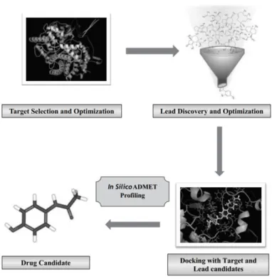
1.5 Drug Design and In Silico Approach
Designing new drug candidates and validation of this candidate is a time- and
money-consuming, exhaustive process. During drug discovery and development
much or less 75% of total process cost is consumed due to failures. More
importantly, approximately 10 to 15 years of hard work may result as failure or
success (Figure 8). In Figure 8, process of drug discovery and drug improvement is
summarized step by step by showing important facts involved in each phase. Before
any potential drug discovery, researchers from universities, associations and
governments carry lots of projects, studies about the disease. Underlying conditions,
signaling cascades, genes encoding for proteins that is involved in these cascades
should be discovered to build the idea. It will take many years to turn what causes
the disease into the idea of treatment of diseases. If it is resulted in success then new
process, therapeutic discovery may begin. Treatments of almost all diseases for
patients depend on effective therapeutics. First of all, after finding a target, a gene or
protein, the target is validated to confirm relatedness of it with illness and to pre-see
possible side effects. And after target validation, lead compound for that target
Figure 8 | Biomedical Research from Idea to Market. IND: Investigational New Drug application; POC = proof of concept; NDA: New Drug Application; Pharma: pharmaceutical
companies.15
There are few ways to obtain this lead compound; nature, de novo,
biotechnology and high-throughput screening. High-throughput screening is very
fashionable way, which is used to find lead structure using compound libraries. In
this approach, power of computers and robotics make possible to test and screen
thousands of lead candidates and to choose promising ones from these compound
libraries by docking and evaluating binding constants and investigating how lead
Figure 9 | In Silico Approach during Drug Discovery Process.16
In silico drug designing is another demanding approach which applies
computational methods for molecular modelling.16 In Figure 9, three major steps of
in silico approach in drug discovery are summarized. There are two methods for lead
generation; ligand-based and structure-based. If only knowledge is about ligands
previously defined ligands as active or as inactive for target enzyme, unknown
models can be aligned to these known ligands and then lead generation can be
achieved. In this study, we applied structure-based in silico methods. To use
structure-based in silico approach, protein-ligand interactions, structure of receptor
a wide library, which is known as Protein Data Bank (PDB). Today, this data bank
contains almost 100 000 structures.
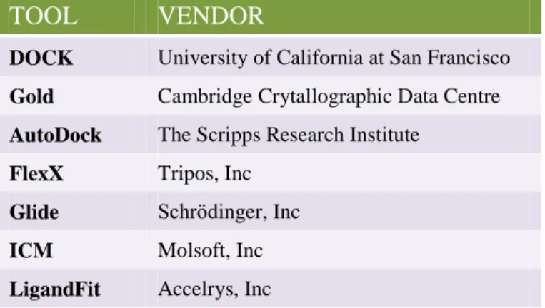
Virtual screening (VS) and de novo design are two paths used in
structure-based in silico method. VS is a version of HTS and in some sources it can be seen
abbreviated as vHTS (Virtual High Throughput Screening).18 Molecular docking is
main method in virtual screening. There are several VS tools commercially
available. Some popular VS tools can be seen in Table 1. 19 Using these tools, drug
candidates are tested by predicting compatibility of ligands with target protein.
Using these predictions, 3D pose of docked ligand in the active site and binding
energy of ligands can be calculated. These tools generate many representations,
which can be varied by conformations, positions and orientations, of a small
molecule and places each representation in the active site of receptor protein. This
process aims to find most energetically favorable pose of a small molecule in the
active site of receptor. To calculate pose predictions from many binding modes of
small molecule and affinity predictions of best posed small molecules, various
algorithms and scoring functions have been developed and applied by different
TOOL VENDOR
DOCK University of California at San Francisco
Gold Cambridge Crytallographic Data Centre
AutoDock The Scripps Research Institute
FlexX Tripos, Inc
Glide Schrödinger, Inc
ICM Molsoft, Inc LigandFit Accelrys, Inc
Table 1 | List of Some Commercially Available VS Tools and Their Vendors. 19
After obtaining promising candidates, they should go through for several tests
to assess early safety of the lead compound. In this step, early tests for safety,
efficacy and potential toxicity of drugs are done. These studies are also needed for
researchers to propose Phase I studies. In Phase I, scientists apply some tests
considering several important properties to obtain important information about
drug’s properties. In which dose drug shows successful impact on therapy with
minimizing its possible side effects? How good are drug’s pharmacokinetic
properties and toxicity? Does drug show potential effects on other biological
molecules rather than its target molecule? Are effects of drug candidate influenced
by another drug? Such queries should be answered at this phase. To come with
pharmacokinetic properties of drug (ADME) and its toxicity (Tox), a successful drug
must be absorbed in bloodstream (Absorption), distributed to target site of molecule
(Metabolism), excreted successfully from the body (Excretion), not show toxicity in
the body (Toxicity). And these ADME/Tox studies can be performed in living cells,
in animals and also by applying computational tools. Phase I is the first phase in
which drug can be tested in human in a small group of healthy volunteers. So this
phase is one of the crucial phases during drug discovery process. 15
Second phase clinical trial is done on a small group of patients. In Phase II,
researchers try to obtain information about drug’s effectiveness on patients with
illness and to examine adverse events and risks showed by drug. After these
examinations, if the drug is found promising, these almost same examinations are
tested in large group of patients as Phase III clinical trials. It is crucial to do these
trials as large as and as diverse as possible group of patients. So many clinics,
regions are involved in this phase. After completing and being successful in all three
phases, New Drug Application (NDA) must be written and FDA approval must be
received. 15
1.6 Selective Inhibition of Neuronal Nitric Oxide Synthase
In mammalian cells, nitric oxide produced by nitric oxide synthases has crucial
different tissues, each NOS isoforms produce NO for separate biological processes
in human body. In addition, we mentioned before that overproduction of NO by
nNOS is mainly related to many neurodegenerative diseases, chronic headache,
stroke, Alzheimer’s, Parkinson’s, Huntington’s diseases. 421 Therefore, achieving
selective nNOS inhibition in the brain without inhibiting eNOS and iNOS is
important focus as therapeutic for neurodegenerative diseases. On the other hand,
that there is a approximately 50% sequence homology and high similarity in heme
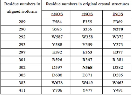
active site of all three isoforms of NOS makes this task very difficult. 22 Residue
differentiation between isoforms in their active sites may be one of major facts that
should be maintained on the way of achieving selectivity. Aligning three isoforms
(Figure 3, Figure 4) reveals these differences as following: S585 in nNOS is N370
in iNOS; D597 in nNOS is N368 in eNOS. 14 Some active site residues in three
Table 2 | Some Residues in Three Isoforms’ Active Sites. Residue differences are indicated in bold.
To conclude, selective inhibition of nNOS in the brain is crucial task
considering neurodegenerative diseases, therefore, many researchers focus on to
discover good selective nNOS inhibitors. On the other hand, due to mentioned
similarity of three isoforms, these researches are exhaustive challenges. That’s why,
no drug, which serves for this purpose as good potential and selective inhibitor has
yet been designed. 6
2 MATERIALS AND METHODS
Collaboratory for Structural Bioinformatics (RSCB) protein databank. Among all
NOS PDB crystal structures, 1OM4 (nNOS with L-arginine bound, resolution
1.75Å), 3DQS ( eNOS with inhibitor C20 H28 Cl N5, resolution 2.03Å), and 1NSI
(iNOS with L-arginine bound resolution 2.55Å) were used for all analysis.
For enzyme preparation, except chain A of all structures, all other chains and
all solvent molecules exist in PDB structures were eliminated. However, heme group
and H4B were left in the active sites of structures since these cofactors should be
involved in energy calculations during docking. Protein Preparation protocol and
then “Clean Geometry” toolkit included in Discovery Studio 3.1 software package
(Accelrys, Inc.) were employed to do energy minimizations and preparations of
enzymes to make them dockable. Missing hydrogen atoms were added based on the
protonation state of the titratable residues at a pH of 7.4. Ionic strength was set to
0.145 and the dielectric constant was set to 10. Inhibitors designed by Richard B.
Silverman [Richard B. Silverman, March 2013] [Richard B. Silverman, June 2013]
and his colleagues and newly designed ligands were drawn and prepared in silico
using Discovery Studio 3.1. “Clean Geometry” toolkit is also used to prepare and
2.2 Generation of Potential Inhibitors 2.2.1 Test Inhibitors
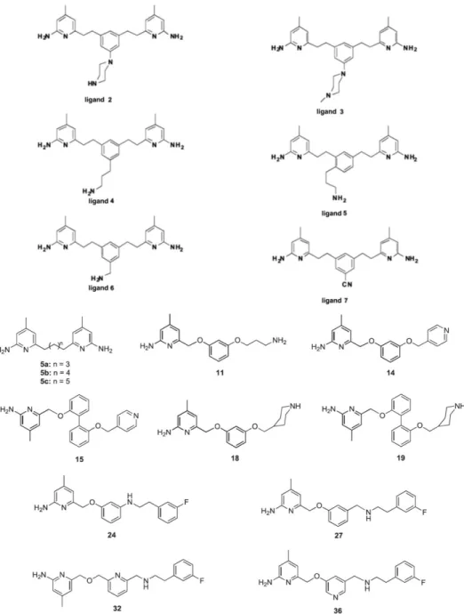
Richard B. Silverman and his colleagues designed and synthesized inhibitors
for selective inhibition of nNOS. 6, 22 To control our in silico docking calculations,
we prepared these ligands on Discovery Studio, can be seen in Figure 10, designed
by Silverman and they are docked into each isoforms of NOS selected by us. And we
compared experimentally calculated inhibition constants with our computationally
Figure 10 | Designed and Synthesized Ligands by Richard B. Silverman. 6, 22
2.2.2 Inhibitor Candidates
Two scaffolds (Figure 11 and 12) were created and used for derivation of our
inhibitor candidates. Table 3 and Table 4 show two groups of potential inhibitors
GROUP A
Lead Scaffold
Figure 11 | First Scaffold Used in this Study.
Designed Inhibitor Candidates R1 R2 R3 KB20 -H -H KB21 -H -H KB22 -F -H KB23 -F -H KB24 -F
Table 3 | Lead IDs of First Group of Potential Inhibitors and Corresponding Functional
Groups.
GROUP B
Lead Scaffold
Figure 12 | Second Scaffold Used in this study
KB26 -F KB27 -H KB28 -F -H KB29 -H -H KB30 -H
Designed Inhibitor Candidates R1 R2 R3 KB31 -H -H KB32 -H -H KB33 -F -H KB34 -F -H KB35 -F KB36 -F KB37 -F KB38 -H KB39 -F -H KB40 -H -H KB41 -H
2.3 Docking
Binding affinities and docking orientations of inhibitor candidates were
calculated applying famous docking virtual tool, AutoDock. Predictions of binding
affinities of flexible ligands into target enzymes, in this project targets are eNOS,
iNOS and nNOS, are obtained via using AutoDock 4.2. For calculations of these
binding affinities, AutoDock applies empirical binding free energy function based on
AMBER force field. 23 As conformational search method, AutoDock uses
Lamarckian genetic algorithm. AutoDock Tool (ADT) was used to set up AutoGrid
parameter files (gpf) and AutoDock parameter files (dpf) which include required
parameters during docking simulations. Beforehand, the charge of Fe atom of heme
in all three enzymes was changed from +2 to +3. The ligands were docked inside a
grid box with 60 Ao x 60 Ao x 60 Ao dimensions and grid spacing 0.375 Ao. For
center of the grid box, centers of ligands found in original PDB structures' active
sites were noted and used.
Docking simulations were performed applying Lamarckian genetic algorithm.
Genetic Algorithm Parameters were set to 10 independent LGA runs, 150 in
population size, 5000000 in energy evaluations and 27000 in generations and all
other parameters were remained as default.
orientations were collected for further analysis.
3 RESULTS AND DISCUSSION
During enzyme inhibition, if any drug can work at low concentration and do
not inhibit or affect other enzymes at this concentration can be counted as strong
selective inhibitors. In this study, our aim is to obtain this kind of potential strong
selective inhibitors for nNOS enzyme applying computational tools. There are other
physiologically important isoforms of NOS enzyme, eNOS and iNOS, and high
homology in the active sites of these three isoforms makes our task challenging.
(Figure 3, Figure 4). Therefore, selectivity of potential inhibitors between three
isoforms becomes main challenge to be considered.
In previous projects held by Prof. Kemal Yelekçi’s group, all PDB structures
account for three isoforms of NOS enzyme were used for comparative validation
studies to select most reliable PDB structures of NOS isoforms to be used in further
projects. In that project, studies such as computational modeling and re-docking of
bound ligands in crystal structures were applied. Also some properties such as
considered to choose most compatible PDB structures among all. As a result, PDB
structures with code 1OM4 for nNOS, 3DQS for eNOS and 1NSI for iNOS were
selected to be used in all docking simulations. 24 Relying on these results, we also
focused and used these three structures for all docking simulations.
At the most beginning, inhibitors designed by Silverman (Figure 10) were
drawn by us on Discovery Studio and re-docked to the prepared enzymes by us and
binding modes were calculated. X-Ray structures of NOS enzymes with bound
inhibitors designed by Silverman, which are placed on publication of Silverman,
were downloaded from PDB website. Those binding modes were compared with
already originally bound poses in downloaded structures. Ultimately, we observed
close binding modes in the active site of all isoforms. Additionally, inhibition
constants obtained from these re-docking results were checked against inhibitor
constants experimentally obtained by Silverman group. To compare these
computationally and experimentally obtained inhibitor values, all constants were
converted to nanomolar units and their logarithmic values were calculated. These
logarithmic values of experimentally and computationally obtained inhibition
constants were plotted for each isoform (Figure 13). By re-docking already
synthesized inhibitors, we aimed to crosscheck reliability of our virtual screening
simulations.6 To comment Figure 13, behaviors of experimental and computational
plot lines are very close to each other. In fact, many results have given almost same
values with experimental values that can be seen as interceptions in plot lines. Since
there are many facts that can affect and change docking and scoring processes during
in silico screening, we cannot wait to obtain exact same values. These results
obtained from our control studies are enough to fulfill our expectations.
0,00 1,00 2,00 3,00 4,00
27 5a lig6 lig7 lig5 18 36 32 14 lig2
L o g a rit m ic K i V a lues Ligands
nNOS (1OM4)
0,00 1,00 2,00 3,00 4,00 5,00 6,005a 5c 5b 32 27 36 lig6 lig4 11 18 lig5 lig2 14 lig3
L o g a rit m ic K i V a lues Ligands
eNOS (3DQS)
Figure 13 | Plots of Experimentally and Computationally Obtained Inhibition
Constants Values (Ki). Docking experimentally synthesized ligands within active sites of
three isoforms resulted inhibition constants (Ki). These plots represent log values of these
computationally obtained Kis and experimentally obtained Ki s for comparison analysis.
Blue lines represent computational values and red lines represent experimental values.
In Figure 11 and 12, two scaffolds that were used in this study were presented.
Structure-based drug design is applied to obtain these scaffolds and leads. More than
several hundred lead compounds in the ZINCv12 lead library considering their
structural and physicochemical properties, which selectively inhibit nNOS isoform,
were scanned. 25 In previous projects, by utilizing ZINC and Accelrys 3.1
fragment-based libraries, which contain about hundred thousand fragments, about fifty
potential candidates were selected out of a few hundred thousand fragments based on
scoring values in the active site of the nNOS isoform using Accelrys’s de Novo
Design method. After docking all analogues into three isoforms of NOS, only
inhibitors that selectively inhibit nNOS were used for further modifications. 24 The
0,00 1,00 2,00 3,00 4,00 5,00 6,00
5a 5b 5c 36 27 11 32 lig7 18 lig5 14 lig4 lig2 lig3
L o g a rit hm ic K i V a lues Ligands
iNOS (1NSI)
structure-based methods were employed manually for the further optimization of the
potential nNOS inhibitors by adding and removing a few fragments on the scaffolds.
22 potential inhibitors (Table 3 and 4) among all inhibitors for nNOS selective
inhibition is designed using mentioned methods on two scaffolds. These 22 derived
leads from two scaffolds were selected to discuss on them. Current designs and
computational evaluations of these 22 potential inhibitors using various docking
tools are listed in
Table 5.
Inhibitors Chemical Structures nNOS
(1OM4) eNOS (3DQS) iNOS (1NSI) Kb20 2730 6710 6600
Kb21 872 11830 1680
Kb22 1930 36770 2120
Kb23 1640 13670 3550
Kb25 1470 30190 372
Kb26 1250 2320 1200
Kb27 949 1760 1200
Kb29 506 1730 324
Kb30 606 1380 74
Kb31 1000 3180 816
Kb33 550 3260 1500
Kb34 589 5150 1730
Kb35 75 11010 594
Kb37 365 1650 857
Kb38 291 1090 490
Kb39 203 971 241
Table 5 | Chemical Structures of the 22 Designed Potential NOS Inhibitors and Their
Inhibition Values Obtained from Docking Simulations. All inhibition values are given in
nanomolar concentration.
Considering derivations of two scaffolds and docking results based on
inhibition constants (Ki) present in Table 5, we can discuss many facts for nNOS
selective inhibition. Referring first scaffold, results and derivations may reveal
several facts. Between Kb20 and Kb21, there is one functional group differentiation.
In Kb20, isopropyl is replaced with propanamide. It can be easily seen that this
functional group differentiation leads increase in affinity of inhibitor for nNOS. In
addition, selectivity of nNOS over eNOS also increased, however, selectivity of
nNOS over iNOS decreased. Fluoro derivative of Kb21 is Kb22. eNOS inhibition
value increased three-fold, inhibition value of nNOS is doubled whereas value for
iNOS is slightly increased.
Comparisons between Kb20 and Kb23 and between Kb21 and Kb22 show that
Additional functional amine group to Kb23 resulted in stronger inhibition of
Kb24 on all three isoforms but selectivity of nNOS isoform over iNOS is
dramatically decreased. And another group addition to Kb23 instead of amine group
drastically increased eNOS inhibition value but undesirably decreased iNOS
inhibition constant.
Using alcohol group as a functional group in the first scaffold does not provide
well for our aim of nNOS selectivity since it reduces Ki for eNOS and approximates
it to Ki of nNOS. In Kb26 and Kb27, slight differences in inhibition values of all
isoforms can be seen. These results do not meet expectations of us.
For Kb28, Kb29 and Kb30, benzene group is preferred as one of major
functional group in lead scaffold. Based on results for these three inhibitors, it can be
said that benzene group in the first scaffold does not provide the desired selectivity
order, since inhibition constants are close to each other and both inhibitors
selectively inhibits iNOS rather than nNOS.
In the second scaffold for functional group R1, it is acquired that using
propanamide group instead of isopropyl group serves better for our purpose. There is
no selectivity of nNOS over iNOS for inhibitor Kb31. However Inhibitor Kb32
achieved two-fold selectivity and reduced nanomolar concentration for inhibition of
Another interesting and promising discrimination was observed with a shift
between Kb34 and Kb35. Between these two inhibitors, only R3 group was changed
from hydrogen to 1-amino methyl group. This change resulted in dramatic selectivity
of nNOS over eNOS and of nNOS over iNOS.
Replacing 1-amino methyl in Kb35 with pyridine, Kb36 is obtained. This shift
causes obvious decline in inhibition value for eNOS. In Kb36, better selectivity of
nNOS over iNOS is achieved, but good selectivity of nNOS over eNOS observed in
Kb35 was lost. eNOS inhibition reached the strongest inhibition comparing other
candidates.
When alcohol group is used for R3 functional group instead of hydrogen, slight
stronger inhibitions were observed for all isoforms (Kb34, Kb37). Eliminating F
atom from Kb37 structure, more decline in inhibition values of isoforms obtained in
docking simulations of Kb38.
Benzene ring is majored as functional group R1 for Kb39, Kb40 and Kb41. In
these three inhibitor candidates, docking calculations resulted in close inhibition
constants for iNOS and nNOS, so selectivity of nNOS over iNOS is almost lost.
Considering derivatives with benzene ring in Group A and B, it can be concluded as
Docking simulation results of all candidates used in this study highlighted
some important facts on the way of obtaining the most promising inhibitor. In all 22
designed candidates it can be seen that nNOS selectivity over eNOS is achieved with
more inhibitors than nNOS selectivity over iNOS. Almost all candidates bind iNOS
more tightly than eNOS. Substitution of benzopyrazole and imidazole ring in
scaffold one with benzopyrrole ring and pyrrole is increased nNOS selectivity and
potency as it can be clearly seen if results of group A candidates and group B
candidates are compared. Attaching 1-amino methyl group to pyrrole ring made us
to obtain best nNOS selectivity over iNOS. Inhibitor Kb35 binds more tightly to
nNOS among all isoforms. Selectivity of nNOS over eNOS (selectivity ratio
nNOS/eNOS) is 146 fold and selectivity of nNOS over iNOS (ratio nNOS/iNOS) is
8 fold. Removing 1-amino methyl group from pyrrole ring, in Kb34, lead decrease in
both selectivity and potency of nNOS. Subsititution of 1-amino methyl with pyridine
ring attached to pyrrole ring, compound Kb36, made us to obtain best selectivity of
nNOS over iNOS, which is 39 however selectivity of nNOS over eNOS, which is 2
in Kb36 and 8 in Kb35, is lost.
To depict detailed binding interactions of docked compounds, analysis of
optimal binding modes are done with compound Kb35. Best docked poses of Kb35
B. 2D interaction diagram of Kb35 in the active site of nNOS.
F. 2D interaction diagram of Kb35 in the active site of iNOS.
Figure 14 | 3- Dimensional and 2- Dimensional Orientation Diagrams of Best-Docked
Poses of Kb35 in Active Sites of Three NOS Isoforms. In 3D diagrams (A, C, E), amino
acid side chains and heme cofactor within the volume of 3,5 Å distance from inhibitor are shown with sticks configuration. Compound Kb35 is shown with scaled ball and stick configuration. Dashed lines represents hydrogen bond interactions. In 2D diagrams (B, D,
F), pink, green, purple, blue and black dashed lines represent electrostatic, van der Waals, covalent, hydrogen and metallic interactions, respectively.
Optimal binding analysis shows that best poses of Kb35 docked in all isoforms
are located in the vicinity of cofactor. (Figure 14) Predicted pose of Kb35 in nNOS
active site cavity interacts with some significant amino acid residues found in the
cavity. (Figure 14 A) Two hydrogen atoms on the 1-methy amino group formed two
2.15 Å) and one of them also formed H-bond with GLU592 (with 2.827 Å distance).
Last hydrogen atom of 1-methyl amino group formed H-bond with TYR588. A
carbon hydrogen bond is formed between carbon atom of pyrrole and carboxylate
group in side chain of GLU592. Best pose of Kb35 is fitted within an active site
cavity seen in Figure 14 A which includes GLY586, PRO565, TYR588, GLU592,
ASP597, ARG603, GLN478, ASN569, TYR706 and VAL567. Polar and
hydrophobic interactions are formed between Kb35 and these amino acids found in
pocket. In Figure 14 B, nNOS isoform and Kb35 is represented with
two-dimensional diagram. In this diagram, 17 amino acid residues closely interact with
Kb35 via electrostatic, VDW, covalent bond interactions that are highlighted.
3D and 2D diagrams in Figure 14 C, D reveal intermolecular interactions
between Kb35 and eNOS isoform. VAL 338, GLN 249, PRO336, ASN368,
GLU363 are amino acid residues which are in close interaction with Kb35 within a
vicinity of 3,5 Å distance. One hydrogen atom of 1-amino methyl group interacts
with propionate side chain of heme cofactor via strong H-bond (distance is 1.892).
GLU363 side chain carboxylate forms another H-bond with 1-amino methyl group.
Carbon atom of this group forms two carbon H-bond with GLU363 and ASN368
Additionally, residues closely in contact with Kb35 within eNOS active site cavity
are not as many as residues found in nNOS active site cavity. All of these facts are
evidential for that affinity of Kb35 to nNOS dominates affinity of Kb35 to eNOS.
And this explains selectivity ratio nNOS/eNOS which is 1/146.
Figure 14 E, F are representations for interactions between Kb35 and active
site residues of iNOS isoform. Active side residues interacting with Kb35 are
TYR373, GLU377, ASP382, TRP363, THR121, GLN263, ASN354, VAL352,
GLY371 and PRO350. Propionate oxygen atom of heme cofactor is in interaction
with two hydrogens; hydrogen atom of pyrrole ring and of benzopyrrole ring
(distances are 2.086 Å and 1.922 Å). Oxygen atoms in carboxylate side chain of
ASP382 form tripod strong H-bonds with 2 hydrogen atoms in 1-amino methyl
group (distances are 2.164 Å, 2.520 Å and 1,695 Å). Carbon atom of pyridyl ring
also form a H-bond with another amino acid residue GLU 377. There are 14 amino
acid residues closely interact via different bond types with Kb35 as they can be seen
in Figure 14 F.
In all binding pose predictions of Kb35 within three isoforms, there is
something interesting that attracted our attention. nNOS and iNOS isoforms contain
ASP597 and ASP382 at the same place in aligned isoforms, however eNOS contains
ASP597 and ASP382 interacted with compound Kb35 via strong hydrogen bonds,
however pose predictions between eNOS and Kb35 did not reveal any strong bonds
between compound Kb35 and ASN368. As it is mentioned selectivity of nNOS over
eNOS is much more than selectivity of nNOS over iNOS. This fact makes us to
question any possible impacts of residue differences on selectivity of inhibitor.
CONCLUSION
There is almost 50% sequence homology between three isoforms of NOS
enzymes and going deeper shows high similarity in the active sites of nNOS, eNOS
and iNOS. That’s why, designing and developing a selective inhibitor for nNOS
become very challenging task. Virtual screening tools were employed in this present
project and these studies highlighted that designed and selected lead scaffolds,
especially the second one could meet the expectations of us. It is proven that these
lead scaffolds are important candidates for further optimization analysis and
modifications to obtain promising inhibitor candidates that would be employed as
potential and selective inhibitors for nNOS. Among 22 selected inhibitors, Kb35 is
affinity and selectivity towards nNOS. Compound Kb34 is modified by removing
1-amino methyl group from pyrrole ring and it is clearly seen that both affinity and
selectivity of compound towards nNOS declined. Designed compound Kb35 inhibits
nNOS 146 fold better than eNOS and 8 fold better than iNOS. These results also
showed us that subtle residue differences in active sites of isoforms could be
important indicatives and determinants for selective and potential inhibitors. In this
present work, promising lead scaffolds and important determinants for selective
inhibition of nNOS were discovered via various computational tools and virtual
screening tools. Further studies with regard to these important findings would direct
REFERENCES
1. Alderton, W. K., Cooper, C. E. & Knowles, R. G. Nitric oxide synthases: structure, function and inhibition. Biochem. J. 357, 593–615 (2001).
2. Knowles, R. G., Palacios, M., Palmer, R. M. & Moncada, S. Formation of nitric oxide from L-arginine in the central nervous system: a transduction mechanism for stimulation of the soluble guanylate cyclase. Proc. Natl. Acad.
Sci. U. S. A. 86, 5159–62 (1989).
3. Stuehr, D. J., Santolini, J., Wang, Z.-Q., Wei, C.-C. & Adak, S. Update on mechanism and catalytic regulation in the NO synthases. J. Biol. Chem. 279, 36167–70 (2004).
4. Lohou, E. et al. New hypotheses for the binding mode of 4- and 7-substituted indazoles in the active site of neuronal nitric oxide synthase. Bioorg. Med.
Chem. 20, 5296–5304 (2012).
5. Förstermann, U. & Münzel, T. Endothelial nitric oxide synthase in vascular disease: from marvel to menace. Circulation 113, 1708–14 (2006).
6. Huang, H. et al. Structure-Guided Design of Selective Inhibitors of Neuronal Nitric Oxide Synthase. (2013).
7. Silverman, R. B. NIH Public Access. Acc Chem Res. 42, 439–451 (2009).
8. Calabrese, V. et al. Nitric oxide in the central nervous system:
neuroprotection versus neurotoxicity. Nat. Rev. Neurosci. 8, 766–75 (2007).
9. Zhou, L. & Zhu, D.-Y. Neuronal nitric oxide synthase: structure, subcellular localization, regulation, and clinical implications. Nitric Oxide 20, 223–30 (2009).
10. Fedorov, R., Vasan, R., Ghosh, D. K. & Schlichting, I. Structures of nitric oxide synthase isoforms complexed with the inhibitor AR-R17477 suggest a rational basis for specificity and inhibitor design. Proc. Natl. Acad. Sci. U. S.
11. Stuehr, D. J. Structure-function aspects in the nitric oxide synthases. Annu.
Rev. Pharmacol. Toxicol. 37, 339–59 (1997).
12. Andrew, P. J. & Mayer, B. Enzymatic function of nitric oxide synthases.
Cardiovasc. Res. 43, 521–31 (1999).
13. Förstermann, U. & Sessa, W. C. Nitric oxide synthases: regulation and function. Eur. Heart J. 33, 829–37, 837a–837d (2012).
14. Trippier, P. C., Jansen Labby, K., Hawker, D. D., Mataka, J. J. & Silverman, R. B. Target- and mechanism-based therapeutics for neurodegenerative diseases: strength in numbers. J. Med. Chem. 56, 3121–47 (2013).
15. National, T. H. E., Press, A., Street, F. & Washington, N. W. Bernard Lo and
Marilyn J . Field , Editors Committee on Conflict of Interest in Medical Research , Education , and Practice Board on Health Sciences Policy.
16. S, C. K. An Insight to Drug Designing by In Silico approach in Biomedical Research. 1, 63–65 (2013).
17. Kroemer, R. T. Structure-Based Drug Design : Docking and Scoring. 312– 328 (2007).
18. Subramaniam, S., Mehrotra, M. & Gupta, D. Bioinformation Virtual high throughput screening ( vHTS ) - A Bioinformation. (2008).
19. Heal, J. H. in silico structure-based drug design – SHIFTING THE BOTTLE NECK. Cheminformatics 57–60 (2003).
20. Lu, T., Max, N. L., Ding, J., Bethel, E. W. & Crivelli, S. N. DockingShop : A Tool for Interactive Molecular Docking.
21. Salerno, L. et al. Novel inhibitors of nitric oxide synthase with antioxidant properties. Eur. J. Med. Chem. 49, 118–26 (2012).
22. Jing, Q. et al. In search of potent and selective inhibitors of neuronal nitric oxide synthase with more simple structures. Bioorg. Med. Chem. 21, 5323–31 (2013).
23. Morris, G. M. et al. Automated Docking Using a Lamarckian Genetic Algorithm and an Empirical Binding Free Energy Function. 19, 1639–1662 (1998).
24. Büyüktürk, A. B. EXPLORATION OF BINDING SITES OF NOS ISOZYMES TO DESIGN SELECTIVE NEURONAL NITRIC OXIDE SYNTHASE INHIBITORS EXPLORATION OF BINDING SITES OF NOS ISOZYMES TO DESIGN SELECTIVE NEURONAL NITRIC OXIDE.
Grad. Thesis (2013).
25. Irwin, J. J. & Shoichet, B. K. ZINC − A Free Database of Commercially Available Compounds for Virtual Screening. J. Chem. Inf. Model. 45, 177– 182 (2004).
Curriculum Vitae
Bahanur Örtmen was born on May 8th 1989 in Bursa. Her B.Sc. degree has
been earned in Molecular Biology and Genetics in 2012 from Bilkent University.
During Summer 2010 and 2011, she was accepted to work as a research assistant and
an intern in Orwar Labarotary at the Department of Chemical and Biological
Engineering at Chalmers University of Technology. And she assisted four projects
focusing on biomembranes, liposomes and phospholipid nanotube networks during
these periods. After completing her undergraduate, she was accepted to Kadir Has
University to study her graduate on Computational Biology and Bioinformatics in
September 2012. Multidisciplinary approach by combining computational biology
and life sciences is her main interest.
Publications:
1| Generation of phospholipid vesicle-nanotube networks and transport of molecules therein
Jesorka, A ; Stepanyants, N ; Zhang, HJ ; Ortmen, B ; Hakonen, B ; Orwar, O NATURE PROTOCOLS ; 6 ( 6 ), 791 - 805 , 2011.
2| Repair of large area pores in supported double bilayers
Gözen, I.; Ortmen, B.; Põldsalu, I.; Dommersnes, P.; Orwar, O.; Jesorka, A. SOFT MATTER; ,9, 2787-2792, JUN 2013
3| Thermal migration of molecular lipid films as a contactless fabrication strategy for lipid nanotube networks
Gözen, I.; Shaali, M.; Ainla, A.; Örtmen, B.; Põldsalu, I.; Kustanovich, K.; Jeffries, G.; Konkoli, Z.; Dommersnes, P.; Jesorka, A. LAB CHIP, 13, 3822-3826, JAN 2013