Introduction
Renal transplantation is associated with several compli-cations, some of which may cause irreversible loss of graft function. Despite reliable pre-transplant screening methods and improvement of immunosuppression therapy, failures of kidney allografts are still occurred because of cellular and/or humoral mediated rejections (1). Several recent studies evalu-ated the prevalence of human leukocyte antigen (HLA)-spe-cific antibodies and the clinical importance of these antibod-ies in acute allograft rejection (2, 3). Chronic rejection is also known to have several immunologic and non-immunologic causes. Acute rejection episode after renal transplantation is also a known risk factor for the development of chronic rejection (4). Antibodies against HLA developed after blood transfusions, pregnancies and graft rejections were generally described as panel reactive antibodies (PRA). After sensitiza-tion antibodies appear against to both HLA class I and HLA class II. Class I and Class II HLA antibodies activate different cells, initiate immune response and contribute to rejection.
Over the past years many studies reported the relevance of various incidences of alloantibodies detected after trans-plantation (5-8).This variability can be attributed to the use of different techniques to detect the antibodies and differ-ences in the time after transplantation that samples are col-lected (6).Post-transplantation detection of HLA antibodies was found to be associated with high rejection rates (7-10). HLA antibodies developed in the early term of transplanta-tion damages allograft more than antibodies developed after 1 year of transplantation (11). Post-transplantation alloanti-body development in the early period may be associated with reperfusion and prolonged cold ischemia time [a chief factor leading to delayed graft function (DGF)] induced activation of endothelium and impaired cytokine gene expression, release of proinflammatory cytokines, and upregulation of HLA and adhesion molecules (1, 12, 13). These events lead to stimula-tion of the immune response in the early post-transplantastimula-tion period and, as a consequence, to HLA antibody production. However, in some instances even in the absence of detect-able pre-transplantation sensitization, reactivation of memory Address for Correspondence: Dr. Tülay Kılıçaslan Ayna, Department of Medical Biology, Faculty of Medicine, İstanbul University, İstanbul, Turkey
Phone: +90 212 635 11 68 e-mail: [email protected]
Long-Term Effects of Antibodies against Human Leukocyte
Antigens Detected by Flow Cytometry in the First Year after Renal
Transplantation
1Department of Medical Biology, Faculty of Medicine, İstanbul University, İstanbul, Turkey
2Department of Internal Medicine, Division of Nephrology, Faculty of Medicine, İstanbul University, İstanbul, Turkey
Tülay Kılıçaslan Ayna1, Yaşar Çalışkan2, Hayriye Şentürk Çiftçi1, Aydın Türkmen2, Mehmet Gürtekin1
ABSTRACT
Objective: In this study, we aimed to investigate the incidence, dynamics and profiles of human leukocyte antigen (HLA)-directed antibodies developed after transplantation and their impact on graft rejection and outcome in kidney recipients.
Study Design: Prospective follow-up study.
Material and Methods: A total of 56 kidney recipients were monitored at 1st, 6th and 12th months for the development of anti-HLA antibodies using bead based flow-cytometry assays (Flow PRA tests).
Results: In 21 (37.5%) patients, panel reactive antibodies (PRA) was positive after transplantation, however, in 35 (62.5%) patients PRA was found nega-tive. Twelve (57.1%) patients with post-transplantation HLA-reactive antibodies [PRA (+)] and 8 (22.9%) patients with no detectable alloantibodies [PRA (-)] were developed allograft rejection (p=0.010). In the PRA positive patient group the rates of early period infection and delayed graft function (DGF) were higher than the PRA negative patient group. Serum creatinine levels of PRA positive group at 6. and 12. months after transplantation were signifi-cantly higher than the PRA negative group (p=0.015 and p=0.048, respectively). The rejection rates of patients who had class I and II HLA antibodies were significantly higher than the patients who had either class I or II HLA antibodies (p=0.011). Acute rejection rates were significantly higher in patients who had class I and II HLA antibodies at the first month (p=0.007).
Conclusion: Higher occurrence of rejection episodes in PRA positive group may show the importance of anti-HLA antibody monitoring using Flow-PRA after renal transplantation as a prognostic marker in terms of graft survival.
Key Words: Anti-HLA antibodies, flow cytometry, renal transplantation
Received: 15.02.2012 Accepted: 10.07.2012
Balkan Med J 2013; 30: 37-45 • DOI: 10.5152/balkanmedj.2012.071
Original Article
Available at www.balkanmedicaljournal.orgB cells from sensitizing events in the patient’s history may facilitate the alloantibody production in the early days after transplantation. Rejections may still occur in the absence of detectable lymphocytotoxic antibodies, suggesting that non-HLA antigenic systems may also play a role in renal allograft rejections (10-16). Despite increasing recognition of the role of posttransplantation humoral alloreactivity in graft outcome, there is still debate regarding the clinical relevance of anti-HLA antibodies detected by sensitive solid-phase assays.
In this study, we aimed to investigate the incidence, dy-namics and profiles of HLA-directed antibodies developed after transplantation and their impact on graft rejection and outcome in kidney recipients using sensitive and specific flow-cytometry bead-based techniques.
Material and Methods
Patients
A total of 56 patients [35 male, 21 female, mean age 38±10 years (range 15-63)], underwent renal transplantation between 2001 and 2007 at the Istanbul Faculty of Medicine Hospital, were included in this observational prospective study. Information on demography, body mass index (BMI), the etiology of end stage renal disease (ESRD), time on di-alysis treatment, viral serology and donor characteristics were collected by reviewing patient files and medical records. Fifty patients underwent living related and 6 patients underwent cadaveric renal transplantation. The living related donors were mother (n=17, 34%), siblings (n=13, 26%), father (n=12, 24%), spouse (n=4, 8%), cousin (n=3, 6%) and maternal aunt (n=1, 2%). Twenty seven (48%) of the patients have a history of pre-transplant sensitization. The sensitizing events were blood transfusion in 21 patients, blood transfusion and pregnancy in 4 patients and solely pregnancy in 2 patients.
The standard immunosuppressive regimen of the patients at the İstanbul Faculty of Medicine included a calcineurin in-hibitor, mycophenolate mofetil (MMF) and prednisone. Target blood levels for cyclosporine A (CsA) were 200-300 ng/mL in the first 3 months, 100-200 ng/mL between 3-12 months and 50-150 after the first year of transplantation. Target blood lev-els for tacrolimus were 10-15 ng/mL in the first 3 months and 5-10 ng/mL after the third month of transplantation. MMF was given as a daily dose of 2 gr in CsA based regimen and 1 gr in Tacrolimus based regimen. All of the patients received prednisone; beginning with an infusion dose of 250 mg per 6 hours on the day before transplantation, 500 mg infusion on the transplant day and 120 mg iv on the next day of transplan-tation with a rapid taper and reaching to maintenance dose of 10 mg daily within the first month. However, doses were individualized according the patients needs. Induction thera-py (ATG Fresenius, 2 mg/kg/day) was used in transplantations from deceased donors.
Delayed graft function (DGF) was defined as the need for dialysis within the first week post-transplant. Recorded acute rejection and chronic rejection episodes were clinical or bi-opsy proven. Humoral rejection (acute or chronic) was defined by the presence of biopsy C4d staining in peritubular capil-laries and de novo donor spesific antibodies (DSA) in serum.
Post-transplant acute tubular necrosis (ATN) was defined as exclusion of other causes of DGF such as acute rejection and technical complications. Chronic allograft nephropathy (CAN), infection,cardiovascular disease, malignancy and bone dis-ease in thelate post-transplant period were also recorded as long-termpost-transplant complications. Our examinations of the patientsconformed to good medical and laboratory practices and to therecommendations of the World Medical
Association Declarationof Helsinki: Recommendations
Guid-ing Medical Doctors in BiomedicalResearch Involving Human
Subjects.
HLA tissue typing and screening of anti-HLA antibodies in serum
Human leukocyte antigen tissue typing was performed in European Federation for Immunogenetics (EFI)-accredited HLA laboratories of Department of Medical Biology. Class I HLA-A,-B typing was performed by complement dependent cytotoxicity (CDC) method, whereas class II HLA-DRB1 typing was performed by low-resolution polymerase chain reaction (PCR)-sequence-specific primer (SSP), as has been described elsewhere (17, 18). In case of an ambiguity in class I typing, PCR-SSP was performed as well. For the current study, an-ti-HLA antibodies and DSA in serum were monitored at 1st, 6th and 12th months using bead based flow-cytometry assays (FlowPRA™ Screening Test, FL12-60; One Lambda, Canoga Park, CA, USA).
Flow-Cytometric Analysis of Alloantibody
Flow-cytometric detection of HLA-specific antibodies was performed by FlowPRA™ Screening Test (FL12-60; One Lambda, Canoga Park, CA, USA). FlowPRA™ specific tests (FL1SP, FL1SP44, FL1HD, FL2SP; One Lambda, Canoga Park, CA, USA) were used for definition of HLA antibody specificity in the sera. The tests were performed according to the instruc-tion of the manufacturer. In brief, 5 µL of FlowPRA microparti-cles were admixed with 20 µL of patient serum and incubated for 30 min. at room temperature. Control non-MHC-coated beads were included in each sample to monitor the nonspe-cific interaction of the testing serum with the beads. After washing, the beads were stained with 100 µL of pretitered FITC-conjugated F(ab)’2 fragment of goat antihuman IgG for an additional 30 minutes. After a final wash, 500 µL of wash buffer was added per tube and analyzed on flow cytometer (EPICS-XL Coulter Corporation, Miami FL, USA). Screening results were recorded as positive when 10% of class I and/ or class II beads exhibited fluorescence above the negative control serum and/or a significant change in the histogram ar-chitecture compared with the negative serum control. Speci-ficity results were scored according to the scheme included in the kits. HLA specificities were determined by referring to the FlowPRA data sheets and software (HLA-fusion software, ver-sion 1.2.1B, One Lambda, Inc. USA).
Statistical Analysis
The statistical analysis was carried out by Statistical Pack-age for Social Sciences for Windows ver. 15.0 (SPSS Inc., Chi-cago, IL, USA). Numerical variables were given as mean±SD,
and were compared by Independent Samples t-test. When distribution was abnormal, non-parametric tests were used. Relationships were determined with Pearson`s correlation co-efficient. Correlations between numerical parameters were analyzed by Spearman’s rho correlation test. p<0.05 was ac-cepted as significant. Survival analysis was carriedout using Kaplan-Meier estimates. For differences in survival,a log-rank test was used.
Results
A total of 54 patients were followed up clinically for a mean time of 73.3±26.7 (12-115) months after transplanta-tion. Two patients were lost to follow up after 12 months of the study period. The demographic and clinical features of study patients are shown in Table 1. During the study period, PRAs were detected in the serum of 21 (37.5%) patients by FlowPRA™ Screening Test and remaining 35 patients had
negative PRA test. Twelve (57%) of the PRA positive patients and 8 (23%) of the PRA negative patients were developed acute allograft rejection. The rejection rate was significantly higher in the PRA positive group than the PRA negative group (p=0.01) (Figure 1). One of the pre-FCXM (+) patients was de-veloped acute allograft rejection. Rejection was confirmed by allograft biopsy in 7 of the 56 patients. Of these 7 biopsies, 3 showed acute (grade 1, grade 2a and 2b due to Banff classifi-cation) and 4 showed chronic rejection. Five (71.4%) of these 7 patients had positive PRA and the other 2 had negative PRA test.
When the PRA results were evaluated with regard to acute rejection episodes in the first month, acute rejection rates in the first month were significantly higher in the PRA positive patient group (47.6%) than the PRA negative patient group (17.1%) (p=0.015) (Table 2).
One patient who had antibodies against HLA class I and II antigens at 1 year after transplantation developed graft failure. Table 2 showed the graft failure rates of PRA positive (14.3%) and PRA negative patients (5.7%) during the clinical follow up period (12-115 months). Kaplan-Meier survival analysis showed overall graft survival rates of 98.2% at 1 year, 94.6% at 5 years and 91.1% at 10 years. During the clinical follow up period, 4 patients died. Kaplan-Meier survival analysis showed an overall patient survival rates of 98.2% at 1 year, 96.4% at 5 years and 92.8% at 10 years. In the PRA positive patient group the rates of early period infection and DGF (47.6% and 20%) were higher than the PRA negative patient group (14.3% and 0). The hospital stay was also longer in the PRA positive group than the PRA negative group. Both groups were similar with regard to immunosuppressive regimens after transplantation (Table 2).
When the serum creatinine levels at 1st, 6th, 12th months and 2nd, 3rd, 4th, 5th, 6th, 7th, 8th and 9th years after transplanta-tion were compared between PRA positive and PRA negative groups, serum creatinine levels of PRA positive group at 6th and 12th months after transplantation were significantly higher than the PRA negative group (p=0.015 and p=0.048, respectively). When the serum creatinine levels were compared between pa-tients who had rejection episodes and papa-tients with no rejection episode, patients who had rejection episodes had statistically
Tx recipients (n=56) Age (years) 38±10 Gender (M/F) 35/21 Donor Characteristics Living related 50 (89.3%) Cadaveric 6 (10.7%)
Donor age (years) 53±15
Donor gender (M/F) 20/36
HLA matching (min-max) 1A-2A2B2DR
Pre-sensitization 27 (48.2%)
Pre-tx FCXM 3 B FCXM (+)
Time of follow up (months) 73.3±26.7 (12-115) Etiologies of ESRD [n (%)] Unknown 23 (41.1%) Chronic pyelonephritis 15 (26.8%) Chronic glomerulonephritis 12 (21.4%) Diabetic nephropathy 3 (5.4%) Amyloidosis 2 (3.6%) Hypertensive nephrosclerosis 1 (1.8%) Pre-tx RRT type Preemptive 4 (7.1%) Hemodialysis 47 (83.9%) Peritoneal dialysis 5 (8.9%)
M: male, F: female, ESRD: end stage renal disease, RRT: renal replacement treatment
Table 1. The demographic and clinical features of study pa-tients
Figure 1. Relation between rejection and the rates of PRA positive and negative groups
Rejection rates %
significant higher serum creatinine levels at 6th, 12th months and 5th, 6th, 7th years after transplantation (Table 3).
There were no statistically significant differences regarding to presence of anti-HLA antibodies between the patients who received different anti-rejection treatments (Table 4).
Twenty seven patients (48%) have a history of pre-trans-plant sensitization. The demographic features of patients with or without a history of sensitizing events were shown in Table 5. The PRA and acute rejection rates were significantly higher in patients with a history of sensitizing events (p=0.001 and p=0.015, respectively) (Table 5). Of 21 (37.5%) PRA posi-tive patients, 11 (19.7%) patients had class I and II HLA an-tibodies, 7 (12.5%) had solely class I HLA antibodies and 3 (5.3%) had class II HLA antibodies. Anti-HLA antibodies were not detected in 35 (62.5%) patients. The rejection rates of pa-tients who had class I and II HLA antibodies were significantly higher than the patients who had either class I or II HLA anti-bodies (p=0.011). The rejection rates were significantly lower in patients with no detected anti-HLA antibodies (p=0.019) (Table 6). The anti-HLA antibody specificity, donors’ mis-matched HLA antigens, time of antibody detection and rejec-tion rates of post-transplant PRA positive patients were shown in Table 7. Class I DSAs were detected only in one patient’s serum in the post-transplant first year.
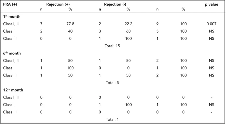
In the PRA positive group antibodies against HLA antigens were detected in 15 (71.4%) patients within the first month, 5 (23.9%) at the 6th month and 1 (4.7%) at the 12th month after transplantation (Table 8). Nine of 15 patients (60%) who had HLA antibodies at the first month after transplantation devel-oped rejection episodes. Three of 5 (60%) patients who had HLA antibodies at the 6th month after transplantation devel-oped rejection episodes. At the 12th month, PRA was positive only in 1 patient and no rejection episode was detected in this patient.
Antibodies against HLA class I and II antigens were detect-ed at the first month in 7 patients who had rejection episodes after transplantation. Acute rejection rates were significantly higher in patients who had class I and II HLA antibodies at the first month (p=0.007). At the 6th month only in one
pa-Serum creatinine levels (mg/dL) PRA (+) PRA (-) p Rejection (+) Rejection (-) p
n=21 n=35 value n=20 n=36 value Post-transplant 1st year 1st month 1.67±1.04 1.34±0.42 0.100 1.59±0.58 1.39±0.76 0.342 6th month 1.58±0.51 1.29±0.33 0.015 1.64±0.48 1.26±0.33 0.002 12th month 1.57±0.67 1.31±0.28 0.048 1.62±0.63 1.29±0.32 0.017 Post-transplant 2nd year 1.38±0.43 1.30±0.34 0.507 1.39±1.04 1.30±0.40 0.418 Post-transplant 3rd year 1.35±0.37 1.32±0.38 0.789 1.41±0.39 1.30±0.36 0.321 Post-transplant 4th year 1.37±0.41 1.39±0.46 0.891 1.45±0.41 1.35±0.45 0.491 Post-transplant 5th year 1.47±0.45 1.46±0.48 0.979 1.78±0.55 1.34±0.37 0.010 Post-transplant 6th year 1.35±0.53 1.55±0.62 0.406 1.95±0.85 1.33±0.36 0.006 Post-transplant 7th year 1.85±1.29 1.51±0.61 0.368 2.24±1.27 1.38±0.54 0.022 Post-transplant 8th year 1.37±0.57 1.24±0.28 0.571 1.27±0.25 1.31±0.46 0.886 Post-transplant 9th year 1.70+0.15 1.27±0.20 0.162 - 1.36±0.26
-PRA: panel reactive antibodies
Table 3. Post-transplantation serum creatinine levels of PRA positive and negative patient groups (mean±SD)
Posttx Complications PRA (+) PRA (-) p
n=21 n=35 value
Acute rejection 12 (57%) 8 (23%) 0.01 Early term rejection 10 (47.6%) 6 (17.1%) 0.015 episode
Graft failure 3 (14.3%) 2 (5.7%) NS
Mortality 2 (9.5%) 2 (5.7%) NS
Infection in early period 10 (47.6%) 7 (20%) 0.030 Acute tubular necrosis 4 (19%) 2 (5.7%) NS Delayed graft function 3 (14.3%) - 0.048 Hospital stay (days) 36.76±33.53 21.20±8.99 0.049 Immunosuppressive treatment regimens
MMFa + Tacb+Predc 10 (33.3%) 20 (66.7%) NS MMF+Cycd+Pred 7 (38.9%) 11 (61.1%) NS
Azae+ Tac+ Pred - 3 (100%) NS
Aza+ Cyc+ Pred 2 (66.6%) 1 (33.4%) NS
Rapaf+ Pred 2 (100%) - NS
PRA: panel reactive antibodies, NS: not significant, MMF: mycophenolate mofetil, Tac: Tacrolimus, Pred: prednisolon, Cyc: Cyclosporine A, Aza: azathiop-rine, Rapa: sirolimus
Table 2. Post-transplant (post-tx) complications and im-munosuppressive treatment regimens of PRA positive and negative patient groups
tient with rejection episode class I and II HLA antibodies were detected. In patients with rejection episodes, no anti HLA an-tibodies detected at the 12th month. At the first month, class I HLA antibodies were detected in 2 patients with rejection episodes and 3 patients with no history of rejection episodes. At the 6th month, class I HLA antibodies were detected only in a patient with a history of post-transplant rejection. At the 12th month, class I HLA antibodies were detected only in a patient without rejection episode. In patients with rejection, class II HLA antibodies were not detected solely at the first month. Only 1 patient without rejection episode developed class II
HLA antibody solely at the first month. At the 6th month, class II HLA antibodies were detected in only one patient in each patient groups with and without rejection episodes. At the 12th month, no HLA antibodies were detected (Table 8).
In the PRA positive patient group, 9 (42.9%) patients had HLA antibodies in all three samplings (1st 6th and 12th months). Among these patients, 5 (24%) patients developed rejection episodes, while no rejection was detected in the remaining 4 (19%) patients. Of patients who had positive PRA in one of the three samplings, 7 (33%) patients developed rejection episodes, while no rejection was detected in the remaining 5 (24%) patients (p>0.05) (Figure 2).
Discussion
The role of HLA antibodies in the late post-transplant period remains an important issue in renal transplantation in general. Some reports indicate that post-transplant detection of HLA antibodies can predate the clinical manifestations of chronic renal allograft rejection, suggesting that allo-antibod-ies may be the cause of CR (4, 19).
The role of sensitive solid-phase assays in the detection of anti-HLA antibodies has been growing recently (1). Here, we investigate the incidence of HLA-directed antibodies de-veloped after renal transplantation and their impact on graft rejection and outcome in kidney recipients using sensitive and specific flow-cytometry bead-based techniques. PRAs were detected in the serum of 21 (37.5%) of the patients by Flow-PRA™ Screening Test and remaining 35 patients had negative PRA test. The rejection rate was significantly higher in the PRA positive group than the PRA negative group. Additionally, the high rate of PRA positive patients in patient group with biopsy confirmed rejection (71.4%) may suggest the important role of anti-HLA antibodies in the rejection.
Previous studies suggested that post-transplant monitor-ing of anti-HLA antibodies is highly useful in predictmonitor-ing pa-tients at risk of acute and/or chronic rejection (4, 16, 19). The results of the present study, which are in conformity with the previous reports, also indicate that development of anti-HLA class I and II antibodies following transplantation is associat-ed with significant rejection (4, 16, 19). Thus the appearance of HLA alloantibody either before or after transplantation is associated with early immunologic complications, which ulti-mately leads to graft loss.
Figure 2. Relation between rejection and the rates of tran-siently or permenantly positive PRAs
Sensitization Sensitization p (+) (-) value (n=27) (48.2%) (n=29) (51.8%) Age (years) 39.81±10.03 34.41±10.35 NS Gender 12/15 9/20 NS (Female/Male) (44.4%/55.6%) (31%/69%) PRA (+) [n (%)] 16 (59.3%) 5 (17.2%) 0.001 Acute rejection 14 (51.9%) 6 (20.7%) 0.015 episode [n, (%)]
PRA: panel reactive antibody, NS: not significant
Table 5. Comparison of age, gender, positive PRA and re-jection rates between patients with and without a history of sensitizing events Anti-HLA antibody n (%) Rejection p status (PRA) [n, (%)] value Class I (-)/Class II (-) 35 (62.5%) 8 (22.9%) 0.019 Class I (-)/Class II (+) 3 (5.3%) 1 (33.3%) 0.599 Class I (+)/Class II (-) 7 (12.5%) 3 (42.9%) 0.691 Class I (+) /Class II (+) 11 (19.7%) 8 (72.7%) 0.011 Table 6. Rejection rates of patients according to anti-HLA antibodies PRA (+)/AR(+) PRA (-)/AR(+) p 21/12 35/8 value Anti-rejection treatment protocols Steroida 3 (30%) 3 (60%) NS ATGb 3 (30%) 1 (20%) NS Steroid+ATG 1 (10%) - NS Steroid+IVIGc 3 (30%) - NS Steroid+ATG+IVIG - 1 (20%) NS
PRA: panel reactive antibody, AR: acute rejection, NS: not significant
aSteroid (500 mg/day for 3 days), bAnti-thymocyte globulin (ATG) (2-5 mg/kg/
day), cIntravenous immunoglobulin (IVIG) [0.5 g/kg (three doses), 0.25 g/kg (4.
ve 5. dose) total 5 doses]
Table 4. Presence of anti-HLA antibodies between the pati-ents who received different anti-rejection treatments
The effect of the time of anti-HLA antibody development on allograft rejection is not still clear (16). In the present study, anti-HLA antibodies were detected in 71.4% patients within the first month, 23.9% patients at the 6th month and 4.7% pa-tients at the 12th month after transplantation. Our results are in conformity with the study by Mihaylova et al. (1), however Abe et al. (20) found no association between HLA anti-bodies developed in the first month and allograft rejection in renal transplant recipients.In the study by Abe et al. (20), sensitive solid-phase assays were not used in the detection of HLA antibodies. This may be the reason why all anti-HLA antibodies might not be detected in this study (20). In PRA studies which CDC method was used for detection of anti-HLA antibodies, it is known that some anti-HLA
antibod-ies could not be detected (21). In the study by Mihaylova et al. (1), post-transplant anti-HLA antibodies were detected in 22% of cadaveric kidney transplant recipients. The 81.2% of these anti-HLA antibodies were detected in the post-transplant first week. Rejection and DGF rates were found higher in the post-transplant PRA positive group. In our study, the rate of DGF in the PRA positive patient group was also higher than the PRA negative group which is in conformity with the study by Mihaylova et al. (1). Anti-HLA antibodies in the early period af-ter renal transplantation may harm the graft endothelium and cause DGF (14). In our study, the rate of graft loss was higher in the PRA positive group than the PRA negative group, how-ever the difference did not reach to significance which may be a result of low patient number.
Patient No Anti-HLA antibody Donors’ mismatched Time of antibody Rejection
specificity HLA antigens detection
1 1C, 5C, DR13, DQ5, DQ6 A2, B51, DR13 1st month +
2 A69, B49 A2, B8, DR03 1st,6th,12th months +
7 7C B55, DR01 6th month +
8 DR1, DR103, DR7 A23, B15, B51, DR14 6th, 12th months
-9 2C, 5C, DR16, DR14, DR15 A24, B51, DR11 1st, 6th months +
10 5C, DR16, DQ7 A24, B35, DR4 1st, 12th months +
11 80% class I A2, B44, DR07 1st,6th,12th months +
12 DR7, DR03 A69, B35, DR03 6th months +
13 A1, DR13, DR03 A3, A68, B7, DR14, DR15 6th months
-14 DR13, DR14, DR16, DR03, DQ7 A1, B51 1st,12th months
-15 7C, DR03 A2, B41, DR03 1st,6th,12th months +
22 12C A2, B55, DR11 1st,12th months
-23 A1, DR53 A3, B7, DR04 1st,6th,12th months
-30 A36, B53, DQ5, DQ6 A1, B57 1st month +
33 12C A24, B35 1st,6th,12th months
-34 A36, B53, B60, B59 - 1st,6th,12th months
-38 B44 A2, B44, DR16 12th month
-52 5C, DR11, DR15, DR16 A3, B35, DR13 6th,12th months +
54 1C, 12C, DR11, DR12, DR8, DQ4 A24, B51, DR14 1st,6th,12th months +
55 90% class I, DR13, DR10, DR1 A31, A33, B14, B51 1st,6th,12th months +
56 A2, 5C, DR8, DR16 A24, B51, DR11 1st,6th,12th months
-1C(10): A25, A26,A34,A66
1C(19): A29, A30, A31, A32, A33, A74 1C: A1, A3, A11,A23, A24, A36, A43,A80 2C:A2, A68, A69, A23, A24
5C: B18, B35, B37, B49, B50, B51, B52, B53, B57, B62, B63, B71, B72, B75, B76, B77, B78 7C: B7, B13, B27, B37, B41, B42, B46, B47, B48, B54, B55, B56, B60, B61, B73, B81. 8C: B8, B18, B38, B39, B59, B64, B65, B67 12C: B41, B45, B48, B49, B50, B60, B61, B82 Table 7. The anti-HLA antibody specificity, donors’ mismatched HLA antigens, time of antibody detection and rejection rates of post-transplant PRA (+) patients
The length of hospital stay after transplantation was also longer in our PRA positive patients than the PRA negative patients. High incidence of rejection in PRA positive patients and the need for antirejection and supportive treatments may cause the long hospital stay in these patients (22).Deka et al. (23) also reported a long hospital stay in PRA positive patients which is in conformity with our results.
Another finding of this study is the higher serum creati-nine levels in PRA positive group at 6th and 12th months after transplantation when compared to PRA negative group. Ad-ditionally, when serum creatinine levels were compared be-tween patients who had rejection episodes and patients with no rejection episode, patients who had rejection episodes had statistically significant higher serum creatinine levels at 6th, 12th months and 5th, 6th, 7th years after transplantation. In their study, Fritsche et al. (24) also reported an association be-tween post-transplant serum creatinine levels at the 6th month and graft failure at the 4th year of transplantation. This study enrolled renal transplant recipients who were transplanted between 1981 and 2004. In this 23 years period, various im-munosuppressive treatments were used and this variability in immunosuppressive treatment may affect the results (24). Cardarelli et al. (19) also reported similar results with our study in their study which suggested the association of anti-HLA an-tibodies and high serum creatinine levels.
In the present study, anti-rejection treatment protocols were also similar between PRA positive and negative pa-tients. In the maintenance treatment two different calcineurin inhibitors were not found to be significantly associated with allograft rejection episodes.
The positive PRA and rejection rates were also found sig-nificantly higher in patients with a history of sensitizing events
in this study. The presence of DSAs after transplantation which were related to pre-transplant sensitizing events is reported to be associated with hyperacute allograft rejection (25, 26). In their study including 4000 patients, Süsal et al. (25) suggested that presensitization of first kidney transplant recipients against either HLA class I or class II is of no clinical consequence, where-as sensitization against both HLA clwhere-ass I and clwhere-ass II results in increased rejection of HLA mismatched grafts (25).
In the present study, 11 of 21 (52.3%) PRA positive patients had class I and II HLA antibodies, 7 (33.3%) had solely class I HLA antibodies and 3 (14.2%) had class II HLA antibodies. The pres-ent study also demonstrated that the rejection rates of patipres-ents who had class I and II HLA antibodies were significantly higher than the patients who had either class I or II HLA antibodies. In the study by Mihaylova et al. (1), 6 of 16 (37.5%) PRA positive patients had class I and II HLA antibodies, 7 (43.7%) had solely class I HLA antibodies and 3 (18.8%) had class II HLA antibodies (1). Our results were also in conformity with this study (1).
Using the new single-antigen-coated flow-cytometry beads, investigators found that most of the post-transplant anti-HLA antibodies were directly against to cross-reactive groups (CREG) which also included donors mismatched HLAs. This case was similar for both class I and II DSAs (26-28).In the present study, antibodies against CREGs, which also included donor’s mis-matched HLAs or non-DSAs, were detected during the clinical follow up in most of the patients who developed immunologic complications. The cause of this anti-HLA antibody formation is still not known, however previous studies reported that non-donor specific anti-HLA antibodies were developed frequently during the immunization period (9).
Acute rejection rates were significantly higher in patients who had class I and II HLA antibodies at the first month. Pre-PRA (+) Rejection (+) Rejection (-) p value
n % n % n % 1st month Class I, II 7 77.8 2 22.2 9 100 0.007 Class I 2 40 3 60 5 100 NS Class II 0 0 1 100 1 100 NS Total: 15 6th month Class I, II 1 50 1 50 2 100 NS Class I 1 100 0 0 1 100 NS Class II 1 50 1 50 2 100 NS Total: 5 12th month Class I, II 0 0 0 0 0 0 -Class I 0 0 1 100 1 100 NS Class II 0 0 0 0 0 0 -Total: 1
vious studies also reported that DSA or non-donor specific HLA antibodies developed in the first month after transplanta-tion affects graft survival (1, 29). Although why and how this mechanism developed is still not clear, it was suggested that T cell response develops when patients meet with antigenic epi-topes similar to previous sensitizing epiepi-topes such as in blood transfusions and pregnancy (27).
As in the present study, HLA antibodies may not be de-tected in all samplings, however graft damage still continues in this period. The higher serum creatinine levels in 6. month PRA positive patients may be related to subclinical rejection. The high rejection rate in the PRA positive patients suggests the association of allograft rejection with HLA antibody for-mation. However, rejection may also occur in cases with no detected or low levels of HLA antibodies. There are many ex-planations in this issue. DSAs can be held by HLA antigens in the kidney and can not be detected in the circulation. Soluble donor HLA antigens in the serum may also develop a complex with anti-HLA antibodies. This can also prevent the detection of HLA antibodies. Immunological events which were not as-sociated with transplantation may also prevent the detection of anti-HLA antibodies after transplantation (1).
Some patients who did not have pre-transplant anti-HLA antibodies developed anti-HLA antibodies after transplanta-tion. Most of these patients had pre-transplant history of sen-sitizing events. Non-donor specific antibodies may be devel-oped as a result of non specific triggered memory response by inflammation in the post-transplant period (30-32).
The development of anti-HLA antibodies in the post-trans-plantation period is a risk for allograft rejection. The higher occurrence of rejection episodes in PRA positive group may show the importance of anti-HLA antibody monitoring using Flow-cytometric analysis after renal transplantation as a prog-nostic marker in terms of graft survival.
Ethics Committee Approval: Ethics committee approval was
re-ceived for this study.
Informed Consent: Written informed consent was obtained from
pa-tients who participated in this study.
Peer-review: Externally peer-reviewed.
Author contributions: Concept: T.K.A. - Design: T.K.A., H.S.Ç. -
Supervision:M.G., A.T. - Resource: Y.K.Ç. - Materials:T.K.A., H.S.Ç. - Data collections and processing:T.K.A., H.S.Ç. - Analysis or interpreta-tions: T.K.A. - Literature search:T.K.A., H.S.Ç.,Y.K.Ç. - Writing:Y.K.Ç. - Reviews:T.K.A., H.S.Ç.,Y.K.Ç., M.G., A.T.
Conflict of Interest: No conflict of interest was declared by the authors. Financial Disclosure: The present work was supported by the Research
Fund of Istanbul University.
References
1. Mihaylova A, Baltadjieva D, Boneva P, Ivanova M, Penkova K, Ma-rinova D, et al. Clinical relevance of anti-HLA antibodies detected by flow-cytometry bead-based assays--single-center experience. Hum Immunol 2006;67:787-94. [CrossRef]
2. Panigrahi A, Gupta N, Siddiqui JA, Margoob A, Bhowmik D, Guleria S, et al. Post- transplant development of MICA and
anti-HLA antibodies is associated with acute rejection episodes and renal allograft loss. Hum Immunol 2007;68:362-7. [CrossRef]
3. Van der Mast BJ, van Besouw NM, Witvliet MD, de Kuiper P, Smak Gregoor P, van Gelder T, et al. Formation of donor-specific human leukocyte antigen antibodies after kidney transplantation:correlation with acute rejection and tapering of immunosuppression. Transplantation 2003;75:871-7. [CrossRef]
4. Lee PC, Zhu L, Terasaki PI, Everly MJ. HLA-specific antibodies devel-oped in the first year posttransplant are predictive of chronic rejec-tion and renal graft loss. Transplantarejec-tion 2009;88:568-74. [CrossRef]
5. Cardarelli F, Saidman S, Theruvath T, Tolkoff-Rubin N, Cosimi AB, Pascual M. The problem of late allograft loss in kidney transplan-tation. Minerva Urol Nefrol 2003;55:1-11.
6. Saidman S. Significance of anti-HLA and donor-specific antibodies in long-term renal graft survival. Transplant Proc 2007;39:744-6.
[CrossRef]
7. McKenna RM, Takemoto SK, Terasaki PI. Anti-HLA antibodies af-ter solid organ transplantation. Transplantation 2000;69:319-26.
[CrossRef]
8. Fernández-Fresnedo G, Pastor JM, López-Hoyos M, Ruiz JC, Zu-bimendi JA, Gonzalez-Cotorruelo J, et al. Relationship of donor-specific class-I anti-HLA antibodies detected by ELISA after kidney transplantation on the development of acute rejection and graft survival. Nephrol Dial Transplant 2003;18:990-5. [CrossRef]
9. Terasaki PI, Cai J. Humoral theory of transplantation:further evi-dence. Curr Opin Immunol 2005;17:541-5. [CrossRef]
10. Terasaki PI. Humoral theory of transplantation. Am J Transplant 2003;3:665-73. [CrossRef]
11. Terasaki PI, Cai J. Human leukocyte antigen antibodies and chronic rejection:from association to causation. Transplantation 2008;86:377-83. [CrossRef]
12. Stastny P, Ring S, Lu C, Arenas J, Han M, Lavingia B. Role of im-munoglobulin (Ig)-G and IgM antibodies against donor human leukocyte antigens in organ transplant recipients. Hum Immunol 2009;70:600-4. [CrossRef]
13. Lawson C, Holder AL, Stanford RE, Smith J, Rose ML. Anti-inter-cellular adhesion molecule-1 antibodies in sera of heart trans-plant recipients:a role in endothelial cell activation. Transtrans-planta- Transplanta-tion 2005;80:264-71. [CrossRef]
14. Everly MJ, Terasaki PI. Monitoring and treating posttransplant hu-man leukocyte antigen antibodies. Hum Immunol 2009;70:655-9.
[CrossRef]
15. Piazza A, Poggi E, Borrelli L, Servetti S, Monaco PI, Buonomo O, et al. Impact of donor-specific antibodies on chronic rejection occurrence and graft loss in renal transplantation:posttransplant analysis using flow cytometric techniques. Transplantation 2001;71:1106-12. [CrossRef]
16. Costa AN, Scolari MP, Iannelli S, Buscaroli A, D’Arcangelo GL, Brando B, et al. The presence of posttransplant HLA-specific IgG antibodies detected by enzyme-linked immunosorbent as-say correlates with specific rejection pathologies. Transplantation 1997;63:167-9. [CrossRef]
17. Terasaki PI, McClelland JD. Microdroplet assay of human serum cytotoxins. Nature 1964;204:998-1000. [CrossRef]
18. Olerup O, Zetterquist H. HLA-DR typing by PCR amplification with sequence-specific primers (PCR-SSP) in two hours:An al-ternative to serological DR typing in clinical practice including donor-recipient matching in cadaveric transplantation. Tissue An-tigens 1992;39:225-35. [CrossRef]
19. Cardarelli F, Pascual M, Tolkoff-Rubin N, Delmonico FL, Wong W, Schoenfeld DA, et al. Prevalence and significance of anti-HLA and donor-specific antibodies long-term after renal transplanta-tion. Transpl Int 2005;18:532-40. [CrossRef]
20. Abe M, Kawai T, Futatsuyama K, Tanabe K, Fuchinoue S, Te-raoka S, et al. Postoperative production of anti-donor antibody
and chronic rejection in renal transplantation. Transplantation 1997;63:1616-9. [CrossRef]
21. Neylan J, de Smet W, Stijlemans B, Brinker K, Gonwa T, Moran J, et al. Detection of clinically relevant antibodies pretransplant and post-transplant with PRA-STAT. Transplant Proc 1997;29:330-2. [CrossRef]
22. Martin S, Dyer PA, Mallick NP, Gokal R, Harris R, Johnson RW. Posttransplant antidonor lymphocytotoxic antibody produc-tion in relaproduc-tion to graft outcome. Transplantaproduc-tion 1987;44:50-3.
[CrossRef]
23. Deka R, Panigrahi A, Aggarwal SK, Guleria S, Dash SC, Mehta SN, et al. Influence of pretransplant panel reactive antibod-ies on the posttransplant sensitization status. Transplant Proc 2002;34:3082-3. [CrossRef]
24. Fritsche L, Hoerstrup J, Budde K, Reinke P, Neumayer H, Frei U, et al. Accurate prediction of kidney allograft outcome based on creatinine course in the first 6 months posttransplant.Transplant Proc 2005;37:731-3. [CrossRef]
25. Süsal C, Opelz G. Kidney graft failure and presensitization against HLA class I and class II antigens. Transplantation 2002;73:1269-73.
[CrossRef]
26. Patel R, Terasaki PI. Significance of the crossmatch test in kidney transplantation. N Engl J Med 1968;280:735. [CrossRef]
27. Lee PC, Terasaki PI, Takemoto SK, Lee PH, Hung CJ, Chen YL, et al. All chronic rejection failures of kidney transplants were pre-ceded by the development of HLA antibodies.Transplantation 2002;74:1192-4. [CrossRef]
28. Utzig MJ, Blümke M, Wolff-Vorbeck F, Lang H, Kirste G. Flow cy-tometry crossmatch. Transplantation 1997;63:551-4. [CrossRef]
29. Smith RN, Kawai T, Boskovic S, Nadazdin O, Sachs DH, Cosimi AB, et al. Four stages and lack of stable accommodation in chronic alloantibody-mediated renal allograft rejection in Cyno-molgus monkeys. Am J Transplant 2008;8:1662-72. [CrossRef]
30. Suciu-Foca N, Reed E, D’Agati VD, Ho E, Cohen DJ, Benvenisty AI, et al. Soluble HLA antigens, anti-HLA antibodies, and antiid-iotypic antibodies in the circulation of renal transplant recipients. Transplantation 1991;51:593-601. [CrossRef]
31. Schönemann C, Groth J, Leverenz S, May G. HLA class I and class II antibodies:monitoring before and after kidney transplantation and their clinical relevance. Transplantation 1998;65:1519-23.
[CrossRef]
32. Müller-Steinhardt M, Fricke L, Kirchner H, Hoyer J, Klüter H. Monitoring of anti-HLA class I and II antibodies by flow cytom-etry in patients after first cadaveric kidney transplantation. Clin Transplant 2000;14:85-9. [CrossRef]