J Turgut Ozal Med Cent
DOI: 10.5455/jtomc.2017.12.165 2018;25(1):122-4
Original Article
Comparison of platelet indices in papillary thyroid
carcinoma and microcarcinoma
Bulent Guvendi1, Fatih Kara2, Kenan Binnetoglu1, Gulname Findik Guvendi3, Yasemen Adali4, Tugba Toyran5, Mahmut Can Yagmurdur6
1Kafkas University, Faculty of Medicine, Department of General Surgery, Kars, Turkey 2Kafkas University, Faculty of Medicine, Department of Biochemistry, Kars, Turkey
3Recep Tayyip Erdogan University, Faculty of Medicine, Department of Pathology, Rize, Turkey 4Canakkale Onsekiz Mart University, Faculty of Medicine, Department of Pathology, Canakkale, Turkey 5Cukurova University, Faculty of Medicine, Department of Pathology, Adana, Turkey
6Private Clinic, General Surgery, Ankara, Turkey
Abstract
Aim: Papillary thyroid carcinoma (PTC) is the most common primary thyroid malignancy. It is called papillary thyroid microcarcinoma (PTMC) if the nodule is less than 10 mm in diameter. It has been shown that platelets may play an essential role during chronic inflammation, cancer progression, and metastasis. The aim of this study was to examine the value of platelet indices including platelet count, mean platelet volume (MPV), platelet distribution width (PDW), and plateletcrit (PCT) in PTC.
Material and Methods: 26 patients (19 female, 7 male) with PTC diagnosis were included in the study. The patients were divided into two groups: papillary thyroid microcarcinoma (PTMC) and PTC. All patients who were included in the study were examined for complete blood count parameters.
Results: There was no significant difference between the groups in terms of age and gender. Leukocyte count, neutrophil count and percentage, lymphocyte count and percentage, platelet count, plateletcrit, and MPV were not statistically different between groups. PDW were significantly higher in patients with PTC than in those with PTMC.
Conclusion: Our results show that patients with PTC have higher PDW levels compared to patients with PTMC. Elevated PDW in PTC may be useful in diagnosis of the disease and for better understanding of its pathogenesis.
Keywords: Thyroid Neoplasms; Blood Platelets; Biomarkers.
Received: 27.12.2017 Accepted: 16.01.2018
Corresponding Author: Bulent Guvendi, Kafkas University, Faculty of Medicine, Department of General Surgery, Kars, Turkey
E-mail: [email protected]
122
INTRODUCTION
Papillary thyroid carcinoma (PTC) is the most common primary thyroid malignancy, which accounts for 85-90% of all thyroid cancers (1,2). In most countries of the world, thyroid cancer incidence (mainly papillary carcinomas) has been significantly increasing over the last few decades in both sexes (3). Prognostic factors in PTCs associated with poor outcomes are old age, large tumor size or distant metastasis (4). Papillary thyroid microcarcinoma (PTMC) is defined as PTC that is less than 10 mm in diameter and is often multifocal (5,6).
It has been shown that platelets may play an essential role during chronic inflammation, cancer progression, and metastasis (7). The aim of this study was to examine the value of platelet indices including platelet count, mean platelet volume (MPV), platelet distribution width (PDW), and plateletcrit (PCT) in PTC.
MATERIAL and METHODS
This study started with the approval of the ethics committee of our medical faculty (31.05.2017/102). Between 01.03.2014 and 28.02.2017, 26 patients (19 female, 7 male) with PTC diagnosis in the pathology clinic of our hospital were included in the study. The patients were divided into two groups: PTMC and PTC. Exclusion criteria were as follows: chronic obstructive pulmonary disease, asthma, coronary artery diseases, diabetes mellitus, and Hashimoto’s thyroiditis. All patients who were included in the study were examined for complete blood count parameters (leukocyte count, neutrophil count and percentage, lymphocyte count and percentage, platelet count, PCT, PDW, and MPV) by using an autoanalyzer (ABX Pentra 120, HORIBA, France) just before biopsy.
Statistical Analyses
The Kolmogorov-Smirnov test was used to determine whether the variables were distributed normally. Fisher’s exact test were used to determine whether there is a
123 gender difference between groups and Mann-Whitney U test to determine whether there is a difference in terms of age, leukocyte count, neutrophil count and percentage, lymphocyte count and percentage, platelet count, plateletcrit, PDW, and MPV. A p value less than 0.05 was considered statistically significant.
RESULTS
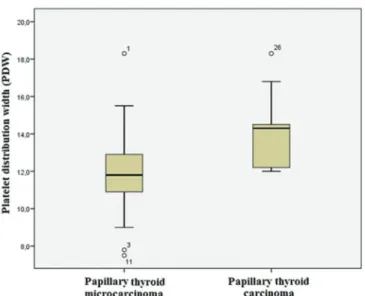
There was no significant difference between the groups in terms of age and gender (Table 1). Leukocyte count, neutrophil count and percentage, lymphocyte count and percentage, platelet count, plateletcrit, and MPV were not statistically different between groups (Table 1). PDW were significantly higher in patients with PTC than in those with PTMC (Figures 1).
Table 1. Comparison of age, gender distribution, and complete blood count parameters between groups
Papillary Thyroid Microcarcinoma (n=17) Papillary Thyroid Carcinoma (n=9) P Age 50.0 ± 13.1 46.1 ± 13.5 0.751 Gender Female 11 8 0.357 Male 6 1 Leukocyte count (x103/mm3) 7.48 ± 2.55 6.47 ± 1.77 0.367 Neutrophil count (x103/mm3) 4.89 ± 2.27 3.82 ± 1.89 0.058 Neutrophil percentage (%) 63.6 ± 8.3 56.9 ± 11.1 0.058 Lymphocyte count (x103/mm3) 1.96 ± 0.56 1.91 ± 0.55 1.000 Lymphocyte percentage (%) 27.2 ± 7.0 35.8 ± 15.6 0.075 Platelet count (x103/mm3) 258 ± 86 272 ± 84 0.560 Plateletcrit (%) 0.288 ± 0.229 0.251 ± 0.075 0.711 Platelet distribution width (%) 11.88 ± 2.62 14.12 ± 2.24 0.045
Mean platelet volume
(fL) 9.8 ± 3.0 9.3 ± 1.0 0.634
DISCUSSION
The extent of thyroid resection for especially low-risk disease in PTCs is a controversial issue (1). Thus, new biomarkers may be useful for the determination of the optimal treatment modality.
Platelet indices recently have started to be defined as beneficial markers in cancers (8). It has been shown that there is a bidirectional interaction between cancer and platelets. Namely, tumor cells have the ability to induce platelet activation and aggregation, and activated platelets participate in each step of cancer development (8). We found that PDW were significantly higher in patients with PTC than in those with PTMC (Figure 1).
Figure 1. Platelet distribution width (PDW) in patients with papillary thyroid microcarcinomaand papillary thyroid carcinoma There are a few studies that investigate platelet indices in PTC. Dincel and Bayraktar found that (9) PDW decreased in patients with PTC when compared with multi-nodular goiter patients and normal healthy subjects. Similarly, Yaylaci et al (10) showed that PDW significantly was lower in the papillary cancer group compared to benign goiter groups. Additionally, it has been determined that PDW was lower in T3 tumors than in T1 and T2 tumors, and there was no significant differences between cases with and without extrathyroidal extension with regard to PDW in patients with PTC (11). However, Li et al. (12) detected that PDW was higher in patients with thyroid cancer compared to the benign cases and moreover higher in patients with papillary carcinoma compared to follicular and medullary carcinoma.
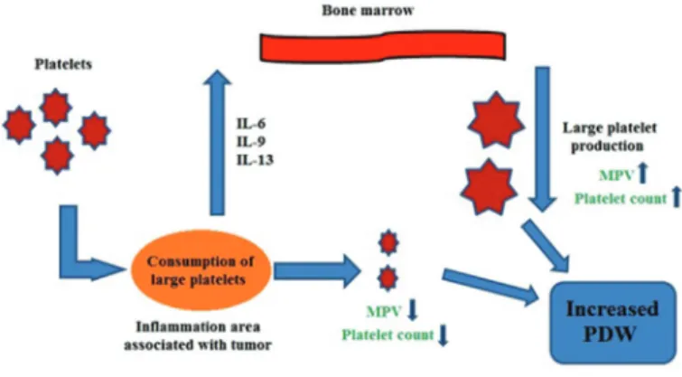
It has been suggested that PTC is a disease associated with chronic inflammation. In PTC, Beksac et al (13) found that IL-6 was higher in presurgical samples and returned to normal following surgery in patients with PTC. Likewise, Kobawala et al. (14) showed that IL-6 levels were remarkably higher in patients with PTC as compared to patients with benign thyroid diseases and that its levels had a significant positive correlation with tumor size, extrathyroidal extension, and distant metastasis. Zivancevic-Simonovic et al. (15) determined that IL-9 and IL-13 were higher in PTC patients than in healthy controls. All these cytokines (IL-6, IL-9, and IL-13) are known to be able to stimulate platelet production (16,17). Increased platelet generation may lead to occurrence of large thrombocytes (18), hence, to elevated PDW levels. We found that platelet count and MPV was not different between study groups. During inflammatory process, there is a recruitment of platelets to the area of inflammation where they are destroyed and their volume decreases (19,20). On this basis, we suggest that in patients with PTC, large platelet production increases in bone marrow, but platelet consumption and conversion of large platelets to small J Turgut Ozal Med Cent 2018;25(1):122-4
124 platelets also enhance in inflammation area; thus platelet count and MPV may remain unchanged and PDW may increase (Figure 2).
Figure 2. The possible mechanism of increased PDW
CONCLUSION
Our results show that patients with PTC have higher PDW levels compared to patients with PTMC. Elevated PDW in PTC may be useful in diagnosis of the disease and for better understanding of its pathogenesis. However, further and larger studies are needed to confirm this.
Conflicts of Interest, There are no any conflicts of interest.
REFERENCES
1. Miccoli P, Bakkar S. Surgical management of papillary thyroid carcinoma: an overview. Updates Surg 2017;69(2):145-50. 2. Pusztaszeri M, Auger M. Update on the cytologic features
of papillary thyroid carcinoma variants. Diagn Cytopathol 2017;45(8):714-30.
3. La Vecchia C, Malvezzi M, Bosetti C, Garavello W, Bertuccio P, Levi F, et al. Thyroid cancer mortality and incidence: a global overview. Int J Cancer 2015;136(9):2187-95.
4. Vuong HG, Duong UN, Altibi AM, Ngo HT, Pham TQ, Tran HM, et al. A meta-analysis of prognostic roles of molecular markers in papillary thyroid carcinoma. Endocr Connect 2017;6(3):R8-R17.
5. Dideban S, Abdollahi A, Meysamie A, Sedghi S, Shahriari M. Thyroid Papillary Microcarcinoma: Etiology, Clinical Manifestations,Diagnosis, Follow-up, Histopathology and Prognosis. Iran J Pathol 2016;11(1):1-19.
6. Sakorafas GH, Giotakis J, Stafyla V. Papillary thyroid microcarcinoma: a surgical perspective. Cancer Treat Rev 2005;31(6):423-38.
7. Menter DG, Kopetz S, Hawk E, Sood AK, Loree JM, Gresele P, et al. Platelet “first responders” in wound response, cancer,
and metastasis. Cancer Metastasis Rev 2017;36(2)199-213. 8. Mezouar S, Frere C, Darbousset R, Mege D, Crescence
L, Dignat-George F, et al. Role of platelets in cancer and cancer-associated thrombosis: Experimental and clinical evidences. Thromb Res 2016;139:65-76.
9. Dincel O, Bayraktar C. Evaluation of platelet indices as a useful marker in papillary thyroid carcinoma. Bratisl Lek Listy 2017;118(3):153-5.
10. Yaylaci S, Tosun O, Sahin O, Genc AB, Aydin E, Demiral G, et al. Lack of variation in inflammatory hematological parameters between benign nodular goiter and papillary thyroid cancer. Asian Pac J Cancer Prev 2016;17(4):2321-3.
11. Machairas N, Kostakis ID, Prodromidou A, Stamopoulos P, Feretis T, Garoufalia Z, et al. Trends in white blood cell and platelet indices in a comparison of patients with papillary thyroid carcinoma and multinodular goiter do not permit differentiation between the conditions. Endocr Res 2017:1-7.
12. Li N, Fu S, Cui MM, Niu Y, Li B, Liu ZP, et al. Platelet distribution width and serum albumin levels for discrimination of thyroid cancer from benign thyroid nodules. Asian Pac J Cancer Prev 2017;18(7):1773-7.
13. Beksac K, Sonmez C, Cetin B, Kismali G, Sel T, Tuncer Y, et al. Evaluation of proinflammatory cytokine and neopterin levels in women with papillary thyroid carcinoma. Int J Biol Markers 2016;31(4):e446-e50.
14. Kobawala TP, Trivedi TI, Gajjar KK, Patel DH, Patel GH, Ghosh NR. Significance of interleukin-6 in papillary thyroid carcinoma. J Thyroid Res 2016;2016:1-12.
15. Zivancevic-Simonovic S, Mihaljevic O, Majstorovic I, Popovic S, Markovic S, Milosevic-Djordjevic O, et al. Cytokine production in patients with papillary thyroid cancer and associated autoimmune Hashimoto thyroiditis. Cancer Immunol Immunother 2015;64(8):1011-9.
16. Cortin V, Garnier A, Pineault N, Lemieux R, Boyer L, Proulx C. Efficient in vitro megakaryocyte maturation using cytokine cocktails optimized by statistical experimental design. Exp Hematol 2005;33(10):1182-91.
17. Shi X, Cai W, Zhou Y, Zhang X, Xiong L, Li R, et al. IL-13 upregulates GPIIb expression in megakaryocytic cell lines via STAT6. Anat Rec (Hoboken) 2010;293(9):1470-6. 18. Fox JG, Barthold S, Davisson M, Newcomer CE, Quimby
FW, Smith A. The mouse in biomedical research: normative biology, husbandry, and models. Amsterdam: Academic Press; 2006:156.
19. Danese S, Motte Cd Cde L, Fiocchi C. Platelets in inflammatory bowel disease: clinical, pathogenic, and therapeutic implications. Am J Gastroenterol 2004;99(5):938-45.
20. Kapsoritakis AN, Koukourakis M, Sfiridaki A, Potamianos SP, Kosmadaki MG, Koutroubakis IE, et al. Mean platelet volume: a useful marker of inflammatory bowel disease activity. Am J Gastroenterol 2001;96(3):776-81.