İstanbul Bilim University, Department of Obstetrics and Gynecology, İstanbul, Turkey Correspondence: Nilgün Güdücü,
İstanbul Bilim University, Department of Obstetrics and Gynecology, İstanbul, Turkey Email: [email protected] Received: 03.09.2012, Accepted: 24.12.2012
Copyright © JCEI / Journal of Clinical and Experimental Investigations 2013, All rights reserved RESEARCH ARTICLE
Serial third trimester ultrasound examinations in predicting fetal weight
Ardışık üçüncü trimester ultrason incelemeleri ile fetal ağırlığın tahmin edilmesi
Nilgün Güdücü, Gökçenur Gönenç, Herman İşçi, Alin Başgül Yiğiter, İlkkan Dünder
ÖZET
Amaç: Bu çalışmanın amacı, üçüncü trimesterin değişik haftalarında ve doğumdan hemen önce yapılan ultraso-nografik ölçümlerin tahmini fetal ağırlığı tespit etmedeki doğruluğunu belirlemektir.
Yöntemler: Toplam 664 hastanın üçüncü trimesterde ya-pılmış 3 adet ultrasonografik verisi elde edildi ve Hadlock formülü ile tahmini doğum ağırlığı ve persentili hesaplan-dı. Daha sonra tahmini fetal ağırlık persentilleri, doğum-daki gerçek ağırlık persentili ile karşılaştırıldı.
Bulgular: Ortalama anne yaşı 30±4 (18-42) yıl, ilk üçüncü trimester ultrasonunun yapıldığı hafta ortalaması 32±0.5 hafta (30.4-33.6 hafta), ikinci ultrasonun yapıldığı hafta ortalaması 36±0.4 hafta (35-36.9 hafta), üçüncü ultraso-nun yapıldığı hafta ortalaması 38.6±1 hafta (36.3– 42 haf-ta). Doğumun gerçekleştiği ortalama gebelik haftası 39±1 hafta (36-42 hafta), ortalama doğum kilosu 3422±423 gram (1900-4900 gram). Birinci, ikinci, üçüncü ultrason ölçümlerinde elde edilen persentillerin doğum ağırlığı persentili ile korelasyonu (sırasıyla, r=0.556, p<0.001), (r=0.623, p<0.001 ), (r=0.747, p<0.001) istatistiksel ola-rak anlamlı bulundu.
Sonuç: Üçüncü trimesterin sonunda yapılan ultrasonog-rafik ölçümler fetal ağırlığı daha iyi tahmin ettirir, ancak üçüncü trimesterin başında yapılan ultrasonografik öl-çümlerin de fetal ağırlıkla korelasyonu iyidir ve gebelik yaşına göre küçük ve büyük bebeklerin yönetimi için daha avantajlı olabilir.
Anahtar kelimeler: Tahmini doğum ağırlığı, üçüncü tri-mester ultrasonu, Hadlock
ABSTRACT
Objective: The aim of this study was to compare the ac-curacy of estimated fetal weights obtained by ultrasonog-raphies performed at different weeks in the third trimester and just before delivery.
Methods: A total of 664 patients underwent three serial ultrasonographic examinations in the third trimester. The sonographically estimated fetal weight was calculated us-ing the Hadlock formula and then converted into percen-tiles. The estimated fetal weight percentiles were com-pared with the actual birth weight percentiles at delivery. Results: Mean maternal age was 30±4 years (18-42 years), mean gestational age at first ultrasound was 32±0.5 weeks (30.4-33.6 weeks), mean gestational age at second ultrasound was 36±0.4 weeks (35-36.9 weeks), mean gestational age at third ultrasound was 38.6±1 weeks (36.3– 42 weeks). Mean gestational age at deliv-ery was 39± 1 weeks (36-42 weeks), mean birth weight was 3422±423 grams (1900-4900 grams). Correlation coefficients of estimated fetal weights percentiles at first, second and third ultrasounds were significantly correlated with birth weight percentiles (r=0.556, p<0.001), (r=0.623, p<0.001), (r=0.747, p<0.001, respectively).
Conclusion: Ultrasonographic fetal weight estimations correlate with the actual birth weight better when per-formed in the late third trimester, but ultrasonographic fe-tal weight estimation early in the third trimester may allow for better follow up and planning of delivery both in small and large for gestational age fetuses. J Clin Exp Invest 2013; 4 (1): 28-33
Key words: Fetal weight estimation, third trimester ultra-sound, Hadlock formula
INTRODUCTION
Accurate prediction of birth weight is of paramount importance to reduce the chance of fetal morbidity and mortality. A fetus with growth restriction is at in-creased risk of hypoxia and perinatal death, on the other hand a macrosomic fetus is associated with an increased risk of cesarean section, fetal injury
and maternal complications.1,2 For the prediction of birth weight some authors developed formula by using only maternal characteristics,3 some authors compared equations depending on ultrasound mea-surements4 and some others compared ultrasound measurements with clinical estimation5 or com-bined clinical and ultrasonographic parameters in an equation.6
Previously it was shown that ultrasonographic fetal weight estimations between 34 and 37 weeks of gestation allow for more accurate prediction of birth weight than sonograms obtained just before birth and at term.7 Others recommended ultraso-nographic fetal weight estimation before delivery.8 The aim of our study was to investigate the accu-racy of estimated fetal weights (EFW) obtained by ultrasonographies performed at different weeks in the third trimester and just before delivery. We used Hadlock’s equations and assumed that the fetuses did not cross the growth percentiles from the day of the sonogram until delivery.9 To the best of our knowledge this is the first study that compared three serial ultrasonographic weight estimations in the third trimester.
METHODS
This was a retrospective study conducted by searching the data of pregnant women attending to our obstetrics policlinic between January 2006 and April 2011. In our clinic, routine ultrasonography was performed every month after 30 weeks of ges-tation. The study protocol was in line with the Dec-laration of Helsinki. All measurements were carried out by two obstetricians (NG, Hİ) using the 5-mega-hertz (MHz) curvilinear abdominal transducer, GE Electric Voluson 730 Expert. Gestational age was determined by the last menstrual period and only patients with a known first trimester crown-rump length (CRL) were included. Ultrasound documen-tations of the measurements of biparietal diameter (BPD), head circumference (HC), abdominal cir-cumference (AC) and femoral length (FL) were ob-tained. Exclusion criteria were pregnancies with in-complete information, known abnormal fetal karyo-type, congenital malformations, multiple pregnancy, maternal illnesses as diabetes, hypertension, gas-trointestinal malabsorption and also smoking. We did not exclude any case on the basis of abnormal fetal biometry or birth weight. We did not take the gender of the fetus, amniotic fluid volume and fe-tal presentation at the time of ultrasonography into consideration.
The study included 664 patients that underwent 1992 ultrasonographic measurements. Patients with serial fetal weight estimations between 30+1 and 33+6 weeks of gestation, between 35+1 and 36+6 weeks of gestation and then within 10 days before delivery were taken into consideration. EFW was calculated in all cases using the formula of Hadlock, which incorporates fetal BPD, HC, AC and FL10 and then converted into percentiles to compare with the birth weight percentiles, as this formula was
shown to be the most consistent throughout the studies performed in the normal pregnant popula-tion.11 The EFW percentiles were compared with the actual birth weight percentiles at delivery. Small for gestational age (SGA) was defined as below and equal to the 10th percentile for that gestational age and large for gestational age (LGA) was defined as above and equal to the 90th percentile for that ges-tational age.
Statistical analysis
For statistical analysis we used NCSS (Number Cruncher Statistical System) 2007 and PASS (Pow-er Analysis and Sample Size) 2008 statistical Soft-ware (Utah, USA). Data showing anthropometric parameters were presented as mean plus minus standard deviation. Data showing normal distribu-tion of parameters were compared with Paired sam-ples t-test, data showing non-normal distribution of parameters were compared with Wilcoxon test. For categorical analysis, we used McNemar test, Odds Ratio and Pearson’s correlation coefficient Within 95% confidence interval p values <0.05 were con-sidered as statistically significant. The results were considered statistically significant when the p-value was calculated less than 0.05 at a confidence inter-val of 95%.
RESULTS
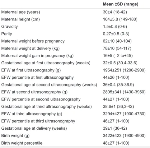
We included 664 patients in our study. The demo-graphic features of the patients were shown in Ta-ble 1. Mean maternal age was 30±4 years (18-42 years), mean maternal height was 164±5.8 cm (149-180cm), mean maternal weight before pregnancy was 62±10 kg (40-104 kg), mean maternal weight at delivery was 78±10 kg (54-117 kg), mean mater-nal weight gain during pregnancy was 16±5 kg (-2 to +45 kg), mean gestational age at first ultrasound was 32±0.5 weeks (30.4-33.6 weeks), mean gesta-tional age at second ultrasound was 36±0.4 weeks (35-36.9 weeks), mean gestational age at third ultrasound was 38.6±1 weeks (36.3–42 weeks), mean EFW at first ultrasound was 1954±251 grams (1200-2900), mean EFW at second ultrasound was 2805±341grams (1430-3950), mean EFW at third ul-trasound was 3294±427 grams (1900-4750), mean EFW percentile at first ultrasound was 44±26%, mean EFW percentile at second ultrasound was 44±27%, mean EFW percentile at third ultrasound was 46±27%. Mean gestational age at delivery was 39±1weeks (36-42 weeks), mean birth weight was 3422±423 grams (1900-4900 grams), mean birth weight percentile was 48±27. We used Paired
sam-ples t-test and Wilcoxon test to understand the ef-fects of patient characteristics on birth weight and estimated fetal weight. Maternal age (p<0.005), ma-ternal weight before pregnancy (p<0.001), mama-ternal
weight gain during pregnancy (p<0.01) and gravidi-ty (p<0.01) were statistically significantly associated with the birth weight percentile.
Table 1. Demographic features
of the patients Mean ±SD (range)
Maternal age (years) 30±4 (18-42)
Maternal height (cm) 164±5.8 (149-180)
Gravidity 1.5±0.8 (0-6)
Parity 0.27±0.5 (0-3)
Maternal weight before pregnancy 62±10 (40-104)
Maternal weight at delivery (kg) 78±10 (54-117)
Maternal weight gain in pregnancy (kg) 16±5 (–2 to+45) Gestational age at first ultrasonography (weeks) 32±0.5 (30.4-33.6) EFW at first ultrasonography (g) 1954±251 (1200-2900) EFW percentile at first ultrasonography 44±26 (1-100)
Gestational age at second ultrasonography (weeks) 36±0.4 (35-36.9) EFW at second ultrasonography (g) 2805±341 (1430-3950) EFW percentile at second ultrasonography 44±27 (1-100)
Gestational age at third ultrasonography (weeks) 38.6±1 (36,3-42) EFW at third ultrasonography (g) 3294±427 (1900-4750) EFW percentile at third ultrasonography 46±27 (1-100)
Gestational age at delivery (weeks) 39±1 (36-42)
Birth weight (g) 3422±423 (1900-4900)
Birth weight percentile 48±27 (1-100)
EFW: Estimated fetal weight, cm: centimeter, kg: kilogram, SD: Standard deviation Table 2 showed the correlations between the
first, second and third ultrasound measurements and the birth weight. All of the measurements cor-related with birth weight (r=0.556, r=0.623, r=0.747 at first, second and third ultrasound measurements respectively, p<0.001)
Table 2. Correlation between EFW percentile and birth weight percentile
Birth weight percentile
r p
First measurement 0.556 0.001**
Second measurement 0.623 0.001**
Third measurement 0.747 0.001**
r: Pearson correlation coefficient, **p<0.01
Table 3 showed the sensitivity, specificity, posi-tive (PPV) and negaposi-tive predicposi-tive values (NPV) of ultrasound in detecting SGA infants. Sensitivity of ultrasound measurements in detecting SGA in-fants were 37.5%, 50% and 59.4% and specificity of ultrasound measurements in detecting SGA in-fants were 93.8%, 92.3% and 95.7% respectively at first, second and third ultrasound measurements. PPVs for detection of SGA infants were 39.4%, 41% and 59.4% and NPVs for detection of SGA infants were 93.4%, 94.5% and 95.7% respectively for the first, second and third ultrasound measurements. Odds ratio was calculated as 9.13 (4.98-16.73), 12.04(6.77-21.4), 32.27 (17.09-60.89) respectively for the first, second and third ultrasound measure-ments.
Table 3. Sensitivity, specificity, positive and negative predictive values (PPV and NPV) of ultrasound in detecting SGA infants
Birth weight percentile
First measurement percentile Second measurement percentile Third measurement percentile
SGA Non-SGA SGA Non-SGA SGA Non-SGA
SGA 24 (3.6%) 37 (5.6%) 32 (4.8%) 46 (6.9%) 38 (5.7%) 26 (3.9%) Non-SGA 40 (6.0%) 563 (84.8%) 32 (4.8%) 554 (83.4%) 26 (3.9%) 574 (86.4%) Total 64 (9.6%) 600 (90.4%) 64 (9.6%) 600 (90.4%) 64 (9.6%) 600 (90.4%) OR 9.13 (4.98-16.73) 12.04 (6.77-21.40) 32.27 (17.09-60.89) Sensitivity (%) 37.5 50 59.4 Specificity (%) 93.8 92.3 95.7 PPV 39.4 41 59.4 NPV 93.4 94.54 95.7 Accuracy (%) 88.4 88.3 92.2
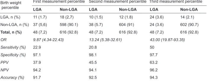
SGA: Small for gestational age, OR: Odds oranı, PPV: Positive predictive values, NPV: Negative predictive values Table 4 showed the sensitivity, specificity,
posi-tive and negaposi-tive predicposi-tive values of ultrasound in detecting LGA infants. Sensitivity of ultrasound mea-surements in detecting LGA infants were 22.9%, 20.8% and 50% and specificity of ultrasound mea-surements in detecting SGA infants were 97%, 98% and 97.7% respectively at first, second and third ultrasound measurements. PPVs for LGA infants
were 37.9%, 45.5% and 63.2% and NPVs for LGA infants were 94.2%, 94.1% and 96.2% respectively for the first, second and third ultrasound measure-ments. Odds ratio was calculated as 9.13 (4.98-16.73), 12.04 (6.77-21.4), 32.27 (17.09-60.89) re-spectively for the first, second and third ultrasound measurements.
Table 4. Sensitivity, specificity, positive and negative predictive values (PPV and NPV) of ultrasound in detecting LGA infants
Birth weight percentile
First measurement percentile Second measurement percentile Third measurement percentile
LGA Non-LGA LGA Non-LGA LGA Non-LGA
LGA, n (%) 11 (1.7) 18 (2,7) 10 (1.5) 12 (1.8) 24 (3.6) 14 (2.1) Non-LGA, n (%) 37 (5,6) 598 (90.1) 38 (5.7) 604 (91) 24 (3.6) 602 (90.7) Total, n (%) 48 (7,2) 616 (92.8) 48 (7.2) 616 (92.8) 48 (7.2) 616 (92.8) OR 9.87 (4.34-22.43) 13.24 (5.38-32.61) 43.00 (19.87-93.35) Sensitivity (%) 22.9 20.8 50 Specificity (%) 97.1 98.1 97.7 PPV 37.9 45.5 63.2 NPV 94.2 94.1 96.2 Accuracy (%) 91.7 92.5 94.3
LGA: Large for gestational age, PPV: Positive predictive values, NPV: Negative predictive values On receiver operating curve analysis areas
un-der the curve for SGA were 0.781, 0.861 and 0.911 for the first, second and third ultrasound measure-ments (p<0.001 for all). On receiver operating curve
analysis area under the curve for LGA were 0.752, 0.791 and 0.881 for the first, second and third ultra-sound measurements (p<0.001 for all).
DISCUSSION
This study showed that ultrasound examinations done in the third trimester to predict fetal birth weight performed accurately. Ultrasound identified both SGA and LGA infants accurately. Sensitivity of ultrasound was better when a SGA fetus was diag-nosed and specificity was better when a LGA fetus was diagnosed at all gestational weeks. Previously Pressman et al announced that ultrasonographic fetal weight estimations predicted birth weight bet-ter when performed between 34 and 36.9 weeks of gestation than ultrasonographic fetal weight esti-mations done at a later gestational age. We com-pared three serial ultrasonographic measurements and the EFWs obtained at these ultrasonographies correlated with birth weight better when performed after 37 weeks of gestation, just before the deliv-ery. Accuracy of birth weight prediction improved as the term was approached, when more than one US estimation of fetal weight is available depend on the last one said a previous study,12 our study con-firmed this. We can expect fetal weight estimation prior to labor to be inaccurate due to low position of the head, molding of the bones, distortion of the abdominal circumference and posterior position of the femur. The low sensitivity and high specificity in LGA and SGA groups proved that the ultrasound overestimated LGA and underestimated SGA fe-tuses. The PPVs for SGA and LGA were low and the NPVs for SGA and LGA were high, the results improved as the gestational age approached term. These results were similar to those reported previ-ously.13-15 The PPVs and NPVs were similar at all measurements and high NPVs are important for the exclusion of SGA and LGA fetuses. Although ultra-sonographies after 37 weeks predicted birth weight better, identification of SGA fetuses at an earlier gestational week might give us the chance of close monitoring and planning of delivery to reduce peri-natal risks. Ultrasound at an earlier gestational age also allows timely identification of placental localiza-tion problems.
A previous study by Nelson et al found the abil-ity of early third trimester ultrasonography enough to predict LGA babies of diabetic mothers.16 We excluded patients with GDM and DM because their fetuses are exposed to increased intrauterine nu-trients that result in extreme fetal adiposity and growth.17 Normal fetuses are expected not to cross the growth curve percentiles,9 therefore patients with diseases that were expected to cause growth abnormalities were excluded.
We presented the results of serial ultrasonog-raphies performed by two clinicians; the knowledge
of previous ultrasound measurements by the so-nographers might have caused bias in the latter measurements.
Amniotic fluid volume may affect the accuracy of the measurements by affecting the image quality and by distorting the abdominal circumference18,19 and we did not take it into consideration. Maternal adiposity may also affect the image quality. We did not consider the gender-related differences in fetal weight calculation proposed by previous studies.20,21 We did not account the effect of breech presenta-tion on the accuracy of birth weight calculapresenta-tion, a recent study found that the presentation had no ef-fect on EFW when the Hadlock formula was used.22 There was no difference in the prediction of the birth weight of the fetus between nullipara and multipara. Prior knowledge that a fetus is macrosomic may be useful in planning induction of labor after fetal lung maturation to avoid cesarean section. It may also help in counseling parents regarding the complications associated with the vaginal delivery of a macrosomic fetus. To rely on the estimated fetal weight completely may also increase over-manage-ment. Previous studies suggested that induction of labor for suspected macrosomia increased the ce-sarean section rates without improving the perinatal outcome.23,24
In conclusion ultrasonographic fetal weight es-timations correlate with actual birth weight better when performed in the late third trimester, but ul-trasonographic fetal weight estimations in the early third trimester may allow for better follow up and planning of delivery both in SGA and LGA fetuses.
REFERENCES
1. Heiskanen N, Raatkainen K, Heinonen S. Fetal macro-somia- a continuing obstetric challenge. Biol Neonate 2006;90:98-103.
2. Medchill MT, Peterson CM, Kreinick C, et al. Prediction of estimated fetal weight in extremely low birth weight neonates (500-1000 g). Obstet Gynecol 1991;78:286-290.
3. Nahum GG, Stanislaw H. Validation of a birth weight prediction equation based on maternal characteris-tics. J Reprod Med 2002;47:752-760.
4. Mongelli M, Benzie R. Ultrasound diagnosis of fetal macrosomia: a comparison of weight prediction mod-els using computer simulation. Ultrasound Obstet Gy-necol 2005;26:500-503.
5. Chauhan SP, Hendrix NW, Magann EF, et al. Limita-tions of clinical and sonographic estimates of birth weight: experience with 1034 parturients. Obstet Gy-necol 1998;91:72-77
6. Halaska MG, Vlk R, Feldmar P, et al. Predicting term birth weight using ultrasound and maternal character-istics. Eur J Obstet Gynecol Biol 2006;128:231-235 7. Pressman EK, Bienstock JL, Blakemore KJ, et al.
Pre-diction of birth weight by ultrasound in the third trimes-ter. Obstet Gynecol 2000;95:502-506.
8. Krantz C, Hitschold T. Impact and limitations of ultra-sound examination immediately before delivery-a prospective evaluation with 1127 pregnant women. Z Geburtshilfe Neonatol 2011;215:69-76.
9. Mongelli M, Gardosi J. Gestation-adjusted projection of estimated fetal weight. Acta Obstet Gynecol Scand 1996;75:28-31.
10. Hadlock FP, Harrist RB, Sharman RS, et al. Estima-tion of fetal weight with the use of head, body, and fe-mur measurements: a prospective study. Am J Obstet Gynecol 1985;151:333-337.
11. Dudley NJ. A systematic review of the ultrasound es-timation of fetal weight. Ultrasound Obstet Gynecol 2005;25:80-89.
12. Larsen T, Greisen G, Petersen S. Prediction of birth weight by ultrasound-estimated fetal weight: a com-parison between single and repeated estimates. Eur J Obstet Gynecol Reprod Biol 1995;60:37-40.
13. Ben-Haroush A, Yogev Y, Bar J, et al. Accuracy of so-nographic estimated fetal weight in 840 women with different pregnancy complications prior to induction of labor. Ultrasound Obstet Gynecol 2004;23:172-176. 14. Colman A, Maharaj D, Hutton J, et al. Reliability of
ultrasound estimation of fetal weight in term singleton pregnancies. N Z Med J 20068;119:2146-2149. 15. Ben-Haroush A, Yogev Y, Hod M, et al. Predictive
value of a single early fetal weight estimete in normal pregnancies. Eur J Obstet Gynecol Biol 2007;130:187-192.
16. Nelson L, Wharton B, Grobman WA. Prediction of large for gestational age birth weights in diabetic mothers based on early third trimester sonography. J Ultrasound Med 2011;30:1625-1628.
17. Kehl RJ, Krew MA, Thomas A, Catalano PM. Fetal growth and body composition in infants of women with diabetes mellitus during pregnancy J Matern Fetal Med 1996;5:273-280.
18. Blann DW, Prien SD. Estimation of fetal weight before and after amniotomy in the laboring gravid woman. Am J Obstet Gynecol 2000 ;182:1117-1120.
19. Townsend RR, Filly RA,Callen PW, et al. Factors af-fecting prenatal sonographic estimation of weight in extremely low birthweight infants. J Ultrasound Med 1988;7:183-187.
20.Schild RL, Sachs C, Fimmers R, et al. Sex-specific fe-tal weight prediction by ultrasound. Ultrasound Obstet Gynecol 2004;23:30-35.
21. Schwarzler P, Bland JM, Holden D, et al. Sex-specific antenatal reference growth charts for uncomplicated singleton pregnancies at 15-40 weeks of gestation. Ultrasound Obstet Gynecol 2004;23:23-29.
22. McNamara JM, Odibo AO, Macones GA, Cahill AG.The effect of breech presentation on the accuracy of estimated fetal weight. Am J Perinat 2012;29:353-360.
23. Sanchez- Ramos L, Bernstein S, Kaunitz AM. Expect-ant management versus labor induction for suspected fetal macrosomia: a systematic review. Obstet Gyne-col 2002;100:997-1002.
24. Weiner Z, Ben-Shlomo I, Beck-Fruchter R, et al. Clini-cal and ultrasonographic weight estimation in large for gestational age fetus. Eur J Obstet Reprod Biol 2002;105:20-24.