Dicle Tıp Dergisi, 2009 OLGU SUNUMU Cilt: 36, Sayı: 1 , (56-58)
Yazışma Adresi: Sultan Ecer Menteş, Dicle Üniversitesi Tıp Fak., Çocuk Sağ. ve Hast. AD, Diyarbakır Tel: 0 412 2488001 Fax:0 412 2488440 E-mail: [email protected]
Geliş Tarihi : 02.11.2007 Yayına Kabul Tarihi : 10.01.2008
Acute hemorrhagic edema of infancy
Sultan Ecer Menteş¹, Mustafa Taşkesen2, Selahattin Katar3, M.Emin Günel4, Sedat Akdeniz5 Departments of 1,2,3,4Pediatrics and 5Dermatology, Dicle University, Faculty of Medicine, Diyarbakır, Turkey
ABSTRACT
Acute hemorrhagic edema of infancy is a rare form of leukocytoclastic vasculitis. Mostly it appears under three years of age and is characterized by purpuric skin lesions, fever and edema. A three years-old boy, who has cough and coryzea was admitted to our clinic for fever and red spots on legs and arms. In physical examination; ecimotic skin lesions on right ear, face, arms, dorsal of the hands, buttocks, legs and dorsal of the feet were found. In the laboratory tests acute phase reactants were elevated and blood coagulation tests were in normal range. Hepatit A,B,C and TORCH markers were negative. Punch biopsy obtained from gluteal area showed leukositoclastic vasculity. Focal fibrinogen accumulation was detected by immun fluorescent microscopy. Regression on lesions was not observed despite supportive therapy, so prednisolone (1 mg/kg/day) therapy was started. On the third day of the steroid therapy, complete recovery was achived.
Key words: Hemorrhagic edema of infancy , leukocytoclastic vasculitis, steroid therapy Bebeklik dönemi akut hemorajik ödemi
ÖZET
Akut infantil hemorajik ödem lökositoklastik vaskülitin bir formu olup, daha çok üç yaş altı çocuklarda ateş, ödem ve purpurik deri lezyonları ile karakterizedir. Üç yaşında öksürük ve soğuk algınlığı olan erkek çocuk kliniğimize bacak ve kollarında kırmızı renkli döküntü, ateş nedeni ile başvurdu. Fizik incelemede sağ kulak üstünde, yüzünde, kollarında ve ekstremitelerinde ekimotik lezyonlar bulundu. Laboratuvar sonuçlarında akut faz reaktanları pozitif, koagulasyon testleri ise normal idi. Hepatit A,B,C ve TORCH belirteçleri negatif idi. Gluteal bölgeden alınan punch biyopsi sonucu lökositoklastik vaskülit ile uyumlu ve immünfloresan incelemede lokal fibrinojen birikimi tespit edildi. Destek tedavisine rağmen lezyonlarında gerileme gözlenmeyen hastaya bu nedenle 1mg/kg/gün prednizolon tedavisi başlandı. Tedavinin üçüncü gününde lezyonlar tamamen düzeldi.
Anahtar kelimeler: infantil hemorajik ödem, lökositoklastik vaskülit, steroid tedavisi INTRODUCTION
Acute hemorrhagic edema of infancy (AHEI) is a leukocytoclastic vasculitis; clinically characterized by purpuric skin lesions, fever and edema1. There is an abrupt
onset of the large cockade, annular, or targetoid lesions involving the face, ears, and extremities. Scrotal lesions have been reported rarely. The edema is nontender and mostly symetric2.The disease must be considered on
discrimination of meningococcal infection, septicemia, purpura fulminans and other purpuric skin eruptions especially Henoch
schönlein purpura3,4. In this case, three years
old, asymetric skin lesions mostly on right and edema, was presented.
CASE REPORT
A three years old boy, had complaint of fever and red spots on legs and arms. The complaints of cough and coryzea had started three days ago, then red spots on the legs were realised in the evening of the next day by his parents. This situation became widespread on face, legs and arms in the morning of the third day. It was reported that the patient had not
S. Ecer, M. Taşkesen, S. Katar, M.E. Günel, S. Akdeniz Dicle Tıp Dergisi, 2009
57
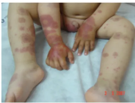
been vaccinated or drugged in earlier time. In physical examination; weight:14 kg, height:96 cm, temp:37.5ºC, blood pressure:100/70 mmHg, there were ecimotic skin lesions on right ear, face, arms, dorsal of the hands, buttocks, legs and dorsal of the feet (Figure 1). The laboratory resulted values: hemoglobin: 10gr/dl, amount of leukocytes in blood: 9.700/mm³, eriythrocytes: 5.1million/mm³,
trombocytes: 360.000/mm³, eriythrocyte
sedimentation rate (ESR): 41mm/h, C-Reactive Protein (CRP): 90mg/dl, ASO 25 todd/U, Romatoid factor: 20 IU/ml, fibrinogen 392mg/dl. Periferic blood smear: 56% polimorf nuvel leukocytes (PNL) 44% lymphocyte, activated partial tromboplastin time (aPTT) 26.1sec, prothrombin time (PT) 12.6sec, INR 1.05, serum biochemical
parameters, immunglobulins, C3, C4,
Antitrombin Ш, protein C, protein S, eozinofil katyonic protein (ECP) in normal values, salmonella and brucella antibodies were negative in serology study. Hepatit A,B,C parameters were negative in elisa test other than depending on vacine antiHbS 1000 IU/L, TORCH markers negative, direct urine examination normal. Abdomen and scrotal ultrasonography, bilateral lower extremity deep vena-arteria system colored doppler research were in normal limits. Punch biopsy resulted from gluteal area cross-section. Under the multi storey epitel, near the vascular or on vascular walls mixed type inflammation cell infiltery includes neutrophils were detected and accommodated with leukositoclastic vasculity. Focal fibrinogen accumulating was detected by immun fluorescent microscopy. AHEI was diagnosed by all these clinical studies and symptoms. Despite the supportive therapy regression on lesions was not observed. On the eighth day, the patient still had not recovered completely and the laboratory studies showed; leukocytes: 10.500/mm³, eriythrocytes: 4.99million/mm³ thrombocytes: 354.000/mm³ , CRP: 8.65mg/dl,
ESR: 32mm/h. Steroid (prednisolone
1mg/kg/day) therapy was given. On the third day of the steroid therapy, complete recovery
was reached. In the follow up no complication was detected.
Figure 1: The asymmetric purpuric lesions and edema which located dominantly on the right side of the body are seen.
DISCUSSION
The etiology of the AHEI is not known exactly today and 12% of leukocytoclastic vasculitis are AHEI, many of them are seen between four month to two years old, not depending on gender and especially the patients who have a past with having cold before5. This disease is defined as an
immunologic vasculity which progresses against different antigenic stimulants. 75% of the cases have a past of having an infection, using drugs or immunization background3.
Patient had not been vaccinated or drugged in earlier time. Especially purpuric skin lesions on face, buttock and extremities medallion like ecimotic purpura with inflammation edema on extremities and face are two important characteristic about AHEI6,7. In our case, there
is an ecimotic purpura on scrotal area which is unusual. Beside this, the disease was not in symetric character in clinically, the right side was dominant in spite of its characteristic specialitiy of symetrical progression. There are some data support that immun complexies pathogenesis of leukocytoclastic vasculitis cause derms to be poisoned by settling on vessel walls. The starters of the immun
complex producing on leukocytoclastic
vasculitis are not known well. Furthermore viral and bacterial infection, drugs and chemicals with other proteins are put forward as etiologic factors. The pathogens from
S. Ecer, M. Taşkesen, S. Katar, M.E. Günel, S. Akdeniz Dicle Tıp Dergisi, 2009
58
infections that have the best known connection with leukocytoclastic vasculity are: group A or B hemolytic streptococci, Staphylococcus aureus, Mycobacterium leprae, hepatitis B and C viruses, HIV, and cytomegalovirus. The drugs which cause leukocytoclastic vasculity such as various antibiotics (penicilin), thiazids and some nonsteroid antiinflammatories are reported3. Several reports have described cases
that systemic corticosteroids and
antihistaminics are ineffective on therapy of cutaneous lesions5. Our patient who had
cutaneous lesions which had not been retreating with supportive therapy for eight days has recovered with steroid therapy for three days. In this study we emphasize that steroid might be need on therapy of AHEI and seperative diagnosis must be claimed.
REFERENCES
1. Shah D, Goraya J, Poddar B, Parmar V, Acute Infantile Hemorrhagic Edema and Henoch-Schonlein Purpura Overlap in a Child. Pediatr Dermatol 2002;19:92-3.
2. Odom RB, James WD, Berger TG: Andrew’s Disease of the Skin Clinical Dermatology. Ninth edition. Philadelphia, WB Saunders Company, 2000;1035-1036.
3. Tınaztepe K, Güçer Ş. Lökositoklastik vaskülit. Katkı Pediatri Dergisi 1995;16:152-164.
4. Beselga E, Drolet BA, Esterlag NB. Purpura in infants and childiren. J Am Acad Dermatol 1997;37:673-674.
5. Legrain V, Lejean S, Taieb A, Guillard JM, Battin J, Malaville J. Infantile Acute Hemorrhagic Edema of skin: study of ten cases. J Am Acad Dermatol 1991;24:17-22.
6. Millard T, Haris A, McDonald D. Acute Infantile Hemorrhagic Edema. J Am Acad Dermatol 1999;41:837-839.
7. Cunningham BB, Caro WA, Eramo LR. Neonatal acute hemorrhagic edema of childhood: case report and review of the English-language literature. Pediatr Dermatol 1996;13: 39-44.