Relationships between Abelson tyrosine kinase (c-abl) and spindle
during meiotic division in mouse oocyte in vitro
*Duygu MUTLUAY
1, Hakan ÖNER
11Mehmet Akif Ersoy University, Faculty of Veterinary Medicine, Department of Histology and Embryology, Burdur, Turkey.
Summary: The c-Abl gene is a nonreceptor tyrosine kinase that has roles in cell growth, control of the cell cycle, initiation of
pronuclear movement and actin cytoskeleton dynamics. The present study has examined the localization of c-Abl protein during oocyte activation in mouse in vitro. Confocal immunofluorescence analysis showed that c-Abl originates at the cortex with a crescent shape before the activation and continues up along both sides of the cortex after activation during meiotic division until pronuclear stage. These results demonstrate that c-Abl may play role in asymmetric division of the egg to produce polar body, positioning of the spindle, and may regulate the microtubule cytoskeleton or have some roles in this process.
Keywords: c-Abl, meiotic division, oocyte activation, spindle positioning
Fare oositlerinde mayoz bölünme sırasında Abelson tirozin kinaz (c-abl) ile mekik iplikleri arasındaki
in vitro ilişki
Özet: c-Abl geni, hücre büyümesi, hücre döngüsünün kontrolü, pronüklear hareketin başlatılması ve aktin sitoskeleton
dinamikleri üzerinde rol oynayan bir nonreseptör tirozin kinazdır. Bu çalışmada, fare de oosit aktivasyonu sırasında c-Abl proteininin lokalizasyonu in vitro olarak incelenmiştir. Konfokal immünfloresans analizi, c-Abl'in aktivasyon öncesinde kortekste yarım ay şeklinde lokalize olurken aktivasyon sonrasında mayoz bölünmeden pronükleus evresine kadar korteksin her iki tarafını sararak yukarı doğru devam ettiği gösterilmiştir. Bu sonuçlar, c-Abl'nin yumurta hücresinin kutup cisimciğinin oluşumu için gerekli olan asimetrik bölünmede, mekik ipliği pozisyonlandırılmasında rol oynayabileceğini ve mikrotübül hücre iskeletini düzenleyebileceğini veya bu süreçte bazı rollere sahip olabileceğini göstermektedir.
Anahtar sözcükler: c-Abl, mayoz bölünme, mekik ipliği pozisyonlandırılması, oosit aktivasyonu.
Introduction
The Abl family of nonreceptor tyrosine kinases have members, c-Abl (Abelson tyrosine kinase; Abl1) and Arg (Abl-related gene; Abl2). c-Abl gene was first encountered as an oncogene in the Abelson murine leukaemia virus (1) and later the human ortholog of c-Abl was identified as a part of a mutationally activated fusion oncoprotein Bcr-Abl1 common in human leukemias (3, 8). c-Abl belongs to Src family and contain N-terminal cap, myristoylation site, SRC homology 3 (SH3), SH2 and SH1 (tyrosine kinase) domains. The carboxyl termini contain nuclear localization and export signals, globular and filamentous actin binding domain and a DNA- binding domain (6, 10). Thus, c-Abl is localized at the plasma membrane, cytoplasm, nucleus and is associated with actin filaments (11). It has been suggested that c-Abl has a role in cell growth, epithelial cell-cell adhesion, polarity, migration, invasion and development of female
* This study was prepared from PhD thesis entitled “Localization of c-Abl protein in preimplantation embryo development” of the
first author.
and male mouse germ cells (4, 6, 13, 25). Previous studies showed that c-Abl supports development and has a role on preimplantation embryo development, implantation and placentation throughout the pregnancy (21, 28).
In mammalian oocytes, meiotic maturation is a process in which oocytes undergo two cellular divisions, and is vital for production of a functional gamete,
successful fertilization and subsequent embryo
development (5). During the first meiotic division, asymmetric cell division occurs and the microtubules form the spindle in the center of the oocyte. When the spindle has reached the cell cortex, the metaphase-anaphase transition is triggered and the first polar body forms and is released in actin rich cortical domain. While unfertilized oocytes are arrested in the metaphase II (MII), spindle is located parallel to the surface under a cortical domain. Embryonic development of the mouse is initiated by fertilization of oocytes by sperm. After fertilization, two
cortical bumps form in this cortical domain over the two sets of anaphase chromosomes and the spindle rotates for cleavage to take place and for the release of the second polar body (16). Fertilization is followed by mitotic cell divisions and cleavage divisions to generate blastomers.
In order to understand the role of c-Abl protein during meiotic division, we examined the localization of
c-Abl by using immunofluorescence confocal
microscopy. Significantly, we showed for the first time that c-Abl may play roles on oocyte activation in mouse in
vitro.
Materials and Methods
Animals and collection of oocytes: The studies
included in this project were performed at the University of Hawaii. The protocol for animal handling and use was reviewed and approved by the Institutional Animal Care and Use Committee of the University of Hawaii on July 19, 2012 with a protocol number of 05-029-8. B6D2F1 (C57BL/6 females × DBA/2 males; National Cancer Institute) female mice were used in this study. B6D2F1 females of 6-8 weeks of age were superovulated by intraperitoneal injections of 5 IU of equine chorionic gonadotropin (pregnant mares serum gonadotropin – PMSG) and human chorionic gonadotropin (hCG). The superovulated mice were sacrificed 15 hours after the hCG injection to dissect the oviducts. After the dissection of the oviducts, the oviductal ampulae were broken to release the cumulus-oocyte complexes in FHM HEPES-buffered medium (Millipore). Oocytes were freed from cumulus cells by exposure to 0.5% hyaluronidase (Sigma) in FHM HEPES-buffered medium. Then unfertilized, MII oocytes were cultured in KSOM-AA medium (Millipore) overlaid
with mineral oil at 37ºC in a 5% CO2 incubator for the
experiments (22).
Activation of oocytes by parthenogenesis: MII
oocytes freed from cumulus cells were incubated in activating medium prepared with strontium chloride
(SrCl2) (0.5M) and ethylene glycol-bis (β-aminoethyl
ether)-N,N’-tetraacetic acid (EGTA; Sigma) (0.5M) diluted in KSOM-AA medium under mineral oil in a 5%
CO2 incubator at 37ºC (15).
Fixation and immunofluorescent staining: MII and
artificially activated oocytes were fixed with 4% paraformaldehyde solution in phosphate-buffered saline for 20-30 minutes at various times after treating embryos with 0.5 % Pronase (Roche) in FHM for 10 minutes to remove the zona-pellucida. Randomly chosen oocytes fixed at MII oocyte (n=18) stage and the others fixed after (1h, 2h, 3h, 6h) activating the oocytes (n=16/per each) by strontium chloride. Oocytes were washed in PBS containing 0.1% Tween-20 (PBSw) and permeabilized with 0.5% Triton X-100 in PBS for 15 min at 25ºC. After blocking with 5% bovine serum albumin in PBSw
(blocking solution), samples were incubated overnight with primary antibody at 4ºC and following day oocytes were incubated in secondary antibody for 2-3 hours at 25ºC. The primary antibodies used were, rabbit anti-c-Abl (c-19) (1:400; Santa Cruz Biotechnology), mouse anti-β-tubulin (1:10000; Invitrogen). Secondary antibodies (1:1000; Life Technologies) used were conjugated with Alexa Fluor 488 (Green), namely goat anti-mouse, and conjugated with Alexa Fluor 546, namely goat anti-rabbit. Additionally, we did not find any non-specific Ab binding by processing samples as described above in the absence of primary antibody. Stained samples were mounted on a glass slide with ProLong Gold antifade reagent containing
4’,6’-diamidino-2-phenylindole (DAPI; Life
Technologies) (20).
Microscopy and image analysis: Oocytes from the
same experiment were imaged in the same session using a FV1000 confocal laser scanning microscope. For confocal microscopy, serial optical sections were imaged at 1-2 µm intervals under a 40x objective lens with oil.
Results
Expression and distribution of c-Abl proteins during oocyte activation: In order to determine whether the c-Abl
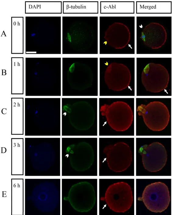
protein is expressed during oocyte activation and the relationships between c-Abl and spindle, we artificially activated zona pellucida-free oocytes by using strontium chloride that induces parthenogenetic activation of oocytes. We also viewed the distribution of c-Abl protein and the spindle by immunostaining with an anti-c-Abl and anti β-tubulin antibody using confocal microscopy. We observed that c-Abl was localized in the cytoplasm and cell periphery but enriched in the cell cortex at MII arrested oocytes and throughout the activation. In the MII arrested oocytes that were collected at 0hr, the c-Abl protein was enriched at the cell cortex while it was absent above the area where the spindle apparatus positioned. We observed, during MII arrest, that the metaphase spindle was located parallel to the surface under a cortical domain, and was positively stained with c-Abl protein (Figure 1A). We also detected that c-Abl protein was localized at the spindle at 1hr-2hr-3hr after activation (Figure 1B,1C, 1D).
After activation (1hr), two cortical bumps started to form this cortical domain over the two sets of anaphase chromosomes where the c-Abl staining was absent (Figure 1B). In addition to that, distribution results indicate there was no difference in c-Abl localization comparing 0 hr and 1hr (Figure 1B). While the oocyte (2-3hr) triggers a 90° spindle rotation and the second polar body was extruded, c-Abl proteins also started to localize at the cortex that is adjacent to the spindle (the side where c-Abl was absent in 0hr group) (Figure 1C,1D). 6hr after the activation c-Abl enriched at all around the cortex of the egg (Figure E).
Figure 1. Distribution of c-Abl protein at MII and activated oocytes under a confocal microscopy. Zona-free oocytes were artificially activated in vitro and samples were fixed at different times during activation to view different stages of division. Samples presented were fixed at 0 hr (A), 1hr (B), 2hr (C), 3hr (D) and 6hr (E) before and after the activation procedure. (A) At 0 hr (MII arrested oocytes) c-Abl protein was enriched in the egg cortex as a crescent shape (arrow), it was absent above the area where the spindle apparatus positioned (white arrowhead) and spindle apparatus was positively stained with c-Abl protein (yellow arrowhead). (B) At 1hr, localization of the c-Abl protein was like MII arrested oocytes (arrow). Metaphase spindle was stained with c-Abl protein (yellow arrowhead). (C) At 2h and (D) At 3hr, the secondary polar body was extruded (arrowhead) and c-Abl protein started to localize at the cortex that adjacent to the spindle (arrow). (D) 6hr after the activation c-Abl enriched at all around the cortex of the egg (arrow). Confocal images of the samples was stained with c-Abl (red), β-tubulin (showing spindle) (green), chromatin and pronucleus were stained with DAPI (blue). Scale bar represents 20μm. c-Abl: Abelson tyrosine kinase; MII: Metaphase II; DAPI: 4’,6’-Diamidino-2-phenylindole.
Şekil 1: MII ve aktive olmuş oositler de c-Abl proteinin konfokal mikroskop altında dağılımı. Zona içermeyen oositler yapay olarak in
vitro koşullarda aktive edildi ve örnekler aktivasyon sırasında farklı zamanlarda farklı bölünme evrelerini gözlemlemek için tespit
edildi. Tespit edilen örnekler aktivasyon öncesinde ve sonrasında 0 sa (A), 1sa (B), 2sa (C), 3sa (D) ve 6sa (E) şeklinde gösterildi. (A) 0. saatte (MII bekleyen oositler) c-Abl proteinin yumurta korteksinde yarım ay şeklinde yoğunlaşmıştır (ok), mekik ipliğinin yer aldığı alanın üstünde yer almamaktadır (beyaz ok ucu) ve mekik ipliği c-Abl proteini ile pozitif olarak boyanmıştır (sarı ok ucu). (B) 1.saatte c-Abl proteinin lokalizasyonu MII’de bekleyen oositlerde olduğu gibidir (ok). Metafaz ipliği c-Abl proteini ile boyanmıştır (sarı ok ucu). (C) 2. saatte ve (D) 3. saatte ikinci kutup cisimciğinin çıktığı (ok ucu) ve c-Abl proteinin mekik ipliğinin bitişiğinde bulunan korteks kısımlarında da lokalize olmaya başlamıştı (ok). (D) Aktivasyondan 6 saat sonra c-Abl tüm yumurta korteksi boyunca yoğunlaşmıştır. c-Abl (kırmızı), β-tubulin (mekik ipliğini gösteren) (yeşil), kromatin ve pronükleus DAPI (mavi) ile boyanan örneklerin konfokal görüntüleri. Ölçüm barı 20μm’yi göstermektedir. c-Abl: Abelson tirozin kinaz; MII: Metefaz II; DAPI: 4’,6’-Diamidino-2-phenylindole.
Together, these data demonstrate that c-Abl originates at the cortex with a crescent shape before the activation and continues up along both sides of the cortex after activation during meiotic division until pronuclear stage. This finding suggests that c-Abl may play role in “asymmetric division of the egg to produce polar body”. We also observed c-Abl staining on the spindle during meiotic division, suggesting that c-Abl may regulate the microtubule cytoskeleton or have some roles in this process.
Discussion and Conclusion
The current study showed that c-Abl is expressed during the egg activation in mouse. We also analyzed the distribution of c-Abl and showed the relationship between c-Abl tyrosine kinase and spindle during meiotic division which c-Abl protein is considered to be important. We did observe an asymmetric distribution at MII arrested oocytes.
Based on the literature, the c-Abl expression and localization during preimplantation embryo development demonstrated that c-Abl may have a role in embryo development (2, 21). In our previous study, we showed the localization of c-Abl and suggested a role in trofectoderm formation and differentiation that is essential for implantation and placentation (21). c-Abl knock-out mice exhibit neonatal lethality, lymphopenia (23) and reduced fertility (14).
Dynamic organization of the oocyte cytoskeleton during meiotic division is important. Defects in the cytoskeleton organization during divisions can cause chromosome segregation errors (5). Previous studies showed that c-Abl protein was associated with cytoskeletal components (18). The c-Abl kinase is activated by physiological signals that regulate the actin cytoskeleton (27). Walker et al., demonstrated that Abl kinase is related with the cortical cytoskeleton of the fertilized egg (24). It has also been reported that Abl kinases and their distribution to cytoskeletal structures suggest that they have associated functions in cytoskeletal regulation (12). This study demonstrated that c-Abl is localized in the cytoplasm and cell periphery but is enriched in the cell cortex; suggesting that c-Abl may be associated with the cortical cytoskeleton in mouse.
Moore and Kinsey (1995) suggested that c-Abl is activated following fertilization and may play roles in the later events of egg activation. The group also indicated that c-Abl may be effective on the initiation of pronuclear movement and entry into the S phase of the cell cycle (19). We did observe an asymmetric distribution at MII arrested oocytes like we detected in our previous study (21). Before the egg activation c-Abl is distributed in the cortex, but is absent on the side where the spindle apparatus is placed
and c-Abl starts to localize all around the egg cortex as activation progresses (2hr-3hr-6hr).
Our results suggest the possibility that c-Abl may also control or play role in the setting up of the asymmetric division during meiosis. It has been known that loss of asymmetry in the meiotic division and disorganization of the oocyte cytoskeleton are characteristic of ageing or low quality gametes (7, 26). Matsumura et al. (2012) indicated that c-Abl is a regulator of spindle orientation in mouse epidermis and epithelial cells. In addition, loss of c-Abl induces spindle misorientation (17). Spindle orientation is important for asymmetric cell division (9) and spindle microtubules and actin microfilaments control the asymmetry of the meiotic divisions (5). We demonstrated the relationship between c-Abl and spindle by immunofluorescent staining suggesting the possible roles of c-Abl in positioning of the spindle and polar body formation and orientation. Based on our data, we also suggest that c-Abl may regulate the microtubule cytoskeleton or have some roles in this process.
In this study our results indicated that c-Abl might have roles in setting up of the asymmetric division, positioning of the spindle, polar body formation and orientation during meiosis.
Acknowledgements
The first author was financially supported by the Council of Higher Education of Turkey to do this research at John A. Burns School of Medicine, Department of Anatomy, Biochemistry and Physiology Institute for Biogenesis Research, Honolulu, Hawaii, US. We would like to thank Dr. Vernadeth B. Alarcon who provided all the equipment and antibodies used in this research.
References
1. Abelson HT, Rabstein LS (1970): Lymphosarcoma:
virus-induced thymic-independent disease in mice. Cancer Res,
30, 2213-2222.
2. Ahmad K, Naz RK (1994): Protein phosphorylation
pattern and role of products of c-erbB-1 and c-abl proto-oncogenes in murine preimplantation embryonic development. Am J Reprod Immunol, 32, 226–237.
3. Ben-Neriah Y, Daley GQ, Mes-Masson AM, et al. (1986):
The chronic myelogenous leukemia-specific P210 protein is the product of the bcr/abl hybrid gene. Science, 233, 212–
214.
4. Bradley WD, Koleske AJ (2009): Regulation of cell
migration and morphogenesis by Abl-family kinases: emerging mechanisms and physiological contexts. J. Cell
Sci, 122, 3441–3454.
5. Brunet S, Maro B (2005): Cytoskeleton and cell cycle
control during meiotic maturation of the mouse oocyte: integrating time and space. Reproduction, 130, 801-811.
6. Colicelli J (2011): ABL tyrosine kinases: evolution of
7. Diaz H, Esponda P (2004): Ageing-induced changes in the
cortical granules of mouse eggs. Zygote, 12, 95–103.
8. Goff SP, Gilboa E, Witte ON, et al. (1980): Structure of
the Abelson murine leukemia virus genome and the homologous cellular gene: studies with cloned viral DNA.
Cell, 22, 777–785.
9. Gonzalez C (2007): Spindle orientation, asymmetric
division and tumour suppression in Drosophila stem cells.
Nature Rev Genet, 8, 462–472.
10. Greuber EK, Smith-Pearson P, Wang J, et al. (2013):
Role of ABL family kinases in cancer: from leukaemia to solid tumours. Nat Rev Cancer, 13, 559-571.
11. Hantschel O, Superti-Furga G (2004): Regulation of the c
Abl and Bcr-Abl tyrosine kinases. Nature Rev Mol Cell
Biol, 5, 33–44.
12. Hernandez SE, Krishnaswami M, Miller AL, et al. (2004): How do Abl family kinases regulate cell shape and
movement? Trends Cell Biol, 4, 36-44.
13. Iwaoki Y, Matsuda H, Mutter GL, et al. (1993):
Differential expression of the proto-oncogenes abl and c-mos in developing mouse germ cells. Exp Cell Res, 206,
212–219.
14. Li B, Boast S, de los Santos K, et al. (2000): Mice deficient
in Abl are osteoporotic and have defects in osteoblast maturation. Nature Genet, 24, 304–308.
15. Ma SF, Liu XY, Miao DQ, et al. (2005): Parthenogenetic
activation of mouse oocytes by strontium chloride: a search for the best conditions. Theriogenology, 64, 1142-1157.
16. Maro B, Verlhac MH (2002): Polar body formation: new
rules for asymmetric divisions. Nat Cell Biol, 4, E281-3.
17. Matsumura S, Hamasaki M, Yamamoto T, et al. (2012):
ABL1 regulates spindle orientation in adherent cells and mammalian skin. Nat Commun, 3, 626.
18. Moore KL, Kinsey WH (1994): Identification of an
abl-related protein tyrosine kinase in the cortex of the sea urchin egg: possible role at fertilization. Dev Biol, 164,
444–455.
19. Moore KL, Kinsey WH (1995): Effects of protein tyrosine
kinase inhibitors on egg activation and fertilization-dependent protein tyrosine kinase activity. Dev Biol, 168,
1-10.
20. Mutluay D (2016): Distribution of primitive endoderm and
epiblast lineage specific factors in late stage blastocysts.
Slov Vet Res, 53, 211-217.
21. Mutluay D, Oner H (2017): The Abelson tyrosine kinase
(c-Abl) Localization in Preimplantation Mouse Development. Rom J Morphol Embryol, 58, 1385-1391.
22. Nagy A, Gertsenstein M, Vintersten K, et al. (2003):
Collection of zygotes and removal of cumulus cells with hyluronidase. 139-142. In: Manipulating the Mouse
Embryo: A Laboratory Manual. Cold Spring Harbor Laboratory Press, New York.
23. Tybulewicz VL, Crawford CE, Jackson PK, et al. (1991):
Neonatal lethality and lymphopenia in mice with a homozygous disruption of the c-abl proto-oncogene. Cell,
65, 1153–1163.
24. Walker G, Burgess D, Kinsey WH (1996): Fertilization
promotes selective association of the Abl [correction of AbI] kinase with the egg cytoskeleton. Eur J Cell Biol, 70,
165-171.
25. Wang JY (2014): The capable ABL: what is its biological
function? Mol Cell Biol, 34, 1188-1197.
26. Webb M, Howlett SK, Maro B (1986): Parthenogenesis
and cytoskeletal organization in ageing mouse eggs. J
Embryol Exp Morphol, 95, 131-145.
27. Woodring PJ, Litwack ED, O'Leary DD, et al. (2002):
Modulation of the F-actin cytoskeleton by c-Abl tyrosine kinase in cell spreading and neurite extension. J Cell Biol,
156, 879-892.
28. Yaba A, Kayisli UA, Johnson J, et al. (2011): The Abelson tyrosine kinase (c-Abl) expression on the mouse uterus and placenta during gestational period. J Mol Histol, 42, 91-96.
Geliş tarihi : 06.02.2018 / Kabul tarihi : 25.03.2018
Adress for Correspondence:
Assistant Prof. Duygu Mutluay, Mehmet Aki Ersoy University, Faculty of Veterinary Medicine
Department of Histology and Embryology 15030, Burdur, Turkey