Research Article
Kadriye Demirkaya, Birsen Can Demirdöğen*, Zeynep Öncel Torun,
Onur Erdem and Yaşar Meriç Tunca
The effects of hydraulic calcium silicate
containing endodontic materials on oxidative
stress in erythrocytes and liver
Hidrolik kalsiyum silikat içerikli endodontik
materyallerin eritrositler ve karaciğerdeki oksidatif
stres üzerindeki etkileri
DOI 10.1515/tjb-2016-0263
Received December 7, 2016; accepted April 11, 2017 Abstract
Objective: The aim of this study was to evaluate the effects of hydraulic calcium silicate endodontic cements, MTA Angelus, MTA Fillapex, and Theracal LC, on erythrocyte and liver oxidative stress parameters of rats.
Methods: Right upper incisor of each rat was extracted and polyethylene tubes containing the dental cements, or left empty for the control group, were inserted into the extraction socket. Blood and liver samples of each animal were obtained after 7, 30, or 60 days. Thiobarbituric acid reactive substances (TBARS) levels and catalase (CAT), superoxide dismutase (SOD), and glutathione peroxidase (GPx) activities were determined by spectrophotometry. Results: Erythrocyte and liver TBARS levels, and CAT and SOD enzymatic activities were significantly increased in dental cement applied groups compared with controls on
day 7. The highest erythrocyte and liver TBARS concentra-tions were observed in the MTA Angelus group on day 7 of exposure. On day 30, erythrocyte CAT activity remained markedly high, but the other parameters returned to almost normal levels. On day 60, all parameters were similar between the control and the experimental groups. Conclusions: This is the first study to show that TBARS levels and antioxidant enzyme activities are transiently increased as a result of dental cement application.
Keywords: Catalase; Dental cement; Mineral trioxide aggregate; Oxidative stress; TBARS.
Özet
Amaç: Bu çalışmanın amacı, hidrolik kalsiyum silikat içe-rikli endodontik simanların (MTA Angelus, MTA Fillapex ve Theracal LC), sıçanların eritrositlerinde ve karaciğer-lerindeki oksidatif stres parametreleri üzerine etkisini değerlendirmektir.
Yöntem: Her sıçanın sağ üst kesici dişi çekildi; diş siman-larını içeren ya da kontrol grubu için boş bırakılan iki ucu açık polietilen tüpler diş çekim soketine yerleştirildi. Her hayvanın kan ve karaciğer örnekleri 7, 30 veya 60 gün sonra elde edildi. Tiobarbitürik asit reaktif maddeler (TBARS) seviyeleri ve katalaz (CAT), süperoksit dismutaz (SOD) ve glutatyon peroksidaz (GPx) aktiviteleri spektro-fotometri ile belirlendi.
Bulgular: Eritrosit ve karaciğer TBARS seviyeleri ve CAT ve SOD enzim aktiviteleri 7. günde diş simanı uygulanmış gruplarda kontrollere kıyasla anlamlı şekilde artmıştı. En yüksek eritrosit ve karaciğer TBARS konsantrasyonları, maruziyetin 7. gününde MTA Angelus grubunda gözlendi.
*Corresponding author: Dr. Birsen Can Demirdöğen, Assoc.
Prof., Department of Biomedical Engineering, TOBB University of Economics and Technology, Söğütözü Cad. No: 43 Söğütözü, Ankara, Turkey, Tel.: +90 312 292 42 79, Fax: +90 312 287 19 46, e-mail: [email protected]
Kadriye Demirkaya: Department of Endodontics, Gülhane Education
and Research Hospital, University of Health Sciences, Ankara, Turkey
Zeynep Öncel Torun: Balgat Oral and Dental Health Center, Ankara,
Turkey
Onur Erdem: Department of Toxicology, Gülhane Education and
Research Hospital, University of Health Sciences, Ankara, Turkey
Yaşar Meriç Tunca: Department of Endodontics, Near East
30. günde, eritrosit CAT aktivitesi belirgin olarak yüksek kaldı, ancak diğer parametreler neredeyse normal seviye-lere döndü. 60. günde, tüm parametreler kontrol grubu ile deney grupları arasında benzerdi.
Sonuç: Bu, TBARS düzeyleri ve antioksidan enzim akti-vitelerinin, diş simanı uygulamasının bir sonucu olarak geçici olarak arttığını gösteren ilk çalışmadır.
Anahtar Kelimeler: katalaz; Diş simanı; Mineral trioksit agregat; Oksidatif stres; TBARS.
Introduction
Mineral trioxide aggregate (MTA) is a hydraulic calcium silicate cement that has been successfully used for over 15 years in endodontic therapy for sealing the communi-cation between the root canal system and periodontium [1]. MTA has been used as a root end filling and apical barrier material, root canal perforation repair agent, pulp capping material, and paste for root canal obturation material [2]. The satisfactory biocompatibility and good sealing ability, as well as the capacity to promote miner-alization have contributed to the clinical success of using MTA [3]. As such, dental materials based on MTA have been commercially developed.
MTA Angelus (Angelus, Londrina, PR, Brazil), one of the most commonly used MTA containing calcium silicate dental cements, is a commercial brand of MTA that contains tricalcium and dicalcium silicate, tricalcium aluminate, tetracalcium aluminoferrite, and bismuth oxide [4]. MTA Fillapex (Angelus, Londrina, PR, Brazil), a new endodontic sealer developed in 2010 [5], is composed of MTA, salicylate resin, natural resin, bismuth oxide, and silica [6]. Theracal LC (Bisco, Schaumburg, IL, USA) is a new light-curable, MTA-like pulp-capping material [7]. It contains polymerizable methacrylate monomers, Portland cement type III, polyeth-ylene glycol dimethacrylate, and barium zirconate [8].
Heavy metal release from dental cements is well docu-mented [9–14]. Heavy metals can generate ROS and reac-tive nitrogen species; which can, in turn, cause damage to important biomolecules such as proteins, lipids, and DNA. Moreover, MTA and similar dental cements have dif-ferent setting times [4, 5, 8, 15]; therefore, their element release rates and level of ROS production may also vary. Increased ROS generation leads to a state known as oxi-dative stress if the balance between the production and scavenging of ROS is lost.
One of the most important outcomes of increased oxi-dative stress is lipid peroxidation of the polyunsaturated
fatty acids of cellular membranes, which results in the formation of malondialdehyde (MDA). MDA, commonly measured as thiobarbituric acid reactive substances (TBARS), is an indicator of free radical activity and oxida-tive stress. Superoxide dismutase (SOD; EC 1.15.1.1), cata-lase (CAT; EC 1.11.1.6), and glutathione peroxidase (GPx; EC 1.11.1.9) are the main antioxidant enzymes protecting cells from oxidative damage, by eliminating ROS. The activities of SOD, CAT, and GPx are dysregulated under oxidative stress conditions [16, 17].
In the present study, we evaluated the effects of MTA Angelus, MTA Fillapex, and Theracal LC on erythrocyte and liver oxidative stress parameters such as TBARS level and SOD, CAT, and GPx enzyme activities. We uti-lized rats that had one of these dental cements in the dental extraction socket of their right upper incisor. To the best of our knowledge, this is the first study to evalu-ate these effects.
Materials and methods
Animals and applications
The Animal Ethics Committee of the Gülhane Military Medical Academy approved all experimental procedures (Decision no: 13/103). Experiments were carried out in accordance with the European Community Council Direc-tive issued on November 24, 1986 (86/609/EEC). Male Wistar albino rats (n = 96) weighing 350–400 g (approxi-mately 10–13 months old) were used in this study. All animals were housed in temperature-controlled rooms and adequate measures were taken to minimize pain or discomfort during experiments.
MTA Angelus (Angelus, Londrina, PR, Brazil), MTA Fillapex (Angelus, Londrina, PR, Brazil), and Theracal LC (Bisco, Schaumburg, IL, USA) were prepared accord-ing to the manufacturer’s recommendations. Polyethylene tubes (Ayset, Adana, Turkey) of 1.0 mm internal diameter, 1.6 mm external diameter, and 3.0 mm length were pre-pared so that both ends of the tubes were open. A total of 96 tubes were sterilized with ethylene oxide and then the tubes were divided into four groups of 24 tubes per group. After sealing one end of each tube with a lightly heated 1.0 mm-thick layer of gutta-percha (Diadent Europe, the Netherlands), each group of tubes were filled with 7.6 mg of MTA Angelus, MTA Fillapex, Theracal LC, or left empty for the control group.
Each animal, including the control animals, was anesthetized by intraperitoneal injection of 10% ketamine
HCl (Alfasan, Woerden, the Netherlands) at 40–70 mg/kg and xylazine (Bayer, Munich, Germany) at 7–12 mg/kg. The right upper incisor of each rat was extracted using maxillary anterior forceps. After controlling for bleeding, the polyethylene tubes were inserted into the depth of the extraction socket, so that the dental cements would always face towards the base of the alveolar socket [18] and gingival tissue was sutured over the extraction socket using absorbable silk 4.0 sutures. Each group of rats receiving the same dental cement and control rats was further divided into three groups to test for the effect of exposure time (eight rats in each group) and were euthan-atized 7, 30, and 60 days after the operation. Animals were anesthetized before killing and blood (1 mL) was taken from each rat directly from the heart into EDTA containing tubes. The liver tissue of each animal was also removed and stored at −40°C until analyzed.
Laboratory methods
Each blood sample was centrifuged for 10 min at 4000 × g at 4°C. After removing plasma and buffy coats, the eryth-rocytes were washed three times with two volumes of isotonic saline, lysed with cold distilled water (1:5), and stored at 4°C for 15 min. Cell debris was removed by cen-trifugation (2000 × g for 10 min), and erythrocyte lysates were stored at −70°C until analysis.
The livers were weighed (approximately 0.5 g), and homogenates were prepared with 1.15% KCl using a homogenizer (Schütt Homgen Plus; Schuett-Biotec GmbH, Göttingen, Germany). The homogenates were centrifuged at 4°C at 5000 × g. The supernatants were used for various enzymatic and non-enzymatic biochemical assays.
TBARS levels were determined in erythrocyte lysate and liver tissue supernatant using previously described method [19]. The TBARS levels of erythrocyte and liver samples were expressed as nmol/L and nmol/g, respec-tively. CAT activity in erythrocyte lysate and liver tissue supernatant was measured by the method of Aebi [20]. The CAT activity of erythrocyte and liver samples was expressed as mU/mL and mU/g, respectively. One unit
is equal to 1 μmol of H2O2 decomposed/min. CuZn-SOD
activity in erythrocyte lysate and liver tissue supernatant was measured as previously described [21]. CuZn-SOD activity in erythrocyte and liver samples was expressed as U/mL and U/g, respectively. GPx activity in erythro-cyte lysate and liver tissue supernatant was determined as previously described [21]. The GPx activity of erythro-cyte and liver samples was expressed as U/mL and U/g, respectively.
Data analysis
Data distribution was assessed using the Kolmogorov-Smirnov test. Data with normal distribution were ana-lyzed by one-way analysis of variance (ANOVA) when the equality of variances test (Levene statistic) was satis-fied. Welch’s ANOVA results were given when the Levene statistic was not met. Data which did not show normal distribution were compared using the non-parametric Kruskal-Wallis test. A p < 0.05 was considered statistically significant. Statistical Package for Social Sciences version 15.0 (SPSS, Chicago, IL, USA) was used for these statistical analyses.
Results
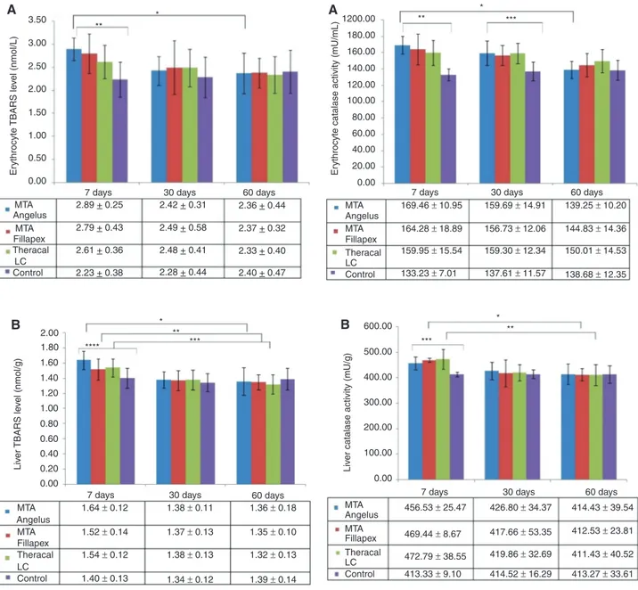
Erythrocyte (Figure 1A) and liver (Figure 1B) TBARS levels were significantly different within the 7 days of exposure groups. The highest erythrocyte and liver TBARS concen-trations were observed in the MTA Angelus group on day 7 of exposure; when compared with the control group, TBARS level in MTA Angelus group increased 29.6% and 17.1% in erythrocytes and liver, respectively. However, this level decreased significantly on day 30 (16.3% in eryth-rocyte; 15.8% in liver) and day 60 (18.3% in erytheryth-rocyte; 17.1% in liver), relative to day 7.
It was observed that, compared with the control group, TBARS level in MTA Fillapex applied rats was 25.1% higher in erythrocytes and 8.6% higher in liver on day 7. There was a considerable decrease in these numbers on the fol-lowing days. Erythrocyte TBARS level decreased around 10% on day 30 and a total of 15% decrease was observed at the end of 60 days. Similarly, liver TBARS level in MTA Fillapex applied rats decreased by almost 10% on day 30 and more than 11% on day 60, relative to day 7.
TBARS level in Theracal LC group was 17% and 10% higher in erythrocytes and liver samples, compared with the control group, on day 7. This level decreased 5% in erythrocytes and 10.4% in the liver on day 30. We deter-mined more than 10% and 14% lower TBARS level in erythrocyte and liver on day 60, compared with day 7, respectively. Hence, TBARS levels did not differ signifi-cantly among the study groups on days 30 and 60.
Erythrocyte and liver (Figure 2A and B) CAT activities were significantly higher in rats with applied calcium silicate cement compared with controls on day 7. In erythrocytes, highest increase in CAT activity was observed in the MTA Angelus group (27%); whereas in the liver highest increase was in the Theracal LC group (14.4%). The erythrocyte CAT
activities were still considerably higher in the experimen-tal groups on day 30. CAT activities decreased with time and on day 60, both erythrocyte and liver CAT levels were similar between the experimental and control groups. The
decrease in erythrocyte CAT activity in MTA Angelus group was more than 17%; while liver CAT activity decreased by 12% in MTA Fillapex group and almost 13% in Theracal LC group throughout the study period.
0.00 MTA A 169.46 ± 10.95 159.69 ± 14.91 139.25 ± 10.20 144.83 ± 14.36 150.01 ± 14.53 138.68 ± 12.35 156.73 ± 12.06 159.30 ± 12.34 137.61 ± 11.57 164.28 ± 18.89 159.95 ± 15.54 133.23 ± 7.01
7 days 30 days 60 days
MTA Angelus Fillapex Theracal LC Control 20.00 40.00 60.00 80.00 Er
ythrocyte catalase activity (mU/mL)
100.00 120.00 140.00 160.00 180.00 1200.00 ** * *** 0.00 MTA 456.53 ± 25.47 426.80 ± 34.37 417.66 ± 53.35 419.86 ± 32.69 414.52 ± 16.29 413.27 ± 33.61 411.43 ± 40.52 412.53 ± 23.81 414.43 ± 39.54 469.44 ± 8.67 472.79 ± 38.55 413.33 ± 9.10
7 days 30 days 60 days
MTA Angelus Fillapex Theracal LC Control 100.00 200.00 Liv
er catalase activity (mU/g)
300.00 400.00 500.00 600.00 *** * ** B
Figure 2: (A) Erythrocyte Catalase (CAT) activity of rats that
received different hydraulic calcium silicate cements in their dental extraction socket and sacrificed after 7, 30 and 60 days post operation. *p < 0.05 among MTA Angelus applied rats on days 7, 30 and 60. **p < 0.05 among the four groups on day 7. ***p < 0.05 among the four groups on day 30. p-Values were determined using one-way ANOVA. (B) Liver CAT Activity of rats that received different hydraulic calcium silicate cements in their dental extraction socket and sacrificed after 7, 30 and 60 days post operation. *p < 0.05 among MTA Fillapex applied rats on days 7, 30 and 60. **p < 0.05 among Theracal LC applied rats on days 7, 30 and 60. ***p < 0.05 among the four groups on day 7. p-Values were determined using one-way ANOVA. 0.00 MTA 2.89 + 0.25 2.79 + 0.43 2.61 + 0.36 2.23 + 0.38 2.28 + 0.44 2.48 + 0.41 2.49 + 0.58 2.42 + 0.31 2.36 + 0.44 2.37 + 0.32 2.33 + 0.40 2.40 + 0.47 MTA Angelus Fillapex Theracal Control LC
7 days 30 days 60 days
0.50 1.00 1.50 2.00 2.50 3.00 3.50 ** * 0.00 7 days MTA 1.64 ± 0.12 1.38 ± 0.11 1.36 ± 0.18 1.35 ± 0.10 1.32 ± 0.13 1.39 ± 0.14 1.37 ± 0.13 1.38 ± 0.13 1.34 ± 0.12 1.52 ± 0.14 1.54 ± 0.12 1.40 ± 0.13 MTA Fillapex Theracal LC Control Angelus 30 days 60 days 0.20 0.40 0.60 0.80 1.00 1.20 1.40 1.60 1.80 2.00 B A **** * *** ** Li ve r TBARS le ve l (nmol/g ) Er ythrocyte TBARS le ve l (nmol/L )
Figure 1: (A) Erythrocyte TBARS levels of rats that received different
hydraulic calcium silicate cements in their dental extraction socket and sacrificed after 7, 30 and 60 days post operation. *p < 0.05 among MTA Angelus applied rats on days 7, 30 and 60. **p < 0.05 among the four groups on day 7. p-Values were determined using one-way ANOVA. (B) Liver TBARS levels of rats that received different hydraulic calcium silicate cements in their dental extraction socket and sacrificed after 7, 30 and 60 days post operation. *p < 0.05 among MTA Angelus applied rats on days 7, 30 and 60. **p < 0.05 among MTA Fillapex applied rats on days 7, 30 and 60. ***p < 0.05 among Theracal LC applied rats on days 7, 30 and 60. ****p < 0.05 among the four groups on day 7. p-Values were determined using one-way ANOVA.
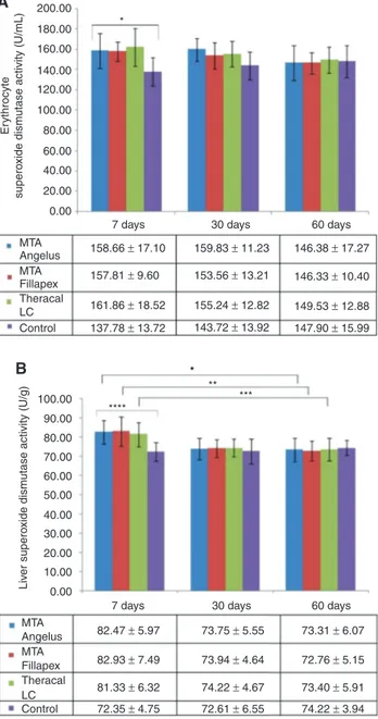
SOD activities in erythrocytes (Figure 3A) and liver (Figure 3B) were substantially higher in the experimental groups compared with controls on day 7. SOD activities of MTA Angelus rats increased 15% and 13% in erythrocyte and liver, respectively, compared to the controls on day 7. MTA Fillapex erythrocyte and liver SOD activities were both 14% higher relative to the controls on the same day. Theracal LC group experienced 17% and 12% increase in erythrocyte and liver SOD activity, respectively, compared to controls. These values returned to almost normal levels seen in controls on day 30 and 60. The decrease in liver SOD activity of dental cement applied rats throughout the study period was significant.
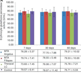
Figure 4A and B show erythrocyte and liver GPx activi-ties, respectively. Statistical tests imply that erythrocyte GPx activities did not change significantly among the study groups throughout the entire study period. On the other hand, liver GPx was significantly higher (p = 0.044); 17%, 23% and 18% increase was observed in MTA Angelus, MTA Fillapex and Theracal LC groups, respectively, on day 7. Liver GPx activity of experimental rats returned to almost control levels on day 30 and 60.
Discussion
In this study, the in vivo effects of three selected hydrau-lic calcium sihydrau-licate cements, MTA Angelus, MTA Fillapex, and Theracal LC, on oxidative stress in the liver and eryth-rocytes of rats were evaluated for the first time. These MTA-based calcium silicate dental cements differ in their setting times. MTA Angelus and MTA Fillapex are self-set-ting. There are different reports regarding the setting time of MTA Angelus, such as 15 min [4] and 165 min [15]. MTA Fillapex sets in 130 min [15]. On the other hand, Theracal LC sets almost immediately, because chemical curing is accomplished by light [6]. Moreover, metals can still leach from dental cements even after setting. Therefore, the rate of element release will vary considerably for these three calcium silicate cements.
In our previous studies using these three dental cements in vivo, we have observed that plasma, liver [14] and brain [22] Al levels increased, indicating that this element was released from the three tested dental cements, most notably from MTA Angelus and MTA Filla-pex, into systemic circulation [14]. Renal Al load most probably exceeded the excretory capacity during the first 7 days, resulting in accumulation in organs, such as liver and brain. It is possible that other metals also leached, however, only Al levels were measured in our study.
Nevertheless, Al itself causes an increase in Fe accumula-tion and thus ROS producaccumula-tion [23].
Because Al and other metals possibly released from the dental cements can lead to oxidative stress elevation,
0.00 MTA 158.66 ± 17.10 157.81 ± 9.60 161.86 ± 18.52 137.78 ± 13.72 143.72 ± 13.92 155.24 ± 12.82 153.56 ± 13.21 159.83 ± 11.23 146.38 ± 17.27 146.33 ± 10.40 149.53 ± 12.88 147.90 ± 15.99 Angelus Fillapex Theracal LC Control MTA
7 days 30 days 60 days
20.00 40.00 60.00 Er ythrocyte supero
xide dismutase activity (U/mL)
80.00 100.00 120.00 140.00 160.00 180.00 200.00 A * 0.00 MTA
7 days 30 days 60 days
82.47 ± 5.97 73.94 ± 4.64 73.75 ± 5.55 73.31 ± 6.07 72.76 ± 5.15 73.40 ± 5.91 74.22 ± 3.94 74.22 ± 4.67 72.61 ± 6.55 82.93 ± 7.49 81.33 ± 6.32 72.35 ± 4.75 MTA Angelus Fillapex Theracal LC Control 10.00 20.00 30.00 40.00 50.00 60.00 Liv er supero
xide dismutase activity (U/g)
70.00 80.00 90.00 100.00 B * **** ** *** *
Figure 3: (A) Erythrocyte Superoxide dismutase (SOD) activity of
rats that received different hydraulic calcium silicate cements in their dental extraction socket and sacrificed after 7, 30 and 60 days post operation. *p < 0.05 among the four groups on day 7. p-Value was determined using one-way ANOVA. (B) Liver SOD activity of rats that received different hydraulic calcium silicate cements in their dental extraction socket and sacrificed after 7, 30 and 60 days post operation. *p < 0.05 among MTA Angelus applied rats on days 7, 30 and 60. **p < 0.05 among MTA Fillapex applied rats on days 7, 30 and 60. ***p < 0.05 among Theracal LC applied rats on days 7, 30 and 60. ****p < 0.05 among the four groups on day 7. p-Values were determined using one-way ANOVA.
we wondered whether oxidative stress levels would be induced in rats that received the dental cements. For this purpose, we employed an in vivo technique originally introduced by Degrood et al. [24] and later used by other researchers [18, 25, 26]. In this method, dental cements within polyethylene tubes are inserted into alveolar sockets of animals so that one end of the tube remains open and the dental cement is in contact with the root apex in the alveolar socket; therefore it simulates what
happens in endodontic therapy. This model was accepted to be adequate for evaluating the tissue response to tubes filled with biomaterials [25, 26]. The recommended method for studying the inflammatory tissue response to dental materials is rat subcutaneous tissue implantation, which includes implanting polyethylene tubes filled with test materials in the subcutaneous tissue in the back of rats [27, 28]. Cintra et al. [18] compared the two methods, subcutaneous and alveolar socket implantation, and observed no differences in tissue responses as far as the implantation site and the studied period were concerned. Thus, alveolar socket implantation methodology can be a suitable alternative to subcutaneous implants [18, 25, 26]. The amount of dental cement placed into the poly-ethylene tubes in this study (7.6 mg) was similar with that used in the subcutaneous implantation method, while the amount used in endodontic therapy of humans will be at least 3–4 times higher than this, depending on the type of therapy applied (pulp capping, pulpotomy, retrograde filling, etc).
Oxidative stress induction in rat liver and erythrocytes was followed by using one of the most frequently used indi-cators of lipid peroxidation, MDA, which was measured as TBARS levels in this study. On day 7 post-exposure, both erythrocyte and liver TBARS levels substantially increased in all three dental cement applied groups compared with the control group. The highest levels were observed in the MTA Angelus group. TBARS levels decreased on days 30 and 60, and were similar in the control and dental cement applied groups. In our previous study, we have shown that Al level was increased in plasma and liver samples of rats that were implanted with one of these dental cements [14]. Al causes an increase in Fe accumulation and thus ROS production [23]. Therefore, increase in TBARS level should be at least in part due to Al that leached from these dental cements.
In our previous study [22], we have measured TBARS level in brain tissue of rats that received the same three dental cements in their alveolar socket. Similarly, we observed a transient increase in TBARS level in brain tissue. Similar to liver, the highest increase was observed in MTA Angelus group; however compared with the liver, the increase in brain was more pronounced. This initial elevation was probably due to higher Al level in brain tissue compared with the liver. Al level reached 16.42 ppm in brain tissue on day 7 [22], whereas the maximum level observed in the liver was 2.17 ppm on day 7 [14]. More-over, different trends were observed for the increase in Al and TBARS levels in the brain and liver. In the brain, there was a sharp increase in Al and TBARS on day 7, which decreased later [22]. In the liver, however, Al level 0.00
Angelus 76.28 ± 5.37
7 days 30 days 60 days
79.21 ± 10.02 79.53 ± 10.00 75.78 ± 7.21 76.71 ± 5.04 78.55 ± 5.48 77.70 ± 7.88 76.08 ± 7.07 78.07 ± 11.42 79.74 ± 7.41 73.83 ± 7.45 79.57 ± 6.41 MTA MTA Fillapex Theracal LC Control 10.00 20.00 30.00 40.00 Er
ythrocyte glutathione pero
xidase activity (U/mL) 50.00 60.00 70.00 80.00 90.00 100.00 A 0.00 MTA 8.82 ± 0.95 8.20 ± 0.48 8.05 ± 0.85 8.20 ± 0.61 8.15 ± 0.50 8.05 ± 0.74 8.29 ± 0.35 8.01 ± 0.64 8.69 ± 1.26 9.23 ± 0.97 8.90 ± 1.39 7.49 ± 0.93
7 days 30 days 60 days
MTA Angelus Fillapex Theracal LC Control 2.00 Liv er glutathione pero xidase activity (U/g) 4.00 6.00 8.00 10.00 12.00 * B
Figure 4: (A) Erythrocyte Glutathione peroxidase (GPx) activity of
rats that received different hydraulic calcium silicate cements in their dental extraction socket and sacrificed after 7, 30 and 60 days post operation. (B) Liver GPx activity of rats that received different hydraulic calcium silicate cements in their dental extraction socket and sacrificed after 7, 30 and 60 days post operation. *p < 0.05 among the four groups on day 7. p-Value was determined using one-way ANOVA.
increased slowly and reached maximum level (3.70 ppm) on day 60 [14]; on the other hand, TBARS level was highest on day 7 and decreased afterwards (the present study). Therefore, this makes us consider that there may be other metal elements, such as lead, cadmium, copper or iron, released from the dental cements on the first days and accumulated in the liver, which caused the TBARS levels to rise in this tissue. Unfortunately, we did not analyze other metal element levels in that study. Future studies should focus on this point.
Even though the increase in TBARS was transient, it may still cause damage in many ways. MDA readily reacts with several functional groups on molecules includ-ing proteins, lipoproteins, and DNA to form a variety of adducts. DNA base adducts are mutagenic and possibly carcinogenic [29], and can result in cytotoxicity. More-over, a variety of chronic diseases including atheroscle-rosis [30], ischemic stroke [31], and kidney disease [32] have been linked to increased levels of lipid peroxidation products.
In the present study, the antioxidant enzymes CAT, SOD, and GPx were measured in both erythrocytes and liver samples after 7, 30, and 60 days. Liver and erythro-cyte CAT and SOD activities and liver GPx activity were sig-nificantly higher in the application groups compared with controls on day 7. This can be attributed to ROS formation due to metal elements released from the dental cements. As stated above, increased liver Al level was observed in the rats which had one of these dental cements in their alveolar socket [14]. Such antioxidant enzymes are known to be upregulated by oxidative stress [16, 17] as a means of compensation. Thus, erythrocyte and liver oxidative stress was transiently increased in rats having one of these hydraulic calcium silicate cements. The activities of CAT, SOD and GPx enzymes were also transiently increased in the brains of rats that had one of these three dental bio-materials in their dental extraction socket [22]. We used separate control groups for each exposure time (7, 30 and 60 days) in an attempt to take into account the effects of normal metabolism, which also causes differences in oxi-dative stress parameters. No statistically significant differ-ence was observed between the control groups in any of the measured parameters.
Potential outcomes of the transient oxidative stress induction should be carefully evaluated. As stated above, increased oxidative stress can result in cytotoxicity. The three studied dental cements were observed to be bio-compatible in several studies [8, 33]; however, cytotox-icity, genotoxicity and lower biocompatibility were also observed due to the presence of various MTA cements [8, 34–42]. For example, Silva et al. [36] found that MTA
Fillapex was more cytotoxic than AH Plus. Tavares et al. [37] observed that upon subcutaneous application, lym-phocytes and macrophages of the MTA Fillapex group scored higher than the control group throughout the experiment (on day 7 and 60). Thus, they concluded that MTA Fillapex did not show the ideal tissue responses. Gomes-Filho et al. [38] evaluated in vivo the subcutane-ous response of rat connective tissue to light-cure MTA and Angelus MTA. The Angelus MTA showed a mild inflammatory response at 30 days and none at 60 days, and light-cure MTA presented a moderate chronic inflam-matory response at 30 days that decreased at 60 days. On the other hand, Mori et al. [39] reported that MTA Angelus did not cause any inflammatory reaction, while Bioden-tine, a MTA containing dental material, caused moderate inflammation at 7 days of contact. However the inflam-matory process was mild or nonsignificant at 14 and 30 days for Biodentine. Khalil and Eid [40] investigated in vivo the systemic toxic effects of DiaRoot BioAggregate and grey ProRoot MTA using subcutaneous application. They observed a significantly more severe inflammatory reaction in both experimental groups in liver and kidney tissues, compared with the control on day 7; the severity decreased after 30 days. They also observed increased liver functions after 7 days, which decreased in the Bio-Aggregate subgroup after 30 days, whilst in the MTA sub-group, a continuous increase in the level of liver function was observed. Working with primary human osteoblasts, Scelza et al. [41] observed that the tested endodontic sealers (Sealapex, Pulp Canal Sealer EWT, Real Seal and MTA Fillapex) all had strong cytotoxicity 24 h after mixing. These findings are in agreement with our current and previous results [22] showing that liver and brain TBARS levels increased substantially on day 7 in the dental cement applied groups and antioxidant enzyme activi-ties were also induced in the same period, probably as a means of compensation. These levels returned to normal levels on later days. The studies mentioned above, includ-ing ours, lasted at most 2 months. The only study that ana-lyzed the long term effects of dental cements was carried out by Bidar et al. [42]. Studying on dogs, they observed that gray and white MTA did not cause acute inflamma-tion, while they caused severe chronic inflammation after 6 months. Distinction between acute and chronic inflam-mation was made depending on the types of inflammatory cells [42].
Further studies are needed to look into the relation-ship between induction of oxidative stress due to metal release and reduced biocompatibility of these dental cements. Induction of oxidative stress transiently can also have systemic effects. For example, it was shown
that repeated exposure of adult rats to transient oxidative stress induces various long-lasting alterations in cogni-tive and behavioral functions [43]. The limitations of the present study include lack of further analysis of adverse effect of oxidative stress on other biomolecules, including DNA.
In conclusion, TBARS level, a biomarker of lipid per-oxidation, transiently increased in all three dental cement application groups. Moreover, antioxidant enzyme activi-ties increased in the same time period. The possible con-sequences of a transient oxidative stress increase should be carefully evaluated. It is likely that oxidative stress accounts for the development of cytotoxicity and reduced biocompatibility of some of the hydraulic calcium silicate containing dental cements.
Ethical considerations
The Animal Ethics Committee of the Gülhane Military Medical Academy approved all experimental procedures (Decision no: 13/103).
Acknowledgements: This study was supported by the Gülhane Military Medical Academy Research Project Fund (ARE-2013/39). We would like to thank the Gülhane Research and Development Center Laboratory Animals Unit for their support in the housing and maintenance of rats.
Conflict of interest statement: The authors declare no conflict of interest.
References
1. Kum KY, Kim EC, Yoo YJ, Zhu Q, Safavi K, Bae KS, et al. Trace metal contents of three tricalcium silicate materials: MTA Angelus, Micro Mega MTA and Bioaggregate. Int Endod J 2014;47: 704–10.
2. Torabinejad M, Parirokh M. Mineral trioxide aggregate: a compre-hensive literature review – part II: leakage and biocompatibility investigations. J Endod 2010;36:190–202.
3. Hakki SS, Bozkurt SB, Hakki EE, Belli S. Effects of mineral trioxide aggregate on cell survival, gene expression associated with min-eralized tissues, and biomineralization of cementoblasts. J Endod 2009;35:513–9.
4. MTA Angelus Reparative Cement Technical Profile. Available at: http://www.angelusdental.com/img/arquivos/mta_techni-cal_profile_download.pdf. Accessed: July 2016.
5. MTA Fillapex Endodontic Sealer Scientific Profile. Available at: http://www.angelusdental.com/img/arquivos/mta_fillapex_tech-nical_profile_download.pdf. Accessed: July 2016.
6. Vitti RP, Prati C, Silva EJ, Sinhoreti MA, Zanchi CH, de Souza e Silva MG, et al. Physical properties of MTA Fillapex sealer. J Endod 2013;39:915–8.
7. Gandolfi MG, Siboni F, Prati C. Chemical-physical properties of TheraCal, a novel light-curable MTA-like material for pulp cap-ping. Int Endod J 2012;45:571–9.
8. Poggio C, Arciola CR, Beltrami R, Monaco A, Dagna A, Lombar-dini M, et al. Cytocompatibility and antibacterial properties of capping materials. ScientificWorldJournal 2014;2014:181945. 9. Michel R. Trace metal analysis in biocompatibility testing. Crit
Rev Biocompat 1987;3:235–317.
10. Geis-Gerstorfer J, Sauer KH, Pässler K. Ion release from Ni-Cr-Mo and Co-Cr-Mo casting alloys. Int J Prosthodont 1991;4:152–8. 11. Wataha JC, Craig RG, Hanks CT. The release of elements of
dental casting alloys into cell-culture medium. J Dent Res 1991;70:1014–8.
12. Bumgardner JD, Lucas LC. Cellular response to metallic ions released from nickel-chromium dental alloys. J Dent Res 1995;74:1521–7.
13. Nejatidanesh F, Savabi O, Yazdanparast A. An investigation on metallic ion release from four dental casting alloys. J Dent (Teh-ran) 2005;2:168–73.
14. Demirkaya K, Can Demirdöğen B, Öncel Torun Z, Erdem O, Çetinkaya S, Akay C. In vivo evaluation of the effects of hydraulic calcium silicate dental cements on plasma and liver aluminium levels of rats. EurJ Oral Sci 2016;124:75–81.
15. Grazziotin-Soares R, Nekoofar MH, Davies TE, Bafail A, Alhaddar E, Hübler R, et al. Effect of bismuth oxide on white mineral triox-ide aggregate: chemical characterization and physical proper-ties. Int Endod J 2014;7:520–33.
16. Perlemuter G, Davit-Spraul A, Cosson C, Conti M, Bigorgne A, Paradis V, et al. Increase in liver antioxidant enzyme activities in non-alcoholic fatty liver disease. Liver Int 2005;25:946–53. 17. Mahoney DJ, Kaczor JJ, Bourgeois J, Yasuda N, Tarnopolsky MA.
Oxidative stress and antioxidant enzyme upregulation in SOD1-G93A mouse skeletal muscle. Muscle Nerve 2006;33:809–16. 18. Cintra LT, Bernabé PF, de Moraes IG, Gomes-Filho JE, Okamoto T, Consolaro A, et al. Evaluation of subcutaneous and alveolar implantation surgical sites in the study of the biological proper-ties of root-end filling endodontic materials. J Appl Oral Sci 2010;18:75–82.
19. Ohkawa H, Ohishi N, Yagi K. Assay for lipid peroxides in animals and tissues by thiobarbituric acid reaction. Anal Biochem 1979;95:351–8.
20. Aebi H. Catalase in vitro. Methods Enzymol 1984;105:121–6. 21. Aydin A, Orhan H, Sayal A, Ozata M, Sahin G, Işimer A. Oxidative
stress and nitric oxide related parameters in type II diabetes mellitus: effects of glycemic control. Clin Biochem 2001;34: 65–70.
22. Demirkaya K, Demirdöğen BC, Torun ZÖ, Erdem O, Çırak E, Tunca YM. Brain aluminium accumulation and oxidative stress in the presence of calcium silicate dental cements. Hum Exp Toxicol 2016. DOI: 10.1177/0960327116679713.
23. Wu Z, Du Y, Xue H, Wu Y, Zhou B. Aluminum induces neurodegen-eration and its toxicity arises from increased iron accumulation and reactive oxygen species (ROS) production. Neurobiol Aging 2012;33:199.e1–199.e12.
24. Degrood ME, Oguntebi BR, Cunningham CJ, Pink R. A com-parison of tissue reactions to Ketac-Fil and Amalgam. J Endod 1995;21:65–9.
25. Nary Filho H, Okamoto T. Evaluation of the hapset implants (Hydroxyapatite/calcium sulphate) biocompatibility in dental extraction wounds. Histological study in rats. J Appl Oral Sci 1996;4:55–64.
26. Gomes-Filho JE, de Moraes Costa MM, Cintra LT, Duarte PC, Takamiya AS, Lodi CS, et al. Evaluation of rat alveolar bone response to Angelus MTA or experimental light-cured mineral trioxide aggregate using fluorochromes. J Endod 2011;37:250–4. 27. American Dental Association. Council on dental material and
devices. Recomended standard practices for biological evalua-tion. J Am Dent Assoc 1972;84:382–90.
28. International Organization for Standardization. Technical report 7405. Dentistry – preclinical evaluation of biocompatibility of medical devices used in dentistry – test methods for dental materials. Genève: The Institution, 1997:22.
29. Niedernhofer LJ, Daniels JS, Rouzer CA, Greene RE, Marnett LJ. Malondialdehyde, a product of lipid peroxidation, is mutagenic in human cells. J Biol Chem 2003;278:31426–33.
30. Esterbauer H, Wäg G, Puhl H. Lipid peroxidation and its role in atherosclerosis. Br Med Bull 1993;49:566–76.
31. Bir LS, Demir S, Rota S, Köseoğlu M. Increased serum malondial-dehyde levels in chronic stage of ischemic stroke. Tohoku J Exp Med 2006;208:33–9.
32. Agarwal R. Chronic kidney disease is associated with oxidative stress independent of hypertension. Clin Nephrol 2004;61: 377–83.
33. Koulaouzidou EA, Economides N, Beltes P, Geromichalos G, Papazisis K. In vitro evaluation of the cytotoxicity of ProRoot MTA and MTA Angelus. J Oral Sci 2008;50:397–402.
34. De Deus G, Ximenes R, Gurgel-Filho ED, Plotkowski MC, Coutinho-Filho T. Cytotoxicity of MTA and Portland cement on human ECV 304 endothelial cells. Int Endod J 2005;38: 604–9.
35. Bin CV, Valera MC, Camargo SE, Rabelo SB, Silva GO, Balducci I, et al. Cytotoxicity and genotoxicity of root canal sealers based on mineral trioxide aggregate. J Endod 2012;38:495–500. 36. Silva EJ, Rosa TP, Herrera DR, Jacinto RC, Gomes BP, Zaia AA.
Evaluation of cytotoxicity and physicochemical properties of calcium silicate-based endodontic sealer MTA Fillapex. J Endod 2013;39:274–7.
37. Tavares CO, Böttcher DE, Assmann E, Kopper PM, de Figueiredo JA, Grecca FS, et al. Tissue reactions to a new mineral trioxide aggregate-containing endodontic sealer. J Endod 2013;39: 653–7.
38. Gomes-Filho JE, de Faria MD, Bernabé PF, Nery MJ, Otoboni-Filho JA, Dezan-Júnior E, et al. Mineral trioxide aggregate but not light-cure mineral trioxide aggregate stimulated mineralization. J Endod 2008;34:62–5.
39. Mori GG, Teixeira LM, de Oliveira DL, Jacomini LM, da Silva SR. Biocompatibility evaluation of biodentine in subcutaneous tis-sue of rats. J Endod 2014;40:1485–8.
40. Khalil WA, Eid NF. Biocompatibility of BioAggregate and mineral trioxide aggregate on the liver and kidney. Int Endod J 2013;46:730–7.
41. Scelza MZ, Linhares AB, da Silva LE, Granjeiro JM, Alves GG. A multiparametric assay to compare the cytotoxicity of endo-dontic sealers with primary human osteoblasts. Int Endod J 2012;45:12–8.
42. Bidar M, Naghavi N, Mohtasham N, Sheik-Nezami M, Fallah-rastegar A, Afkhami F, et al. Mineral trioxide aggregate and port-land cement for direct pulp capping in dog: a histopathological evaluation. J Dent Res Dent Clin Dent Prospects 2014;8:134–40. 43. Iguchi Y, Kosugi S, Nishikawa H, Lin Z, Minabe Y, Toda S.
Repeated exposure of adult rats to transient oxidative stress induces various long-lasting alterations in cognitive and behav-ioral functions. PLoS One 2014;9:e114024.