Molecular Biology
Research Article – 60973
Mısra Nadir, Özlem Tufanlı, Ebru Erbay, Arzu Atalay*
Identification of differentially expressed
microRNAs during lipotoxic endoplasmic reticulum
stress in RAW264.7 macrophages
RAW264.7 makrofajlarında lipotoksik endoplazmik retikulum stres sürecinde ifadesi değişen
mikroRNAların tanımlanması
doi 10.1515/tjb-2016-0031
Received February 20, 2016; accepted April 19, 2016
Abstract: Objective: Increased fatty acids in the
circula-tion and their accumulacircula-tion in non-adipose tissues play a significant role in the development of obesity related metabolic and inflammatory disorders such as insulin resistance, diabetes and atherosclerosis. While fat tissue has the ability to store excess fatty acids, uptake of excess fatty acids to other tissues burdens intracellular metabolic organelles such as mitochondria and endoplasmic retic-ulum (ER), leading to stress response and lipotoxic cell death. Unfolded protein response (UPR) is a key adapta-tion of the ER to stress. It is still not completely clear how lipids engage the UPR and how UPR manages both the adaptive and destructive consequences under its control. Increasing evidence point to the importance of miRNA reg-ulation of the UPR as well as UPR’s role in miRNA biogene-sis. In order to understand how lipids engage the UPR, we set forth to identify microRNAs regulated by lipotoxic ER stress in macrophages.
Methods: We stressed the mouse macrophage cell line (RAW 264.7) with a saturated fatty acid, 500µM palmitate, reflect-ing the levels found in the circulation of obese patients. We analyzed the microRNAome profiles of this cell line using QRT-PCR based miScript miRNA PCR array which con-tained all known mouse microRNAs in miRBase release16 and performed pathway analysis for potential targets.
Results: 227 microRNAs showed altered expression levels; 43 microRNAs above 2 fold difference and 13 microRNAs 3-24 fold difference. Pathway analysis enriched the target mRNAs of these lipotoxic ER stress associated miRNAs. Conclusion: When exposed to high concentrations of sat-urated fatty acids that can induce ER stress, macrophages display a dynamic range of changes in their microRNAome profiles. Our findings reflect the consequences of lipotoxic stress on circulating monocytes and tissue-associated macrophages in obesity. Further studies are needed to deliniate which UPR arm is reponsible for the microRNA changes reported here.
Keywords: Lipotoxic endoplasmic reticulum stress,
microRNA, unfolded protein response, macrophage, QRT-PCR, pathway analysis, RAW264.7
Özet: Amaç: Kan dolaşımındaki serbest yağ asitlerinin
artışı ve adipoz olmayan dokulardaki birikimi, insülin direnci, diyabet ve ateroskleroz gibi obezite ile ilişkili meta-bolik ve emflamatuvar hastalıkların gelişiminde önemli rol oynar. Yağ dokusu, fazla olan yağ asidini depolaya-bilme kabiliyetine sahipken, diğer dokulara ulaşan fazla miktarda yağ asidi, endoplazmik retikulum (ER) ve mito-kondri gibi intraselüler metabolik organelleri zorlayarak stres cevabının oluşmasına ve lipotoksik hücre ölümüne neden olur. Katlanmamış protein yanıtı (KPY) endoplaz-*Corresponding author: Arzu Atalay: Ankara University, Biotechnology
institute, Ankara, Turkey, e-mail: [email protected] Mısra Nadir: Ankara University, Biotechnology institute, Ankara, Turkey, e-mail: [email protected]
Özlem Tufanlı: Bilkent University, Faculty of Science, department of
Molecular Biology and Genetics, Ankara, Turkey, e-mail: ozlemtufanlı[email protected]
Ebru Erbay: Bilkent University, Faculty of Science, department of Molecular Biology and Genetics, Ankara, Turkey,
mik retikulumun strese karşı önemli bir adaptasyonudur. Lipidler ile KPY arasındaki ilişkinin nasıl olduğu ve kat-lanmamış protein yanıtı ile adaptif ve destrüktif sonuçla-rın nasıl yönetildiği halen tam olarak aydınlatılamamıştır. miRNA biyogenezinde KPY’nın rolünün yanı sıra, katlan-mamış protein yanıtını düzenleyen miRNAların önemine işaret eden kanıtlar bulunmaktadır. Bu çalışmada lipidler ile KPY arasındaki ilişkiyi anlamak için, makrofajlardaki lipotoksik ER stresi sürecinde düzenlenen mikroRNA’ların tanımlanması amaçlanmıştır.
Metod: Fare makrofaj hücre hattına (RAW 264.7), serbest yağ asidi olan 500 µM palmitat -obez hastaların kan dola-şımındaki seviyede- uygulanarak stres oluşturulmuştur. miRBase sürüm 16’daki bilinen tüm fare mikroRNAla-rını içeren QRT-PCR temelli miSCRİPT miRNA PCR array sistemi kullanılarak, hücrelerdeki tüm mikroRNAom profili analiz edilmiş ve potansiyel hedefler için yolak analizleri gerçekleştirilmiştir.
Bulgular: Lipotoksik ER stres sonucu, 227 mikroRNA’nın ifade seviyesi 2 kat üzerinde değişmiş ve 43’ü 2 kattan faz-la,13’ü ise 3-24 kat değişim göstermiştir. Yolak analizi ger-çekleştirilerek lipotoksik ER stresi ile ilişkili mikroRNA’la-rın mRNA hedefleri belirlenmiş ve gruplanmıştır.
Sonuç: Makrofajlar, ER stresini indükleyebilen yüksek konsantrasyondaki doymuş yağ asidine maruz bırakıldı-ğında mikroRNAome profillerinde dinamik bir değişim gözlenmektedir. Bulgularımız, obezitedeki doku ilişkili makrofajlar ve kan dolaşımındaki monositlerdeki lipotok-sik stres sonuçlarını yansıtmaktadır. Detaylı çalışmalar gerçekleştirilerek, lipotoksik ER stresi sürecinde ifadesi-nin değiştiğini rapor ettiğimiz mikroRNA değişimlerinden hangi KPY yolağının sorumlu olduğu belirlenebilir.
Anahtar Kelimeler: Lipotoksik endoplazmik retikulum
stres, mikroRNA, katlanmamış protein yanıtı, makrofaj, QRT-PCR, yolak analizi, RAW264.7
Introduction
Endoplasmic Reticulum (ER) functions as a critical met-abolic hub for protein, lipid and calcium metabolism [1]. Accumulation of unfolded proteins in the ER lumen, infections, toxins, hypoxia, excess food and energy depri-vation trigger ER stress and activate the unfolded protein response (UPR) [2]. Upon ER stress, UPR functions as a switch between the adaptation of the cell against stress and the decision for apoptosis.
Chronic ER stress is harmful for cells and tissues and may lead to development of many metabolic diseases such as obesity, diabetes and atherosclerosis. One of the major known causes of these diseases is the accumulation of free fatty acids in non-adipose tissues like pancreas, liver and vascular wall, leading to cellular demise and death known as lipotoxicity [3,4]. The chronic inflammation observed in obesity can also arise from malfunctioning ER as UPR and the organelle itself is intricately linked to many immuno-logical conditions [1]. Under normal conditions, UPR is an essential homeostatic mechanism for the management of stress associated with the accumulation of unfolded proteins in the ER [5]. UPR ensures the signal transfer to the nucleus for chaperone expression [6]. The unfolded or misfolded proteins can also be destined to ER-associated degradation pathways. If the ER cannot restore homeo-stasis in irremediable ER stress, UPR activates apoptotic pathways to initiate cell death [7].
The UPR is mediated by three different stress sensing pathways regulated by pancreatic ER kinase (PERK), inositol-requiring kinase 1 (IRE1) and activat-ing transcription factor 6 (ATF6), all three transmem-brane proteins located in the ER. PERK activation leads to phosphorylation of eukaryotic translation initiation factor 2α (eIF2α) and inhibition of translation [7,8]. IRE-1 is unique in the sense that it has two distinct activi-ties. Its kinase domain mediates autophosphorylation required for further oligomerization and activation. Whether IRE1 has other substrates than itself is not known. Its endoribonuclease activity is responsible for the splicing of XBP-1 (X box binding protein 1) mRNA resulting in production of active transcription factor XBP-1 [9–11]. The third arm is governed by ATF-6 cooper-ating with IRE-1 by upregulcooper-ating the expression of XBP-1 mRNA and moreover, transcriptionally induces similar chaperone targets to XBP1 transcription factor. The expression and activation of XBP1s (XBP-1 spliced) as well as activation and translocation of ATF6 to nucleus leads to a complex transcriptional program that plays a central role in the UPR by upregulating mainly chap-erone proteins promoting protein folding and produc-tion of essential components for protein degradaproduc-tion. Overall, these measures serve to re-establish homeo-stasis in ER [12,13]. In addition to protective responses, these UPR pathways can also induce important inflam-matory signals when induced in cells of the immune system. Moreover, if ER homeostasis is not restored, ER activates apoptotic pathways [7].
ER stress is coupled to inflammation through several mechanisms [1]: IRE1-mediated activation of JNK induces expression of pro-inflammatory genes by directly
influ-encing transcription factor activator protein 1 (AP1). Fur-thermore, activation of PERK triggers the degradation of inhibitor NFkB (IkB), which allows the translocation of NFkB into the nucleus and activation of pro-inflammatory genes. ER stress also leads to cleavage and activation of the transcription factor cyclic-AMP-responsive-element-bind-ing-protein H (CREBH), which induces the production of acute phase proteins like C-reactive protein (CRP) and serum amyloid P-component (SAP). Additionally, reactive oxygen species can be produced during ER stress and lead to oxidative damage and activate many stress and inflam-mation signaling cascades.
Unfolded protein and lipids trigger ER stress through different mechanisms [14]. In order to drive ER stress, saturated fatty acids need to bind intracellular lipid chaperones that shuttle them presumably to intracellu-lar destinations such as the membranes of organelles and to the nucleus [14,15]. Moreover, unlike unfolded proteins, which bind the luminal domains of proximal ER sensors such as IRE1 and PERK to activate UPR sig-naling, saturated fatty acids can trigger UPR signaling in cells expressing luminal domain deletion mutants of IRE1 and PERK. These findings clearly demonstrate important differences in how unfolded and lipid stress signals engage the UPR and represent a window of oppor-tunity for therapeutics designed with an understanding of these molecular differences in order to discriminate between adaptive UPR responses essential for ER homeo-stasis and destructive responses triggered by the excess of lipids in obesity.
MicroRNAs are small RNA molecules, which function during development, organogenesis, maintenance of stem cell status, cancer and stress responses; regulate 30-60% of all protein coding genes. MicroRNAs primarily play important role in post-transcriptional regulation of gene expression, making them potential targets for therapeutic applications.
Many high throughput screening studies have been performed to identify the lipotoxicity associated microR-NAs in different cell types. In 2008, pancreatic beta cells were treated with free fatty acids and significant differ-ences in the expression levels of 132 microRNAs -espe-cially mmu-miR-34a and mmu-miR-146 were detected by microRNA microarray [16]. ER stress was generated in cells by using chimeric tRNA and differences in the expression levels of 200 microRNAs were observed upon activation of UPR [17]. In this study, we identified lipotoxic ER stress associated microRNAs in an immune system cell, namely macrophage. Our goal was to learn more about the role of miRNAs in coupling lipid stress to UPR signaling and outcomes in macrophages.
Materials and Methods
Cell culture and lipotoxicity assay
RAW 264.7 mouse leukemic monocyte macrophage cell line has been cultured in RPMI 1640 medium contain-ing 10% Fetal bovine serum. In order to drive lipotoxic ER stress, cells were treated with 500 µM palmitic acid (Sigma, P0500) for 6 hours. Palmitic acid was dissolved in 1% fatty acid free BSA (Sigma, A8806) containing RPMI at 55°C. After 6 hours of administration, expression of spliced Xbp-1 transcript was detected by QRT-PCR. Cells were treated with 300 µM Thapsigargin as well as positive control for ER stress.
RNA isolation and cDNA synthesis
Total RNA from palmitate treated and untreated RAW 264.7 macrophages were purified according to total RNA isola-tion protocol using Trizol (Invitrogen). cDNA synthesis of microRNAs was done with miScript II Reverse Transcrip-tion kit (Qiagen) according to manufacturer’s protocols.
MicroRNAome profiling assay
“Mouse miRNAome miRNA PCR Array” (Qiagen, MIMM-3216Z) was used in order to identify expression levels of miRNAs in different samples. This array contained all known mouse miRNA (940 miRNAs) primers in the miRBase (Release 16) on 384-well plate format with inter-nal and normalization controls. The expression levels were identified on Roche Light Cycler 480 machine accord-ing to manufacturer’s instructions. Results were analyzed on miScript PCR Array Data Analysis web sofware devel-oped by Qiagen (http://pcrdataanalysis.sabiosciences. com/mirna/arrayanalysis.php). Results were calculated according to ΔΔCT method and displayed in different formats.
Target prediction of differentially expressed
miRNAs and pathway analysis
Available potential mRNA targets of differentially expressed microRNAs were provided from microRNA.org webpage. For pathway analysis, gene lists from microRNA.org were transferred to publicly available GeneCodis 3.0 web soft-ware and genes were grouped according to their biological
functions. Gene Ontology (GO) and KEGG enrichment anal-ysis were performed for available gene lists.
Results
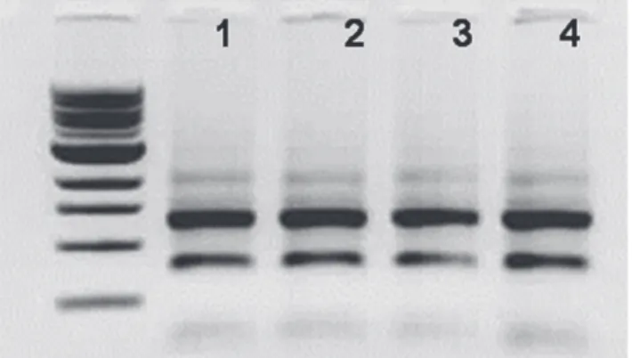
Total RNAs from two biological replicates of palmitate treated and untreated RAW 264.7 macrophages are shown in Figure 1. All ribosomal RNA bands observed to be intact on agarose gel electrophoresis. These biological dupli-cates were used for miRNAome study. The presence of ER stress was checked by detection of the spliced Xbp-1 transcript (Figure 2). Thapsigargin, a potent inducer of ER stressor by way of inhibiting SERCA (SarcoEndoplasmic Reticulum Calcium transport ATPase) was used as positive
control for detection of spliced Xbp-1 transcript (lane 1). 257 bp spliced Xbp-1 transcript was observed in the palmi-tate treated macrophages (lane 3), while 283 bp unsliced Xbp-1 transcript was observed in untreated cells (lane 2).
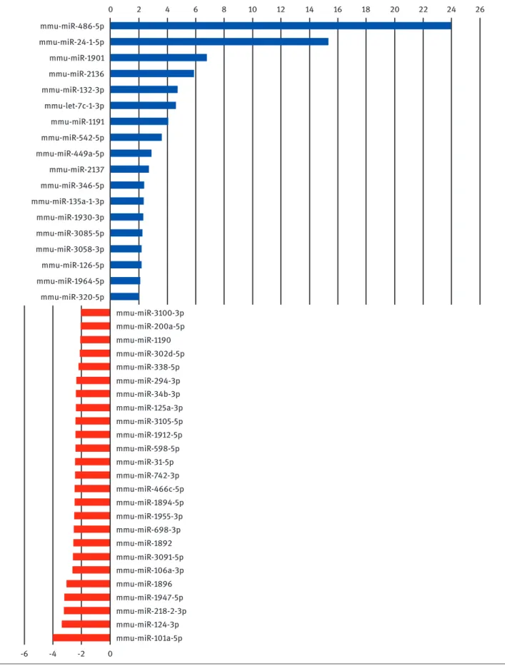
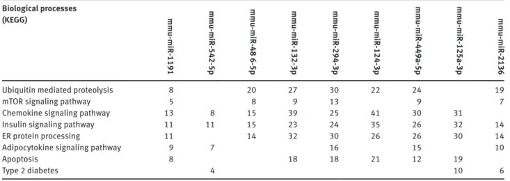
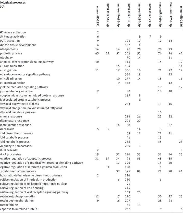
MicroRNAome profiling assay was performed between palmitate-treated (test sample) and untreated macro-phages (control sample) and out of the 940 known mouse mature microRNAs, 43 displayed differential expression above 2 fold (as calculated by miScript PCR Array Data Analysis web sofware). Figure 3 shows the changes in expression levels of these 43 microRNAs during lipotoxic ER stress. The results are mean of two biological repli-cates. Following palmitate treatment, expression levels of 18 microRNAs (Figure 3, blue bars) out of 940 mouse microRNAs increased at least 2 fold. Among these we were able to reach potential target mRNA lists of 486-5p, 2136, 132-3p, mmu-miR-1191, mmu-miR 542-5p and mmu-miR-449-5p. In addition, expression levels of 25 miRNAs decreased (Figure 3, red bars) at least two fold. Among these, we were able to reach predicted target mRNA lists of miR-294-3p, mmu-miR-125a-3p and mmu-mir-124-3p. A partial pathway analy-sis list of potential target mRNA numbers is shown in Table 1. For each microRNA, there were thousands of potential mRNA targets and by pathway analysis these genes were enriched in hundreds of groups according to their bio-logical functions. Table 2 is a longer version of pathway analysis, genes were grouped according to gene ontology (GO). Out of approximately 350 GO terms, 45 of them are included which are related with ER stress, UPR, lipotoxic-ity, inflammation, fatty acid metabolism and so on.
Discussion
Since microRNAs play important roles in post-transcrip-tional regulation of gene expression during stress as well as many biological events, they have been very attractive potential targets for therapeutic approaches. To learn more about the role of microRNAs in coupling lipid stress to UPR signaling and outcomes in an immune cell, we stressed RAW264.7 mouse macrophages with a free fatty acid -thapsigargin- and analyzed the global microRNAome profiles with PCR array. Out of 940 known mouse miRNAs, expression levels of 18 miRNAs increased at least 2 fold and expression levels of 25 miRNAs decreased at least two fold. We were able to find downstream targets of 9 and performed pathway analysis in order to enrich predicted targets in gene groups according to their biological func-tions. Although we did not provide detailed gene lists here, Figure 1: Total RNAs used in the study. 28S, 18S and 5S RNA bands are
intact on agarose gel electrophoresis 1-3) Raw264.7, 2-4) Raw264.7+ Palmitate. Lanes 1- 2 and 3-4 are independent biological duplicates used in the miRNAome study. 1 kb ladder is from NEB (N3232).
Figure 2: Xbp-1 splicing assay. Unspliced Xbp-1 gives 283 bp and spliced Xbp-1 gives 257 bp fragment. 1) Raw264.7 + Thapsigargin treatment, 2) Raw264.7 3) Raw264.7+ 500 µM palmitic acid.
mmu-miR-486-5p mmu-miR-24-1-5p mmu-miR-1901 mmu-miR-2136 mmu-miR-132-3p mmu-miR-1191 mmu-miR-542-5p mmu-miR-449a-5p mmu-miR-2137 mmu-miR-346-5p mmu-miR-135a-1-3p mmu-miR-1930-3p mmu-miR-3085-5p mmu-miR-3058-3p mmu-miR-126-5p mmu-miR-1964-5p mmu-miR-320-5p mmu-miR-3100-3p mmu-miR-200a-5p mmu-miR-1190 mmu-miR-302d-5p mmu-miR-338-5p mmu-miR-294-3p mmu-miR-34b-3p mmu-miR-125a-3p mmu-miR-3105-5p mmu-miR-1912-5p mmu-miR-598-5p mmu-miR-31-5p mmu-miR-742-3p mmu-miR-466c-5p mmu-miR-1894-5p mmu-miR-1955-3p mmu-miR-698-3p mmu-miR-1892 mmu-miR-3091-5p mmu-miR-106a-3p mmu-miR-1896 mmu-miR-1947-5p mmu-miR-218-2-3p mmu-miR-124-3p mmu-miR-101a-5p mmu-let-7c-1-3p 0 2 4 6 8 10 12 14 16 18 20 22 24 26 -6 -4 -2 0
Figure 3: differentially expressed miRNAs following lipotoxic ER stress. 43 miRNAs which are upregulated (blue bars, positive logarithmic fold change values) and downregulated (red bars, negative logarithmic fold change values) during lipotoxic ER stress. Palmitate treated Raw264.7 macrophage is the test sample and untreated Raw264.7 macrophage is the control sample
we were able to see enrichment of predicted target genes in lipotoxic ER stress related biological processes both in Gene Ontology (Table 2) and KEGG (Table 1) enrichments.
Expression of mmu-miR-486-5p was upregulated by 23.97 fold upon lipotoxic ER stress in RAW264.7 macro-phages. Enriched GO terms in the predicted target genes of this miRNA were anti-apoptosis, apoptosis, cell com-munication, cell migration, cell-cell adhesion, cell-matrix adhesion, innate immune response, mRNA processing, negative regulation of apoptotic process, oxidation-reduc-tion process, positive regulaoxidation-reduc-tion of interleukin produc-tion, protein autophosphorylation and dephosphoryla-tion (Table 2). According to our KEGG enrichment, some potential targets were clustered in biological pathways such as mTOR signaling pathway, ubiquitin mediated pro-teolysis, insulin signaling pathway, ER protein processing and chemokine signaling pathway (Table 1). These results were consistent with literature; although upregulation of mmu-miR-486-5p was not previously reported, this miRNA was shown to regulate many pathways in some diseases. During a study on global microRNA expression profile in myostatin knockout mice, mmu-miR-486 was identified as a positive regulator of IGF-1/Akt pathway, as a novel target of myostatin targeting [18]. Myostatin, also known as growth and differentiation factor-8, is a pivotal negative regulator of skeletal muscle mass and reduces muscle protein synthesis by inhibiting the insulin-like growth factor-1 (IGF-1)/Akt/mammalian target of rapa-mycin (mTOR) pathway. In myostatin knockout mice, the expression level of miR-486 in skeletal muscle was signifi-cantly increased. This study indicated miR-486 as one of the intermediary molecules connecting myostatin signal-ing and the IGF-1/Akt/mTOR pathway in the regulation of skeletal muscle size. In another study, stable expression
of miR-486 ameliorated the disease progression in dys-trophin deficient skeletal muscle of mice. Skeletal mus-cle-specific miR-486 overexpression in Dmdmdx-5Cv animals
decreased levels of DOCK3, reduced PTEN expression, and subsequently increased levels of phosphorylated AKT, resulting in an overall beneficial effect [19]. Third study demonstrated FoxO1 to be a dominant mediator of chronic kidney disease-induced muscle wasting and miR-486 coordinately decreases FoxO1 and PTEN to protect against this catabolic response [20]. Chronic kidney disease accel-erates muscle protein degradation by stimulating the ubiquitin proteasome system through activation of the E3 ligases. FoxO1 has a role in controlling ubiquitin pro-teasome system-related proteolysis. miR-486 decreased FoxO1 protein translation and increased FoxO1 phosphor-ylation by down-regulation of PTEN phosphatase, a nega-tive regulator of p-Akt. Finally expression of the E3 ligases was suppressed and muscle mass increased despite the disease. Since our KEGG analysis enriched genes related with these biological processes, further studies are needed to understand the exact function of miR-486-5p during lipotoxic ER stress.
mmu-miR-2136 expression was upregulated by 5.89 fold upon lipotoxic ER stress. Pathway analysis identified GO terms such as apoptotic process, canonical wnt recep-tor signaling pathway, cell communication, cell migration, cell-cell adhesion, cytokine-mediated signaling pathway, lipid metabolic process, MAPK cascade, oxidation-reduc-tion process, protein autophosphorylaoxidation-reduc-tion and response to unfolded protein for mmu-miR-2136 (Table 2). Besides, KEGG enrichment identified biological processes such as ubiquitin mediated proteolysis, mTOR signaling pathway, insulin signaling pathway, ER protein processing, adipo-cytokine signaling pathway and finally type 2 diabetes. Table 1: KEGG enrichment analysis of number of potential mRNA targets of the lipotoxic ER stress associated miRNAs (Partial list).
Biological processes (KEGG) mmu-miR -1191 mmu-miR -542-5p mmu-miR -48 6-5p mmu-miR -132-3p mmu-miR -294-3p mmu-miR -124-3p mmu-miR -449a-5p mmu-miR -125a-3p mmu-miR -2136
Ubiquitin mediated proteolysis 8 20 27 30 22 24 19
mToR signaling pathway 5 8 9 13 9 7
Chemokine signaling pathway 13 8 15 39 25 41 30 31
insulin signaling pathway 11 11 15 23 24 35 26 32 14
ER protein processing 11 14 32 30 26 26 30 14
Adipocytokine signaling pathway 9 7 16 15 10
Apoptosis 8 18 18 21 12 19
This study is the first one indicating mmu-miR-2136 to associate with a biological function.
mmu-miR-132-3p was upregulated 4.73 fold following lipotoxic ER stress in macrophages. According to our KEGG
analysis, predicted target genes were enriched in groups such as ubiquitin mediated proteolysis, mTOR signaling pathway, chemokine signaling pathway, insulin signaling pathway, ER protein processing and apoptosis (Table 1). Table 2: Pathway analysis of number of potential mRNA targets of the lipotoxic ER stress associated miRNAs according to Gene ontology (Partial list). Biological processes (GO) mmu-miR -1191 mmu-miR -542-5p mmu-miR -486-5p mmu-miR -132-3p mmu-miR -294-3p mmu-miR -124-3p mmu-miR -449a-5p mmu-miR -125a-3p mmu-miR -2136
JAK kinase activation 2
JUN kinase activation 6 9 7 9
MAPK activation 9 125 12 12 13
Adipose tissue development 4 187 6
Anti-apoptosis 14 14 28 20 20 29
Apoptotic process 43 22 52 364 93 76 94 42
Autophagy 70 14 11
Canonical Wnt receptor signaling pathway 10 314 15 12
Cell communication 15 184 11
Cell migration 11 17 356 18 21 22 12
Cell surface receptor signaling pathway 336 19 22
Cell-cell adhesion 10 277 14 18 11
Cell-matrix adhesion 9 348 12
Cytokine-mediated signaling pathway 19 12
Cytoskeleton organization 7 30 18 18
Endoplasmic reticulum unfolded protein response 189 8
ER-associated protein catabolic process 9
Fatty acid biosynthetic process 283 13 16
Fatty acid elongation, polyunsaturated fatty acid 2
Fatty acid metabolic process 16
immune response 214 26 25 22
inflammatory response 201 27
innate immune response 14 58 27
JNK cascade 5 5 14 8
Lipid biosynthetic process 59 18 21 21
Lipid catabolic process 41 15
Lipid metabolic process 238 35 23
Lymphocyte homeostasis 269 3
MAPK cascade 7 9
mRNA processing 19 32 226 33 32 46 25
Negative regulation of apoptotic process 31 19 34 94 55 48 65
Negative regulation of canonical Wnt receptor signaling pathway 11 126 13 20
Negative regulation of interferon-gamma production 3 178 6
oxidation-reduction process 39 325 84 74 90 44
Phosphatidylethanolamine biosynthetic process 2 116
Positive regulation of interleukin- production 6 280 6
Positive regulation of NF-kappab import into nucleus 91
Positive regulation of RNA splicing 245
Positive regulation of Wnt receptor signaling pathway 359 20
Protein autophosphorylation 12 17 290 30 27 23
Protein dephosphorylation 9 16 207 28 24
Protein folding 56 13
Associated GO terms for the predicted targets of mmu-miR-132-3p were numerous, as shown in Table 2. Among the groups it is not surprising to observe ER unfolded protein response, fatty acid biosynthetic process, lipid meta-bolic process, lipid catameta-bolic process and inflammatory response genes. In a study investigating the expression levels of inflammation related miRNAs in white blood cells after 8 weeks of healthy diet, miR-132-3p expression was found to be associated with healthy diet [21]. In another study, mmu-miR-132-3p inhibited osteoblast differentiation by directly targeting EP300 (E1A binding protein p300) a type of histone acetyl transferase necessary for the acetyl-ation of osteoblast differentiacetyl-ation factor Runx2, in simu-lated microgravity [22]. We also identified EP300 (Gene ID: 328572) with a mir-SVR score of -1.48, among predicted targets during our pathway analysis (data not shown).
mmu-miR-1191 expression was upregulated by 4 fold in thapsigargin treated macrophages. Our KEGG enrich-ment, grouped high number of genes into different bio-logical process groups except type 2 diabetes (Table 1). Among the associated GO terms with this microRNA (Table 1) most remarkable ones were activation of kinases, adipose tissue development and apoptotic process.
mmu-miR-542-5p expression was upregulated by 3.6 fold upon lipotoxic ER stress. While our KEGG enrich-ment grouped genes related to chemokine signaling pathway, insulin signaling pathway, adipocytokine sig-naling pathway and type 2 diabetes (Table 1); GO terms were apoptotic process, JNK cascade, negative regulation of apoptosis and negative regulation of interferon gam-ma-production (Table 2), which are consistent with the previously reported tumor suppressor function. miR-542-5p was found to be directly targeting EGFR in non-small lung cancer and inhibiting growth of cancer cells [23]. More evi-dence demonstrated that mmu-miR-542-5p is a novel tumor suppressor in neuroblastoma [24]. This is the first report for the role of mmu-miR-542-5p during lipotoxic ER stress.
mmu-miR-449a-5p was upregulated 2.91 fold upon lipotoxic ER stress. Previous studies demonstrated that hsa-miR-449a inhibited expression of MAP2K1 directly by targeting its 3’UTR and its expression was downregu-lated in non-small cell lung carcinoma which suggested the use of this miRNA as a potential therapeutic target [25]. hsa-miR-449a inhibited liver cancer cell proliferation and induced apoptosis via suppression of Calpain6 and POU2F1 [26]. In another study, which also suggested the use of hsa-449a as a therapeutic target in cancer, miR-449a suppressed epithelial-mesenchymal transition and metastasis of hepatocellular carcinoma by multiple targets [27]. GO terms for the predicted targets not only identified cell proliferation and apoptosis associated groups
con-sistent with literature, but also grouped these genes into cell migration, cell-cell adhesion, cytokine mediated sig-naling pathway, fatty acid biosynthetic process, fatty acid metabolic process, immune response, lipid biosynthetic process, lipid catabolic process, lipid metabolic process, oxidation-reduction process and response to unfolded protein that are related to inflammation, immune response, UPR, fatty acid and lipid metabolism as expected during lipotoxic ER stress in macrophages (Table 2). KEGG enrich-ment of predicted targets identified the role of these target genes in many biological processes as well (Table 1).
For pathway analysis, we were able to reach pre-dicted mRNA targets of 124-3p, mmu-miR-125a-3p and mmu-miR-294-3p which are downregulated in thapsigargin treated RAW264.7 macrophages, 3.39 fold, 2.38 fold and 2.34 fold respectively. Previous reports identified that hsa-miR-124-3p targeted ROCK1 and inhib-ited cell migration and invasion [28]. Its downregula-tion was also implicated in gastric cancer tumorigenesis [29]. By targeting STAT3, this miRNA inhibited growth and metastasis [30]. Although pathway analysis did not reveal remarkable gene groups related to ER stress with mmu-124-3p, we have noticed the consistency of miR-125a-3p involvement in such biological processes both with pathway analysis and literature. KEGG enrichment of predicted targets for this miRNA clustered some targets in biological processes such as ER protein processing, insulin signaling pathway, type 2 diabetes, apoptosis and chemokine signaling pathway, all being consistent with previous reports (Table 1). GO terms for mmu-miR125a-3p were adipose tissue development, apoptotic process, auto-phagy, cell migration, cytoskeleton organization, fatty acid biosynthesis, immune response and lipid biosynthe-sis (Table 2). rno-miR-125a-3p directly binds to the 3’UTR of p38 MAPK [31]. hsa-miR-125a-3p regulates the insulin signaling pathway and increased hsa-miR-125a-3p expres-sion in omental adipose tissue might be a characteristic feature of insulin resistance in obese men [32]. Expression of rno-miR-125a-3p was upregulated during high fat diet following early leptin blockade [33]. hsa-miR-125a-3p pro-moted adipogenesis via suppressing the RhoA/ROCK1/ ERK1/2 pathway and authors suggested novel therapies for obesity since they found the upregulation of this miRNA multiple symmetric lipomatosis patients [34].
mmu-miR-294-3p was found to be expressed in mouse embryonic stem cells [35]. It promoted induced plurip-otency during reprogramming with Oct4, Klf4 and Sox2 [36]. miR-294/miR302 family promoted proliferation, sup-pressed G1-S restriction point and inhibited embryonic stem cell differentiation through Rb-dependent and -inde-pendent pathways [37]. mmu-miR-294-3p predicted targets
were nearly in all biological processes in our KEGG enrich-ment results (Table 1). GO terms for this miRNA were acti-vation for kinases, apoptosis, autophagy, cell migration, cell-cell adhesion, cell surface receptor signaling pathway, ER unfolded protein response, ER-associated catabolic process, protein folding, immune response, inflammatory response and lipid biosynthetic process (Table 2).
In this study, we have identified 43 microRNAs with at least 2 fold changes in expression level upon lipotoxic ER stress in macrophages. We restricted our pathway analysis results and mainly tried to focus on biological processes such as fatty acid and lipid metabolism, apoptosis, immu-nity, chemotaxis, cell migration and endoplasmic retic-ulum unfolded protein response. Among 43 miRNAs we were able to get the predicted target gene lists of 9 miRNAs. Our results were consistent with previous reported func-tions for mmu-miR-486-5p, mmu-132-3p, mmu-miR-542-5p, mmu-miR- 125a-3p and mmu-miR-294-3p. Although most information mentioned above originated from cancer studies, a few global microRNA profiling studies revealed functions for these miRNAs for ER protein processing, insulin signaling pathway, apoptosis, and inflammation and lipid metabolism. Since one of our questions was to figure out miRNA regulation of UPR as well as its role in miRNA biogenesis, it was not surprising to see “mRNA processing” in our GO term enrichment table for almost all differentially regulated miRNAs.
Our global miRNAome study identified expression level changes of many miRNAs during lipotoxic ER stress in an immune cell for the first time. High concentrations of saturated fatty acids - reflecting the levels found in the circulation of obese patients- induced ER stress, and mac-rophages displayed a dynamic range of changes in their microRNAome profiles. Our findings reflect the conse-quences of lipotoxic stress on circulating monocytes and tissue-associated macrophages in obesity. Further studies are needed to delineate which UPR arm is responsible for the microRNA changes reported in our study.
Acknowledgements: This study is funded by “The
Sci-entific and Technological Research Council of Turkey” (Grant number 212T171).
Conflict of Interest: The authors have no conflict of interest.
References
[1] Hotamisligil GS, Erbay E. Nutrient sensing and inflammation in metabolic diseases. Nat Rev immunol 2008; 8(12):923–34. [2] Xie Y, Luo J, Kennedy S, davidson No. Conditional intestinal
lipotoxicity in Apobec-1-/- Mttp-iKo mice: a survival advantage for mammalian intestinal apolipoprotein B mRNA editing. J Biol Chem 2007; 282(45):33043–51.
[3] Schaffer JE. Lipotoxicity: when tissues overeat. Curr opin Lipidol 2003; 14(3):281–7.
[4] Borradaile NM, Han X, Harp Jd, Gale SE, ory dS, Schaffer JE. disruption of endoplasmic reticulum structure and integrity in lipotoxic cell death. J Lipid Res 2006; 47(12):2726,37. [5] Kaufman RJ, Scheuner d, Schröder M, Shen X, Lee K, Liu CY,
et al. The unfolded protein response in nutrient sensing and differentiation. Nat Rev Mol Cell Biol 2002; 3(6):411–21. [6] Bernales S, Papa FR, Walter P. intracellular signaling by the unfolded protein response. Annu Rev Cell dev Biol 2006;22:487–508.
[7] Gregor MF, Hotamisligil GS. Thematic review series: Adipocyte Biology. Adipocyte stress: the endoplasmic reticulum and metabolic disease. J Lipid Res 2007; 48(9):1905–14.
[8] deng J, Lu Pd, Zhang Y, Scheuner d, Kaufman RJ, Sonenberg N, et al. Translational repression mediates activation of nuclear factor kappa B by phosphorylated translation initiation factor 2. Mol Cell Biol 2004; 24(23):10161–8.
[9] Cox JS, Shamu CE, Walter P. Transcriptional induction of genes encoding endoplasmic reticulum resident proteins requires a transmembrane protein kinase. Cell 1993;73(6):1197–206. [10] Mori K, Ma W, Gething MJ, Sambrook J. A transmembrane
protein with a cdc2+/CdC28-related kinase activity is required for signaling from the ER to the nucleus. Cell 1993;74(4):743–56.
[11] Urano F, Wang X, Bertolotti A, Zhang Y, Chung P, Harding HP, et al. Coupling of stress in the ER to activation of JNK protein kinases by transmembrane protein kinase iRE1. Science 2000; 287(5453):664–6.
[12] Todd dJ, Lee AH, Glimcher LH. The endoplasmic reticulum stress response in immunity and autoimmunity. Nat Rev immunol 2008; 8(9):663–74.
[13] Ron d, Walter P. Signal integration in the endoplasmic reticulum unfolded protein response. Nat Rev Mol Cell Biol 2007; 8(7):519–29.
[14] Erbay E, Babaev VR, Mayers JR, Makowski L, Charles KN, Snitow ME, et al. Reducing endoplasmic reticulum stress through a macrophage lipid chaperone alleviates atherosclerosis. Nat Med 2009; 15(12):1383–91.
[15] Hotamisligil GS. Endoplasmic reticulum stress and athero-sclerosis. Nat Med 2010; 16(4):396–9.
[16] Lovis P, Roggli E, Laybutt dR, Gattesco S, Yang JY, Widmann C, et al. Alterations in microRNA expression contribute to fatty acid-induced pancreatic beta-cell dysfunction. diabetes 2008; 57(10):2728–36.
[17] Geslain R, Cubells L, Bori-Sanz T, Alvarez-Medina R, Rossell d, Martí E, et al. Chimeric tRNAs as tools to induce proteome damage and identify components of stress responses. Nucleic Acids Res 2010; 38(5):30.
[18] Hitachi K, Nakatani M, Tsuchida K. Myostatin signaling regulates Akt activity via the regulation of miR-486 expression. int J Biochem Cell Biol 2014; 47:93–103.
[19] Alexander MS, Casar JC, Motohashi N, Vieira NM, Eisenberg i, Marshall JL, et al. MicroRNA-486-dependent modulation of doCK3/PTEN/AKT signaling pathways improves muscular dystrophy-associated symptoms. J Clin invest 2014; 124(6):2651–67.
[20] Xu J, Li R, Workeneh B, dong Y, Wang X, Hu Z. Transcription factor Foxo1, the dominant mediator of muscle wasting in chronic kidney disease, is inhibited by microRNA-486. Kidney int 2012; 82(4):401–11.
[21] Marques-Rocha JL, Milagro Fi, Mansego ML, Zulet MA, Bressan J, Martínez JA. Expression of inflammation-related miRNAs in white blood cells from subjects with metabolic syndrome after 8 wk of following a Mediterranean diet-based weight loss program. Nutrition 2016; 32(1):48–55.
[22] Hu Z, Wang Y, Sun Z, Wang H, Zhou H, Zhang L, et al. miRNA-132-3p inhibits osteoblast differentiation by targeting Ep300 in simulated microgravity. Sci Rep 2015; 5:18655. [23] Yamaguchi G, Takanashi M, Tanaka M, Fujita K, ohira T,
Kuroda M, et al. isolation of miRNAs that target EGFR mRNA in human lung cancer. Biochem Biophys Res Commun 2012; 420(2):411–6.
[24] Bray i, Tivnan A, Bryan K, Foley NH, Watters KM, Tracey L, et al. MicroRNA-542-5p as a novel tumor suppressor in neuroblastoma. Cancer Lett 2011; 303(1):56–64. [25] You J, Zhang Y, Li Y, Fang N, Liu B, Zu L, Zhou Q. MiR-449a
suppresses cell invasion by inhibiting MAP2K1 in non-small cell lung cancer. Am J Cancer Res 2015; 5(9):2730–44.
[26] Liu Y, Wang Y, Sun X, Mei C, Wang L, Li Z, et al. miR-449a promotes liver cancer cell apoptosis by down-regulation of Calpain6 and PoU2F1. oncotarget 2015.
[27] Chen SP, Liu BX, Xu J, Pei XF, Liao YJ, Yuan F, et al. MiR-449a suppresses the epithelial-mesenchymal transition and metastasis of hepatocellular carcinoma by multiple targets. BMC Cancer 2015; 15:706.
[28] Xu X, Li S, Lin Y, Chen H, Hu Z, Mao Y, et al. MicroRNA-124-3p inhibits cell migration and invasion in bladder cancer cells by targeting RoCK1. J Transl Med 2013; 11:276.
[29] Li H, Xie S, Liu M, Chen Z, Liu X, Wang L, et al. The clinical significance of downregulation of mir-124-3p, mir-146a-5p,
mir-155-5p and mir-335-5p in gastric cancer tumorigenesis. int J oncol 2014; 45(1):197–208.
[30] Xu S, Zhao N, Hui L, Song M, Miao ZW, Jiang XJ. MicroRNA-124-3p inhibits the growth and metastasis of nasopharyngeal carcinoma cells by targeting STAT3. oncol Rep 2016; 35(3):1385–94.
[31] dong Y, Li P, Ni Y, Zhao J, Liu Z. decreased microRNA-125a-3p contributes to upregulation of p38 MAPK in rat trigeminal ganglions with orofacial inflammatory pain. PLoS one 2014; 9(11):111594.
[32] Yeh CL, Cheng iC, Hou YC, Wang W, Yeh SL. MicroRNA-125a-3p expression in abdominal adipose tissues is associated with insulin signalling gene expressions in morbid obesity: observations in Taiwanese. Asia Pac J Clin Nutr 2014; 23(2):331–7.
[33] Benoit C, ould-Hamouda H, Crepin d, Gertler A, Amar L, Taouis M. Early leptin blockade predisposes fat-fed rats to overweight and modifies hypothalamic microRNAs. J Endocrinol 2013; 218(1):35–47.
[34] Chen K, He H, Xie Y, Zhao L, Zhao S, Wan X, et al. miR-125a-3p and miR-483-5p promote adipogenesis via suppressing the RhoA/RoCK1/ERK1/2 pathway in multiple symmetric lipomatosis. Sci Rep 2015; 5:11909.
[35] Hanina SA, Mifsud W, down TA, Hayashi K, o’Carroll d, Lao K, et al. Genome-wide identification of targets and function of individual MicroRNAs in mouse embryonic stem cells. PLoS Genet 2010; 6(10):e1001163.
[36] Judson RL, Babiarz JE, Venere M, Blelloch R. Embryonic stem cell-specific microRNAs promote induced pluripotency. Nat Biotechnol 2009; 27(5):459–61.
[37] Wang Y, Melton C, Li YP, Shenoy A, Zhang XX, Subramanyam d, et al. miR-294/miR-302 promotes proliferation, suppresses G1-S restriction point, and inhibits ESC differentiation through separable mechanisms. Cell Rep 2013; 4(1):99–109.