Contents lists available atScienceDirect
Journal of Autoimmunity
journal homepage:www.elsevier.com/locate/jautimmPAM3 supports the generation of M2-like macrophages from lupus patient
monocytes and improves disease outcome in murine lupus
Begum Horuluoglu
a,b,1, Defne Bayik
a,1, Neslihan Kayraklioglu
a, Emilie Goguet
a,
Mariana J. Kaplan
c, Dennis M. Klinman
a,∗aCancer and Inflammation Program, National Cancer Institute, NIH, Frederick, MD 21720, USA bDepartment of Molecular Biology and Genetics, Bilkent University, Ankara, Turkey
cSystemic Autoimmunity Branch, National Institute of Arthritis and Musculoskeletal and Skin Diseases, NIH, Bethesda, MD 20892, USA
A R T I C L E I N F O Keywords: SLE Treatment M2 macrophages TLR2/1 PAM3 NZB/W A B S T R A C T
Systematic Lupus Erythematosus (SLE) is an autoimmune syndrome of unclear etiology. While T and B cell abnormalities contribute to disease pathogenesis, recent work suggests that inflammatory M1-like macrophages also play a role. Previous work showed that the TLR2/1 agonist PAM3CSK4 (PAM3) could stimulate normal human monocytes to preferentially differentiate into immunosuppressive M2-like rather than inflammatory M1-like macrophages. This raised the possibility of PAM3 being used to normalize the M1:M2 ratio in SLE. Consistent with that possibility, monocytes from lupus patients differentiated into M2-like macrophages when treated with PAM3 in vitro. Furthermore, lupus-prone NZB x NZW F1 mice responded similarly to weekly PAM3 treatment. Normalization of the M2 macrophage frequency was associated with delayed disease progression, decreased autoantibody and inflammatory cytokine synthesis, reduced proteinuria and prolonged survival in NZB x NZW F1 mice. The ability of PAM3 to bias monocyte differentiation in favor of immunosuppressive macrophages may represent a novel approach to the therapy of SLE.
1. Introduction
Systemic lupus erythematosus (SLE) is an autoimmune syndrome characterized by periods of intense inflammation affecting multiple organ systems (most notably the kidneys, skin and CNS) in association with the production of autoantibodies targeting nucleic acids and pro-teins that bind to nucleic acids [1,2]. Lupus is a multifactorial disease whose onset and severity are influenced by both genetic and environ-mental factors. Current treatments include antimalarials, corticoster-oids and various immunosuppressive drugs [3]. These are largely but not universally effective at controlling disease flares and their pro-longed use has undesirable side effects. The development and testing of novel therapies for lupus is facilitated by the availability of spontaneous murine models of the disease. In particular, NZB x NZW F1 mice (hereafter NZB/W) develop lupus-like disease whose symptoms and gender bias are similar to those seen in humans [4,5]. The immune abnormalities manifested by female NZB/W mice also mimic those of lupus patients including autoantibody production, lymphocyte activa-tion and changes in monocyte/macrophage frequency [6].
Monocytes constitute 5–10% of peripheral blood leukocytes and
function to protect the host from infection by differentiating into macro-phages and dendritic cells [7]. Monocytes are typically classified into 3 subtypes based on their phenotype and function. Those that express CD14 but not CD16 are termed ‘classical’ monocytes. These are phagocytic but do not support inflammatory responses. ‘Non-classical’ monocytes express a CD14lowCD16+ phenotype and are inflammatory. An ‘intermediate’ or transitional monocyte population also exists that is CD14+CD16+and has both phagocytic and inflammatory properties [7]. In response to stimula-tion, monocytes differentiate into macrophages with diverse phenotypes and functions. These range from ‘classical’ pro-inflammatory M1-like mac-rophages that protect the host from infection to ‘alternatively activated’ M2-like macrophages that are immunosuppressive, support tissue remodeling, and phagocytose apoptotic cells [8–10].
Recent studies suggest that monocytes and macrophages can contribute to the pathogenesis of SLE. Indeed, lupus flares are associated with a relative increase in the frequency of M1 vs M2 macrophages [11]. The frequency of inflammatory monocytes is also elevated in SLE patients and the con-centration of serum cytokines that promote macrophage differentiation (such as CSF-1 and MCP-1) correlates with disease severity [12]. Normal-izing the M1:M2 ratio by adoptive transfer of M2 macrophages is of benefit
https://doi.org/10.1016/j.jaut.2019.01.004
Received 13 November 2018; Received in revised form 16 January 2019; Accepted 17 January 2019 ∗Corresponding author. Bldg 567, Rm 205 NCI in Frederick, Frederick, MD 21702, USA.
E-mail address:[email protected](D.M. Klinman). 1These authors contributed equally to this work.
Available online 22 January 2019 0896-8411/ Published by Elsevier Ltd.
in the treatment of murine lupus [13]. Hence, efforts to increase the fre-quency of immunosuppressive M2-like macrophages represents a poten-tially useful strategy for the treatment of SLE.
Our lab previously showed that the TLR2/1 agonist PAM3 was un-ique among TLR ligands in promoting the generation of M2-like mac-rophages from normal human monocytes [14,15]. This work extends those findings to show that PAM3 similarly induces monocytes from lupus patients to preferentially differentiate into M2-like macrophages. Weekly in vivo treatment of female NZB/W mice with PAM3 normalized the M1:M2 ratio in those animals. Treatment was accompanied by immunologic changes including a reduction in IgG anti-DNA autoAb and inflammatory cytokine levels and clinically by decreased protei-nuria and prolonged survival. The ability of PAM3 to preferentially support the generation of immunosuppressive M2 rather than in-flammatory M1 macrophages represents a novel approach to the therapy of SLE.
2. Materials & methods
2.1. Isolation and differentiation of human monocytes
Blood was obtained from SLE patients and healthy volunteers after obtaining IRB-approved written consent (NIDDK/NIAMS IRB, Bethesda, MD). PBMC were isolated by density centrifugation over Histopaque-1077 (Millipore Sigma, Merck) at 400 g for 30 min. Monocytes were sorted using human CD14 Microbeads (MACS, Miltenyi Biotec) ac-cording to the manufacturers’ protocol. Briefly, 20 μl of CD14 mi-crobeads were added to 107cells suspended in 80 μl of MACS buffer. After 15 min incubation in the dark at 4 °C cells were washed, re-suspended in 500 μl of MACS buffer and loaded onto magnetized LS columns. The columns were extensively washed and CD14+cells col-lected by removing the magnet and flushing with 5 ml of MACS buffer. 105purified monocytes/well were seeded into 96-well flat bottom plates (Corning) and stimulated for 5 days at 37 °C with 1 μg/ml PAM3CSK4 (PAM3) (Invivogen), 250 ng/ml MCSF (Miltenyi Biotec), 250 ng/ml IFNγ (Miltenyi Biotec) or 1 μl/ml R848 (Invivogen). Previous studies established that these were the optimally stimulatory con-centrations of each agent ([14,15] and data not shown). Cells were maintained in medium consisting of RPMI 1640 (Lonza) supplemented with 2% heat-inactivated fetal calf serum 25 nmol/L HEPES, 1 mmol/L sodium pyruvate, non-essential amino acids (NEAAs) and 0.0035% 2-mercaptoethanol (2-ME).
2.2. In vivo experiments
Female Balb/C, Female NZB and male NZW mice purchased from The Jackson Laboratory (Bar Harbour, ME) were bred. Female NZB/W F1 offspring were housed under pathogen-free conditions in the NCI animal facility. All experiments were reviewed and approved by the NCI Animal Care and Use Committee. Lupus-prone NZB/W mice were injected weekly with 100 μg of PAM3 (Invivogen) i.p. starting early (7 wk of age) or late (13 wk).
The protein content of early morning urine was monitored using Albustix test strips (Siemens Heathineers). BUN and creatinine levels were measured in serum of 6 month old mice by ELISA (Bioassay sys-tems and Thermofischer, respectively) according to the manufacturers’ protocol.
Two long term studies were performed in NZB/W mice. In the first, 5 mice/group were treated with PAM3 starting at either 7 or 13 weeks of age. Surviving mice were sacrificed at 6 months of age to assess the phenotype of macrophages and cytokine production. In the second experiment, 6 mice/group were treated as described above and survival monitored through 45 weeks. An excess of controls (NZB/W mice treated with PBS group) were included in these experiments. Survival was monitored until 45 weeks of age. Mice were sacrificed only when
Single cell suspensions were prepared from peritoneal and spleen cells of sacrificed mice and cultured in medium consisting of RPMI 1640 (Lonza) supplemented with 5% heat-inactivated fetal calf serum, 100 U/ml penicillin, 100 mg/ml streptomycin 25 nmol/L HEPES, 1 mmol/L sodium pyruvate, NEAAs and 0.0035% 2-ME. Mice were bled to obtain sera for the evaluation of cytokine and auto-antibody levels.
2.3. Kidney histology
Kidney damage was evaluated in 2–3 μm formalin fixed paraffin embedded sections using Periodic Acid Shiff (PAS) and Masson tri-chrome staining. Pathological changes evaluated included glomerular activity (i.e., glomerular proliferation, karyorrhexis/fibrinoid necrosis, cellular crescents and hyaline deposits), tubulointerstitial activity (i.e., tubular dilatation and interstitial inflammation) and chronicity of the lesions (i.e., glomerulosclerosis, tubular atrophy, interstitial fibrosis) as previously reported [16]. Glomerular activity was evaluated on 30 glomeruli/sample and scored using 0–3 scale in which 0 = no lesions, 1 = lesions in < 25% of glomeruli, 2 = lesions in 25–50% of glomeruli and 3 = lesions in > 50% of glomeruli. Tubular activity and chronicity indices were based on a 0–4 scale as 0 = no lesions, 1 = lesions in 1–10%, 2 = 11–25%, 3= > 25–50% and 4= > 50%. Glomerulone-phritis was calculated using the mean scores for each pathological feature. The infiltration of M2 macrophages into these kidneys was determined by immunohistochemical staining of formalin fixed sections stained with antibodies specific for F4/80 and CD206.
2.4. Flow cytometry
Human cells were stained with fluorochrome-conjugated Abs spe-cific for human CD16, 25F9 (eBioscience), CD163, CD40 and/or CD14 (Biolegend) for 20 min in PBS – 2% BSA on ice. Murine cells were first incubated with Fc Block (Biolegend) for 15 min and then stained with fluorochrome conjugated Abs specific for murine CD45, CD11b, F480 and CD206 under the same conditions. Stained cells were washed and fixed with Fix & Perm Medium A (Invitrogen).
To detect intracellular IL-12, cells were cultured overnight with a 1: 1000 dilution of GolgiPlug (BD Biosciences), stained in Fix & Perm Medium B for 20 min with fluorochrome-conjugated anti-IL-12 (BDBiosciences). All stained cells were washed, resuspended in PBS -2% BSA and analyzed using an LSR Fortessa (BD Biosciences).
2.5. Endocytosis assay
Human macrophages that were stimulated for 5 days, were then incubated with 25 μg/ml of pH-Rodo Zymosan (Invitrogen) particles or medium (as background control) for 45 min at 37 °C. Cells were then washed and stained with 25F9 and CD163. Uptake was calculated by subtracting background from pH-Rodo Zymosan levels determined by LSR Fortessa.
2.6. ELISA
Immunol 2HB-microtiter plates were coated with human anti-TNFα (R&D Systems), mouse anti-IL12 (BD Biosciences), mouse anti-TNFα (R &D Systems) or mouse anti-IL-10 (R&D Systems) and then blocked with 2% BSA-PBS for 2 h. After washing, culture supernatants or serially diluted sera were added overnight at 4 °C. Plates were washed, in-cubated with biotin-labeled secondary Ab and developed by adding phosphatase-conjugated streptavidin (AKP) followed by p-nitrophenyl phosphatase (pNPP) substrate (Southern Biotech). Optical density was measured using a SpectraMax M5 microplate Reader and SoftMax Pro Acquisition and Analysis software (Molecular Devices).
IgG anti-dsDNA was detected by coating Immunol 2HB-microtiter plates with 4 μg/ml of CT-DNA (Sigma) in DNA binding solution
incubated with diluted mouse serum as above. Bound Ab was detected with biotin labeled IgG (Invitrogen) followed by phosphate-conjugated streptavidin and pNPP substrate.
2.7. Statistical analysis
Statistical analysis was done using 2-tailed unpaired Student t-tests or Dunnett corrected One-way ANOVA multiple comparison tests (GraphPad Software Inc.).
3. Results
3.1. Frequency of inflammatory monocytes in lupus patients
Human monocytes are conventionally classified into 3 subsets based on their activity and phenotype (see Introduction). The literature in-dicates that an imbalance in the relative frequency of monocyte sub-populations supports the development and persistence of autoimmune disease [6]. To clarify the role of monocytes in patients with SLE, PBMC from patients with both active and inactive disease (Table 1) were ex-amined. Consistent with previous reports, the absolute number of monocytes in the peripheral blood of patients did not differ from normal controls [6] although the frequency of inflammatory monocytes (intermediate and non-classical) was significantly elevated in SLE pa-tients (Fig. 1A and B). The frequency of inflammatory versus classical monocytes did not differ between patients with active versus inactive lupus (Supplemental Fig. 1).
3.2. PAM3 induces lupus monocytes to preferentially mature into phenotypically M2-like macrophages
Human monocytes can be stimulated to differentiate into macro-phages that vary in both phenotype and function depending upon the nature of the stimulus [8,17]. While the best way to categorize these different types of macrophages remains controversial, for simplicity the scheme originally developed to describe murine macrophages will be used in this work [18]. Macrophages that are inflammatory, pro-mote Th1 responses and/or support anti-tumor immunity will be re-ferred to as classically activated “M1-like” whereas those that down-modulate over-exuberant inflammatory responses, clear apoptotic cells and/or support tissue remodeling will be referred to as alternatively activated “M2-like” [19].
Previous studies showed that normal human monocytes pre-ferentially matured into M1-like macrophages when stimulated by most
TLR agonists [8,20]. The one exception to this rule was PAM3 which uniquely triggered human monocytes to mature into im-munosuppressive M2-like macrophages [14,15]. Given the excess of inflammatory monocytes detected in the blood of lupus patients cou-pled with evidence from lupus-prone mice that boosting the frequency of M2 macrophages can reduce disease severity [13,21], experiments were undertaken to determine whether PAM3 could stimulate mono-cytes from lupus patients to mature into M2-like macrophages.
Preliminary experiments identified the optimal conditions for trig-gering the differentiation of freshly isolated human monocytes ([14,15] and data not shown). The maturation process was characterized by acquisition of 25F9, a surface marker uniquely expressed by mature human macrophage [22]. Consistent with earlier studies of normal human donors, monocytes from lupus patients preferentially differ-entiated into 25F9+ macrophages when cultured with PAM3 (p < .001,Fig. 2A, B). Other agents that support the generation of M2-like (MCSF) or M1-M2-like (IFNγ or R848) macrophages were included as controls [14]. The majority of macrophages generated by PAM3 and MCSF expressed an M2-like phenotype characterized by the up-reg-ulation of CD163 (a scavenger receptor preferentially expressed by M2-like macrophage) as well as 25F9 (p. < 0.01,Fig. 2A–D). In contrast, the macrophages generated by R848 and IFNγ predominantly expressed the M1-like CD163- 25F9+ phenotype (p. < 0.05,Fig. 2A–D). There was no difference in the response of monocytes isolated from lupus patients vs normal controls in their response to PAM3, MCSF, R848 or IFNγ. As previously reported, M1-like macrophages also selectively up-regulated the co-stimulatory molecule CD40 (p < .001,Fig. 2E) [23]. It is noteworthy that although unstimulated monocytes generally die during in vitro culture, those from lupus patients survived 3-fold better and these preferentially differentiated into M1-like macrophages (p = .031,Fig. 2C and data not shown).
Table 1
Characteristics of SLE patients.
Number of patients 35 Age 46 ± 13 Gender (N and %) Female 32 (91%) Male 3 (9%) SLEDAI-2K < 6 30 (85%) > 6 5 (15%) Medications HCQ 27 (77%) Prednisone 26 (74%) AZA 7 (20%) MMF 9 (26%)
The SLEDAI-2K scoring system was used to evaluate disease severity. Those with scores > 6 were considered to have active disease [60,61]. Medications included HCQ (hydroxychloroquine), AZA (azathioprine), and MMF (mycophenolate mofetil).
Fig. 1. Characterization of monocytes from patients with SLE. Data show A) the percent of monocytes in PBMC and B) the percent of each monocyte sub-population in samples from 24 lupus patients and 10 healthy controls. *p < .05; **p < .01; ***p < .001.
3.3. PAM3 treatment induces lupus monocytes to preferentially mature into functionally M2-like macrophages
An important feature of M2-like macrophages is their ability to endocytose apoptotic debris [10]. Indeed, defective dead cell clearance reportedly contributes to the inflammation and autoAb production characteristic of lupus patients [24–27]. The ability of PAM3 to gen-erate macrophage with endocytic activity was therefore evaluated. Results show that lupus monocytes treated with PAM3 acquired en-docytic activity (reflected by their ability to internalize zymosan par-ticles) whereas those treated with M1 stimuli did not (Fig. 3A). This mirrored the effect of PAM3 on monocytes from normal volunteers. In both lupus patients and controls, this gain in function correlated with increased expression of CD163 by PAM3 treated monocytes (R2= 0.92, Fig. 3B).
The production of pro-inflammatory cytokines such as TNFα and IL-12 is characteristic of inflammatory M1-like macrophages [8,28]. The production of these cytokines reportedly worsens disease in lupus pa-tients [29]. The effect of PAM3 on cytokine production was monitored. As expected, macrophage generated by treatment with M1 stimuli se-creted large amounts of TNFα and gained the ability to produce IL-12. Those cultured with PAM3 showed no increase in the production of those cytokines when compared to unstimulated controls (Fig. 3C, D). However, they did acquire the ability to produce the im-munosuppressive cytokine IL-10 (Fig. 3E). In toto, these findings
support the possibility that PAM3 might be useful for generating M2-like macrophages for the treatment of SLE. As a precursor to human clinical evaluation, studies were initiated to examine the effect of PAM3 in a relevant murine model.
3.4. The imbalance of M1:M2 macrophages in lupus-prone NZB/W mice is normalized by treatment with PAM3
Initial experiments examined whether administering PAM3 to fe-male NZB/W mice (a widely accepted model of human lupus) elicited the same alteration in macrophage phenotype in vivo as was observed using human cells in vitro. Towards that end, mice were injected weekly with 100 μg of PAM3 i.p., a schedule and dose previously reported to be effective in the treatment of allergic asthma and cardiac dysfunction [30–32]. Therapy was initiated at either 7 or 13 wk of age, timepoints that were either before or after the onset of detectable kidney damage (increased proteinuria) [33].
To monitor the effect of treatment on the M1:M2 ratio, macrophages were obtained from the peritoneal cavity of 6 month old mice. Murine macrophages were identified by their expression of the F480 surface marker and M2 macrophages by the co-expression of the CD206 man-nose receptor [22,34]. The ratio of M1:M2 macrophage was sig-nificantly elevated in untreated NZB/W mice when compared to control BALB/c mice (Fig. 4). This is consistent with previous reports showing that disease in NZB/W mice is worsen by an excess of inflammatory Fig. 2. Effect of PAM3 on the phenotype of lupus monocytes. Monocytes were MACS purified from the peripheral blood of lupus patients and normal controls and stimulated in vitro for 5 days with optimal concentrations of PAM3, M-CSF, IFNγ or R848. A) Representative histogram showing the expression of 25F9 and CD163 by cultured cells. Note that < 1% of purified monocytes expressed either marker before being placed in culture (day 0). B) Percent of cultured cells that acquired 25F9 expression, C) percent of 25F9+cells that co-expressed the CD163 M2-like macrophage marker and D) ratio of CD163+25F9+: CD163-25F9+cells. E) Mean fluorescence intensity of CD40 expression. Data derive from independent studies of 23 lupus samples (solid bars) and 9 normal controls (open bars) where R848 or IFNγ were used as the M1 stimuli. **p < .01; ***p < .001; ****p < .0001 compared to unstimulated cells from the same donors, whereas ++++ p < .0001 compared to Day 0.
macrophages [35]. The M1:M2 ratio was normalized in adult mice following PAM3 treatment starting at either 7 or 13 weeks of age (p. < 0.01 for the early and < 0.05 for the late treatment group).
3.5. Effect of PAM3 treatment on the immune milieu of NZB/W mice
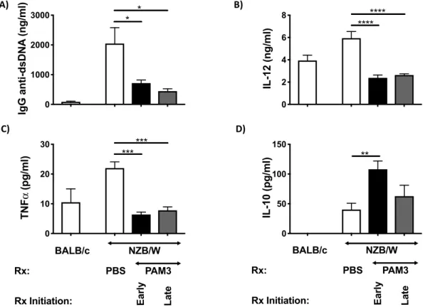
Additional immune manifestations of lupus in NZB/W mice in-cluded the production of autoAbs and inflammatory cytokines [5]. Consistent with the hypothesis that PAM3 might be useful in the treatment of SLE, serum IgG anti-DNA autoAb levels were 3–4 fold lower in the PAM3 vs PBS treated groups (p. < 0.04 and < 0.01 in the early and late treatment groups respectively,Fig. 5A). This was ac-companied by a significant decline in serum IL-12 levels and the amount of TNFα produced by spleen cell cultures (p < .001,Fig. 5B, C). Conversely, splenocytes from mice treated from 7 wk of age with PAM3 produced significantly more IL-10 (an immunosuppressive cy-tokine derived from M2 macrophages) than did untreated controls (p < .01,Fig. 5D). Spleen cells from mice whose treatment was de-layed until wk 13 also produced more IL-10 but this effect did not reach statistical significance.
3.6. Clinical effect of PAM3 treatment
The effect of PAM3 treatment initiated either before or after the onset of proteinuria was monitored in two independent experiments. PAM3 reduced proteinuria in both experiments and both groups (p < .005 in the early and < 0.01 in the late treatment groups, Fig. 3. Effect of PAM3 on the function of lupus monocytes. MACS purified monocytes were cultured as described inFig. 2. A) The uptake of zymosan-labeled particles after 5 days of culture (N = 17 lupus samples and 6 controls). B) Correlation between the expression of CD163 and uptake of zymosan particles. C) Concentration of TNFα in culture supernatants. D) Percent of cultured cells staining for intracytoplasmic IL-12. E) Concentration of IL-10 in culture supernatants (N = 12 lupus samples and controls). **p < .01; ***p < .001; ****p < .0001 compared unstimulated cells from the same donors.
Fig. 4. Effect of PAM3 treatment on the M1:M2 ratio of NZB/W mice. Female NZB/W mice were treated weekly with 100 μg of PAM3 starting either early (7 wk) or late (13 wk). Controls included littermates treated with PBS and normal (BALB/c) mice. The M1:M2 ratio was calculated based on the ratio of CD206-:CD206+F480+macrophages collected from the peritoneal cavity at 6 months of age. N = 5 NZB/W mice/group, N = 2 BALB/c controls. *p < .05; **p < .01; ***p < .001 for PAM3 vs PBS treated NZB/W mice.
Fig. 6A). It also normalized the BUN:creatinine ratio that reached ab-normally high levels as glomerulonephritis developed in control NZB/ W mice (Fig. 6B).
The natural history of disease was examined by monitoring kidney histology and animal survival. Immunohistochemical staining of kid-neys from control NZB/W mice showed that number of CD206+M2 macrophages remained low as disease progressed (Fig. 7A and B). By comparison, the number of M2 macrophages in the kidneys of PAM3 treated mice increased over time consistent with the normalization of M1:M2 ratio observed inFig. 4. Histological analysis of these kidneys confirmed that glomerular and tubular infiltration by inflammatory cells was delayed by PAM3 treatment (Fig. 7C–G). Of greatest import, PAM3 treatment significantly prolonged survival, with > 60% of mice whose treatment starting at 7 wk of age surviving through 11 months of age vs 5% in the control group (p = .0037,Fig. 8). Late initiation of therapy was less effective at slowing disease progression.
4. Discussion
This work establishes that the TLR 2/1 agonist PAM3 induces monocytes from lupus patients to preferentially differentiate into M2-like macrophages capable of suppressing autoimmune disease. The therapeutic potential of PAM3 was examined in the NZB/W model of lupus. Weekly treatment with PAM3 normalized the M2:M1 ratio and shifted the cytokine profile towards the production of suppressive ra-ther than inflammatory factors. Serum levels of anti-dsDNA auto-antibodies fell, glomerulonephritis was delayed, and survival was pro-longed by PAM3 therapy.
The conventional treatment of SLE relies on a combination of im-munosuppressive drugs, steroids, and agents that deplete/inhibit au-toreactive B cells [3,36,37]. Novel treatments designed to inhibit in-terferon or lymphocyte signaling are being evaluated in phase III
macrophages present in SLE patients provides an additional target for therapeutic intervention [11,12,38–40]. In the MRL/lpr model, de-pleting macrophages by knocking out CSF-1, its receptor, or by systemic administration the selective CSF-1R inhibitor GW2580 reduced disease severity [12,41–44]. Proteinuria and glomerular damage (but not au-toAb levels) were also reduced when monocyte infiltration was in-hibited by treating MRL/lpr or NZB/W mice with agents that antag-onized macrophage differentiation/proliferation [44,45].
The dominance of M1-like macrophages in lupus patients is likely driven by the increased level of pro-inflammatory cytokines including IL-12, TNFα, IFNγ and type I IFNs present in that disease. These support the generation of M1-like macrophages via pathways involving JAK/ Stat-1, NFKB and interferon regulatory factors [46–48]. Chemokines that promote monocyte infiltration (e.g. MCP-1, CX3CL1) and macro-phage differentiation/ proliferation are also elevated in mice and pa-tients with lupus and may contribute to this process [12,41].
While murine studies show that reducing overall macrophage abundance slows disease progression, Li et al. showed that increasing the frequency of M2 macrophages accomplished the same goal [13]. That group adoptively transferred M2 macrophages into lupus prone mice and documented a significant reduction in disease severity [13]. Unfortunately, adoptive M2 macrophage transfer is not a practical clinical strategy. We therefore examined whether treatment with PAM3, a TLR agonist that induces normal human monocytes to differ-entiate into M2-like macrophages [14], might be used to normalize the M1:M2 ratio in SLE patients. As shown inFig. 2C, that outcome was obtained in studies of cultured lupus monocytes. In the absence of exogenous stimulation, lupus monocytes preferentially mature into M1-like macrophages (Fig. 2D). This may reflect their in vivo exposure to factors that support the generation of inflammatory macrophages, as described above. Just the opposite is observed in cancer patients, whose monocytes respond to suppressive serum factors by preferentially ma-Fig. 5. Effect of PAM3 treatment on the immune manifestations of lupus. Mice were treated as described inFig. 4. Serum collected at 6 months was analyzed for (A) IgG anti-dsDNA and (B) IL-12. Spleen cells were cultured ex vivo without further stimulation for 24 h and culture supernatants tested for (C) TNFα and (D) IL-10 levels by ELISA (N = 5/group of lupus mice and 2 for BALB/c controls). **p < .01; ***p < .001; ****p < .0001 PAM3 vs PBS treated NZB/W mice.
The potential of PAM3 to increase M2 macrophage frequency re-presents a novel approach to the treatment of autoimmunity. In contrast to agents that non-specifically deplete all types of macrophages (thereby increasing host susceptibility to bacterial infections) PAM3 selectively supports the generation of immunosuppressive macrophages (Fig. 2C).
The patients evaluated in this study were treated with a variety of conventional agents including hydroxychloroquine, prednisone, myco-phenolate mofetil (MMF) and azathioprine (AZA) (Table 1). Hydroxy-chloroquine is an antimalarial that also interferes TLR7 and TLR9 sig-naling [3,51,52]; prednisone is a glucocorticoid that inhibits T and B cell responses [53]; MMF is an immunosuppressive agent that inhibits lymphocyte proliferation and antibody production [3,54]; while AZA regulates DNA synthesis thereby inhibiting cell proliferation [3]. None of these drugs is known to selectively act on monocytes/macrophages or influence monocyte differentiation.
The relative excess of M1-like macrophages in lupus patients con-tributes to the increased level of inflammatory cytokines and reduced apoptotic capacity found in that disease [24–27]. When apoptotic cells are not removed by phagocytosis, they leak DAMPS and self-antigens in
a process known as secondary necrosis. These can trigger inflammation and stimulate B cells to produce autoantibodies [55]. Treatment with PAM3 reversed these abnormalities (Fig. 3A), generating M2-like macrophages with good endocytic activity that preferentially secreted the immunosuppressive cytokine IL-10 rather than the inflammatory cytokines IL-12 and TNFα (Fig. 3C–E). Monocytes from patients with active lupus express elevated levels of the co-stimulatory molecule CD40. This improves their ability to activate B cells (which express the CD40 ligand) and supports autoAb production [6,23]. Current findings confirm that lupus monocytes express high levels of CD40. Treatments that support the differentiation of monocytes into M1-like (IFNγ and R848) but not M2-like (PAM3) macrophages correlated with increased CD40 expression (Fig. 2E).
To evaluate the effect of PAM3 in vivo, NZB/W mice were treated weekly starting either before or after abnormalities in glomerular function (proteinuria) arose (at 7 and 13 weeks of age, respectively). PAM3 normalized the M1:M2 ratio, inhibited the progression of pro-teinuria, reduced the BUN:creatinine ratio and prolonged survival (Figs. 4, 6 and 8). Initiating PAM3 treatment at 7 weeks of age max-imized its survival benefit. Starting at 13 weeks had less of an effect on the magnitude of proteinuria and survival, indicating that treatment could not reverse pre-existing kidney damage. Histologic evaluation of kidneys from these animals confirmed that glomerular and tubular in-filtration were reduced by PAM3 treatment while the frequency of CD206+M2 macrophages rose (Fig. 7). While human lupus is char-acterized by periods of relative inactivity punctuated by disease flares, glomerulonephritis in NZB/W mice is genetically determined and ad-vances inexorably. Thus, the significant delay in disease progression observed in this murine model may translate into even greater benefit for patients. In that context, treatment with PAM3 significantly altered the immune milieu of NZB/W mice. The production of the pro-in-flammatory cytokines IL-12 and TNFα was reduced (Fig. 3C and D; 5 B, C). Previous reports showed that neutralizing IL-12 could prevent the development of Th1-mediated autoimmune disease [56]. Similarly, TNFα levels in the sera of lupus patients correlates with disease activity and there are reports that anti-TNF therapy improves disease outcome [29,57] (Figs. 3C and 5C). IL-10 is an immunosuppressive cytokine produced by M2 macrophage. IL-10 impedes APC activation and alle-viates disease in NZB/W mice (although its effect in human lupus re-mains controversial) [29,58,59]. Human monocytes stimulated with PAM3 and splenocytes from PAM3 treated mice produced IL-10 (Figs. 3E and 5D), an effect linked with a reduction in autoAb levels in NZB/W mice (Fig. 5A).
The findings in this report establish that PAM3 supports the pre-ferential generation of M2-like macrophages from lupus monocytes. That activity altered the internal milieu, increasing levels of suppressive versus inflammatory cytokines, reducing autoantibody production, de-laying disease progression and significantly prolonging survival. We propose that PAM3 mediated modulation of macrophage differentiation be evaluated for efficacy in human SLE.
5. Conclusions
Current findings support the conclusion that macrophage contribute to the pathogenesis of SLE. Our results demonstrate that the TLR2/1 agonist PAM3 induces monocytes from lupus patients to preferentially differentiate into M2-like macrophages. The same effect is observed when PAM3 is administered to lupus-prone NZB/W mice, culminating in the normalization of the M1:M2 balance. Disease progression is de-layed and survival significantly prolonged by weekly PAM3 treatment in this murine model. By regulating macrophage differentiation, PAM3 thus represents a potentially novel approach to the therapy of SLE. Fig. 6. Effect of PAM3 treatment on lupus nephritis. NZB/W mice were
treated with PAM3 or PBS as described inFig. 4. A) Proteinuria was measured at 6 months (N = 11 for each PAM3 treated group and 21 for PBS treated mice). B) The serum BUN:creatinine ratio was measured at 6 months of age (N = 5 for NZB/W, N = 3 BALB/c). *p < .05; **p < .01 PAM3 vs PBS treated NZB/W mice.
Acknowledgements
The authors thank Debra Tross, Fatima Haidar and Madhivanan Elango for their support of the animal experiments, Luz Blanco for or-ganizing the transfer of patient samples and Engin Kayraklioglu for the analysis of kidney sections. This work was supported by the Intramural Research Program of the NIH, NCI. The content of this publication does not necessarily reflect the views or policies of the Department of Health and Human Services, nor does mention of trade names, commercial
products, or organizations imply endorsement by the United States government. The funders had no role in study design, data collection and analysis, decision to publish, or preparation of the manuscript.
Appendix A. Supplementary data
Supplementary data to this article can be found online athttps:// doi.org/10.1016/j.jaut.2019.01.004.
References
[1] A. Kaul, C. Gordon, M.K. Crow, Z. Touma, M.B. Urowitz, V.R. van, G. Ruiz-Irastorza, G. Hughes, Systemic lupus erythematosus, Nat.Rev.Dis.Primers. 2 (2016) 16039. [2] G.C. Tsokos, M.S. Lo, R.P. Costa, K.E. Sullivan, New insights into the
im-munopathogenesis of systemic lupus erythematosus, Nat. Rev. Rheumatol. 12 (2016) 716–730.
[3] C. Yildirim-Toruner, B. Diamond, Current and novel therapeutics in the treatment of systemic lupus erythematosus, J. Allergy Clin. Immunol. 127 (2011) 303–312. [4] J.B. Rottman, C.R. Willis, Mouse models of systemic lupus erythematosus reveal a
complex pathogenesis, Vet. Pathol. 47 (2010) 664–676.
[5] D. Perry, A. Sang, Y. Yin, Y.Y. Zheng, L. Morel, Murine models of systemic lupus erythematosus, J. Biomed. Biotechnol. 2011 (2011) 271694.
[6] Y. Li, P.Y. Lee, W.H. Reeves, Monocyte and macrophage abnormalities in systemic lupus erythematosus, Arch. Immunol. Ther. Exp. 58 (2010) 355–364.
[7] R. Mukherjee, B.P. Kanti, T.P. Kumar, R. Tripathy, D.B. Kumar, B. Ravindran, Non-classical monocytes display inflammatory features: validation in sepsis and systemic lupus erythematous, Sci. Rep. 5 (2015) 13886.
[8] F.O. Martinez, S. Gordon, The M1 and M2 paradigm of macrophage activation: time for reassessment, F1000Prime.Rep. 6 (2014) 13.
[9] S. Gordon, A. Pluddemann, Macrophage clearance of apoptotic cells: a critical as-sessment, Front. Immunol. 9 (2018) 127.
[10] A.W. Roberts, B.L. Lee, J. Deguine, S. John, M.J. Shlomchik, G.M. Barton, Tissue-resident macrophages are locally programmed for silent clearance of apoptotic cells, Immunity 47 (2017) 913–927.
Fig. 7. Effect of PAM3 treatment on kidney pathology and the accumulation of M2 macrophages. NZB/W mice were treated as described inFig. 6A,B) The number of M2 macrophages present in their kidneys was monitored over time by immunohistochemical staining of PBS and PAM3 treated mice. C) The severity of glomerulonephritis was evaluated histologically. N = 9 for PBS and 5 for PAM3 treated animals. D-G) Representative PAS stained kidney sections from 28 week old NZB/W mice treated with PBS (D, E) or PAM3 (F, G). Note the increased interstitial inflammation (arrows) in panel D and hyaline deposits (–), cellular crescent formation (−) and karyorrhexis (arrow) in panel E vs the slight increase in endocapillary proliferation in panel G. Panels D, F were 100x and panels E, G were 800x magnification.
Fig. 8. Effect of PAM3 on survival. Survival was monitored through 45 weeks of age in mice treated with PAM3 starting at 7 or 13 weeks of age (N = 6/ group) or PBS treated controls (N = 21). **p = .0037 when compared to PBS group, Log rank test.
[11] S.C. Funes, M. Rios, J. Escobar-Vera, A.M. Kalergis, Implications of macrophage polarization in autoimmunity, Immunology 154 (2018) 186–195.
[12] S.A. Chalmers, V. Chitu, M. Ramanujam, C. Putterman, Therapeutic targeting of macrophages in lupus nephritis, Discov. Med. 20 (2015) 43–49.
[13] F. Li, Y. Yang, X. Zhu, L. Huang, J. Xu, Macrophage polarization modulates de-velopment of systemic lupus erythematosus, Cell. Physiol. Biochem. 37 (2015) 1279–1288.
[14] D. Bayik, D. Tross, L.A. Haile, D. Verthelyi, D.M. Klinman, Regulation of the ma-turation of human monocytes into immunosuppressive macrophages, Blood Adv 1 (2017) 2510–2519.
[15] J. Wang, Y. Shirota, D. Bayik, H. Shirota, D. Tross, J.L. Gulley, L.V. Wood, J.A. Berzofsky, D.M. Klinman, Effect of TLR agonists on the differentiation and function of human monocytic myeloid-derived suppressor cells, J. Immunol. 194 (2015) 4215–4221.
[16] G.S. Hill, M. Delahousse, D. Nochy, J. Bariety, Class IV-S versus class IV-G lupus nephritis: clinical and morphologic differences suggesting different pathogenesis, Kidney Int. 68 (2005) 2288–2297.
[17] S. Gordon, P.R. Taylor, Monocyte and macrophage heterogeneity, Nat. Rev. Immunol. 5 (2005) 953–964.
[18] C.D. Mills, K. Kincaid, J.M. Alt, M.J. Heilman, A.M. Hill, M-1/M-2 macrophages and the Th1/Th2 paradigm, J. Immunol. 164 (2000) 6166–6173.
[19] C.D. Mills, M1 and M2 macrophages: oracles of Health and disease, Crit. Rev. Immunol. 32 (2012) 463–488.
[20] D. Bayik, D. Tross, D.M. Klinman, Factors influencing the differentiation of human monocytic myeloid-derived suppressor cells into inflammatory macrophages, Front. Immunol. 9 (2018) 608.
[21] L. Liu, W.R. Allman, A.S. Coleman, K. Takeda, T.L. Lin, M. Akkoyunlu, Delayed onset of autoreactive antibody production and M2-skewed macrophages contribute to improved survival of TACI deficient MRL-Fas/Lpr mouse, Sci. Rep. 8 (2018) 1308.
[22] D. Pilling, T. Fan, D. Huang, B. Kaul, R.H. Gomer, Identification of markers that distinguish monocyte-derived fibrocytes from monocytes, macrophages, and fi-broblasts, PLoS One 4 (2009) e7475.
[23] C.G. Katsiari, S.N. Liossis, V.L. Souliotis, A.M. Dimopoulos, M.N. Manoussakis, P.P. Sfikakis, Aberrant expression of the costimulatory molecule CD40 ligand on monocytes from patients with systemic lupus erythematosus, Clin. Immunol. 103 (2002) 54–62.
[24] S.W. Tas, P. Quartier, M. Botto, L. Fossati-Jimack, Macrophages from patients with SLE and rheumatoid arthritis have defective adhesion in vitro, while only SLE macrophages have impaired uptake of apoptotic cells, Ann. Rheum. Dis. 65 (2006) 216–221.
[25] J.Y. Jung, C.H. Suh, Incomplete clearance of apoptotic cells in systemic lupus er-ythematosus: pathogenic role and potential biomarker, Int. J. Rheum. Dis. 18 (2015) 294–303.
[26] I. Baumann, W. Kolowos, R.E. Voll, B. Manger, U. Gaipl, W.L. Neuhuber, T. Kirchner, J.R. Kalden, M. Herrmann, Impaired uptake of apoptotic cells into tingible body macrophages in germinal centers of patients with systemic lupus er-ythematosus, Arthritis Rheum. 46 (2002) 191–201.
[27] M. Herrmann, R.E. Voll, O.M. Zoller, M. Hagenhofer, B.B. Ponner, J.R. Kalden, Impaired phagocytosis of apoptotic cell material by monocyte-derived macrophages from patients with systemic lupus erythematosus, Arthritis Rheum. 41 (1998) 1241–1250.
[28] P.J. Murray, T.A. Wynn, Protective and pathogenic functions of macrophage sub-sets, Nat. Rev. Immunol. 11 (2011) 723–737.
[29] D.Y. Yap, K.N. Lai, Cytokines and their roles in the pathogenesis of systemic lupus erythematosus: from basics to recent advances, J. Biomed. Biotechnol. 2010 (2010) 365083.
[30] M. Patel, D. Xu, P. Kewin, B. Choo-Kang, C. McSharry, N.C. Thomson, F.Y. Liew, TLR2 agonist ameliorates established allergic airway inflammation by promoting Th1 response and not via regulatory T cells, J. Immunol. 174 (2005) 7558–7563. [31] M.C. Tunis, B. Dawod, K.R. Carson, L.L. Veinotte, J.S. Marshall, Toll-like receptor 2
activators modulate oral tolerance in mice, Clin. Exp. Allergy 45 (2015) 1690–1702. [32] T. Ha, C. Lu, L. Liu, F. Hua, Y. Hu, J. Kelley, K. Singh, R.L. Kao, J. Kalbfleisch,
D.L. Williams, X. Gao, C. Li, TLR2 ligands attenuate cardiac dysfunction in poly-microbial sepsis via a phosphoinositide 3-kinase-dependent mechanism, Am. J. Physiol. Heart Circ. Physiol. 298 (2010) H984–H991.
[33] P.H. Lambert, F.J. Dixon, Pathogenesis of the glomerulonephritis of NZB/W mice, J. Exp. Med. 127 (1968) 507–513.
[34] T. Roszer, Understanding the mysterious M2 macrophage through activation mar-kers and effector mechanisms, Mediat. Inflamm. 2015 (2015) 816460. [35] L. Schiffer, R. Bethunaickan, M. Ramanujam, W. Huang, M. Schiffer, H. Tao,
M.P. Madaio, E.P. Bottinger, A. Davidson, Activated renal macrophages are markers of disease onset and disease remission in lupus nephritis, J. Immunol. 180 (2008) 1938–1947.
[36] J. Liao, C. Chang, H. Wu, Q. Lu, Cell-based therapies for systemic lupus er-ythematosus, Autoimmun. Rev. 14 (2015) 43–48.
[37] Z. Touma, D.D. Gladman, Current and future therapies for SLE: obstacles and
recommendations for the development of novel treatments, Lupus Sci.Med 4 (2017) e000239.
[38] J.C. Byrne, G.J. Ni, E. Lazzari, R. Mahony, S. Smith, K. Stacey, C. Wynne, C.A. Jefferies, Genetics of SLE: functional relevance for monocytes/macrophages in disease, Clin. Dev. Immunol. (2012) 582352 (2012).
[39] A. Laria, A. Lurati, M. Marrazza, D. Mazzocchi, K.A. Re, M. Scarpellini, The mac-rophages in rheumatic diseases, J. Inflamm. Res. 9 (2016) 1–11.
[40] J. Orme, C. Mohan, Macrophage subpopulations in systemic lupus erythematosus, Discov. Med. 13 (2012) 151–158.
[41] J. Menke, W.A. Rabacal, K.T. Byrne, Y. Iwata, M.M. Schwartz, E.R. Stanley, A. Schwarting, V.R. Kelley, Circulating CSF-1 promotes monocyte and macrophage phenotypes that enhance lupus nephritis, J. Am. Soc. Nephrol. 20 (2009) 2581–2592.
[42] S.A. Chalmers, J. Wen, J. Shum, J. Doerner, L. Herlitz, C. Putterman, CSF-1R in-hibition attenuates renal and neuropsychiatric disease in murine lupus, Clin. Immunol. 185 (2017) 100–108.
[43] D.M. Lenda, E.R. Stanley, V.R. Kelley, Negative role of colony-stimulating factor-1 in macrophage, T cell, and B cell mediated autoimmune disease in MRL-Fas(lpr) mice, J. Immunol. 173 (2004) 4744–4754.
[44] A. Inoue, H. Hasegawa, M. Kohno, M.R. Ito, M. Terada, T. Imai, O. Yoshie, M. Nose, S. Fujita, Antagonist of fractalkine (CX3CL1) delays the initiation and ameliorates the progression of lupus nephritis in MRL/lpr mice, Arthritis Rheum. 52 (2005) 1522–1533.
[45] H. Hasegawa, M. Kohno, M. Sasaki, A. Inoue, M.R. Ito, M. Terada, K. Hieshima, H. Maruyama, J. Miyazaki, O. Yoshie, M. Nose, S. Fujita, Antagonist of monocyte chemoattractant protein 1 ameliorates the initiation and progression of lupus ne-phritis and renal vasculitis in MRL/lpr mice, Arthritis Rheum. 48 (2003) 2555–2566.
[46] N. Wang, H. Liang, K. Zen, Molecular mechanisms that influence the macrophage m1-m2 polarization balance, Front. Immunol. 5 (2014) 614.
[47] A. Midgley, Z. McLaren, R.J. Moots, S.W. Edwards, M.W. Beresford, The role of neutrophil apoptosis in juvenile-onset systemic lupus erythematosus, Arthritis Rheum. 60 (2009) 2390–2401.
[48] L. Bennett, A.K. Palucka, E. Arce, V. Cantrell, J. Borvak, J. Banchereau, V. Pascual, Interferon and granulopoiesis signatures in systemic lupus erythematosus blood, J. Exp. Med. 197 (2003) 711–723.
[49] S. Hallam, M. Escorcio-Correia, R. Soper, A. Schultheiss, T. Hagemann, Activated macrophages in the tumour microenvironment-dancing to the tune of TLR and NF-kappaB, J. Pathol. 219 (2009) 143–152.
[50] T. Chanmee, P. Ontong, K. Konno, N. Itano, Tumor-associated macrophages as major players in the tumor microenvironment, Cancers 6 (2014) 1670–1690. [51] D. Kyburz, F. Brentano, S. Gay, Mode of action of hydroxychloroquine in
RA-evi-dence of an inhibitory effect on toll-like receptor signaling, Nat. Clin. Pract. Rheumatol. 2 (2006) 458–459.
[52] S. Piconi, S. Parisotto, G. Rizzardini, S. Passerini, R. Terzi, B. Argenteri, P. Meraviglia, A. Capetti, M. Biasin, D. Trabattoni, M. Clerici, Hydroxychloroquine drastically reduces immune activation in HIV-infected, antiretroviral therapy-treated immunologic nonresponders, Blood 118 (2011) 3263–3272.
[53] N. Auphan, J.A. DiDonato, C. Rosette, A. Helmberg, M. Karin, Immunosuppression by glucocorticoids: inhibition of NF-kappa B activity through induction of I kappa B synthesis, Science 270 (1995) 286–290.
[54] P.F. Yong, D.P. D'Cruz, Mycophenolate mofetil in the treatment of lupus nephritis, Biologics 2 (2008) 297–310.
[55] I.K. Poon, M.D. Hulett, C.R. Parish, Molecular mechanisms of late apoptotic/ne-crotic cell clearance, Cell Death Differ. 17 (2010) 381–397.
[56] A. Nakajima, S. Hirose, H. Yagita, K. Okumura, Roles of IL-4 and IL-12 in the de-velopment of lupus in NZB/W F1 mice, J. Immunol. 158 (1997) 1466–1472. [57] L.J. Zhu, X. Yang, X.Q. Yu, Anti-TNF-alpha therapies in systemic lupus
er-ythematosus, J. Biomed. Biotechnol. (2010) 465898 (2010).
[58] A.M. Beebe, D.J. Cua, M.R. de Waal, The role of interleukin-10 in autoimmune disease: systemic lupus erythematosus (SLE) and multiple sclerosis (MS), Cytokine Growth Factor Rev. 13 (2002) 403–412.
[59] L.F. Hou, S.J. He, X. Li, C.P. Wan, Y. Yang, X.H. Zhang, P.L. He, Y. Zhou, F.H. Zhu, Y.F. Yang, Y. Li, W. Tang, J.P. Zuo, SM934 treated lupus-prone NZB x NZW F1 mice by enhancing macrophage interleukin-10 production and suppressing pathogenic T cell development, PLoS One 7 (2012) e32424.
[60] J. Romero-Diaz, D. Isenberg, R. Ramsey-Goldman, Measures of adult systemic lupus erythematosus: updated version of British isles lupus assessment group (BILAG 2004), European consensus lupus activity measurements (ECLAM), systemic lupus activity measure, revised (SLAM-R), systemic lupus activity questionnaire for po-pulation studies (SLAQ), systemic lupus erythematosus disease activity index 2000 (SLEDAI-2K), and systemic lupus international collaborating clinics/American col-lege of rheumatology damage index (SDI), Arthritis Care Res. 63 (Suppl. 11) (2011) S37–S46.
[61] D.D. Gladman, D. Ibanez, M.B. Urowitz, Systemic lupus erythematosus disease ac-tivity index 2000, J. Rheumatol. 29 (2002) 288–291.