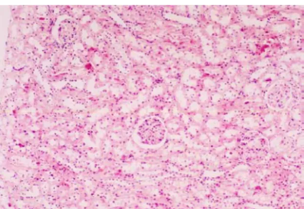
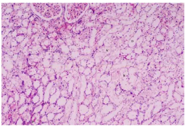
The Effects of nifedipine on renal perfusion pressure and kidney during cisplatin-induced nephrotoxicity in rats
Tam metin
Şekil
Benzer Belgeler
The group of paclitaxel + cisplatin combination therapy, was not found to exhibit a statistically significant difference regarding reduction in the number of primary follicles
CP and protective agents against CP-induced organ damages are current issues in cancer treatment in patients of reproductive age, but none of the studies in literature have
Results: CP group administered cisplatin had significantly increased blood, serum, and cardiac tissue malondialdehyde (MDA), interleukin 1 beta (IL-1β), tumor necrosis factor
maddesiyle yabancı yatırımcıların (yabancı gerçek.. kişiler ile yabancı ülkelerin kanunlarına göre kurulmuş tüzel kişilerin) Türkiye’de kurdukları ya da iştirak
İnşaat sektöründe en sık kullanılan yapı malzemesi olan beton; çimento, agrega, su ve kimyasal katkılardan meydana gelmektedir. Bu bileşenlerin özellikleri ve miktarı,
Abdullah Nazırlı bu kitabında; hafız yetiştiren bir hocanın öğrencilere karşı tutumunun nasıl olması gerektiği, hafız olmak isteyen öğrencilerde bulunması gereken
Table (2): Specific activity levels of different nuclides, radium equivalent Raeq, risk index for gamma rays (Iγ) and absorbed dose rate in air (Dγ) in surface soil models in
HSP70 immunoreactivity was observed in the tubular epithelium of both Group III and Group IV tissue sections, mostly in Group II. Sperm transport, maturation and storage is the
