17
Case Study
Simple Bone Cyst on The Mandibular Ramus: A Case Report Özlem Okumuş 1*, Altan Varol 2, Semih Özbayrak 1
1 Department of Oral and Dentomaxillofacial Radiology, Faculty of Dentistry, Altınbaş University, Istanbul, Turkey.
2 Department of Oral and Dentomaxillofacial Surgery, Faculty of Dentistry, Marmara University, Istanbul, Turkey.
Submitted: March 28, 2018; Accepted: June 04, 2018
Abstract: The simple bone cyst, also called traumatic bone cyst or solitary bone cyst, is an uncommon cavity of
the jaws and occurs on the mandibular symphysis and body, but rarely on the mandibular condyle. It is generally asymptomatic, usually detected incidentally during routine radiological assessment. This article presents a bone cystic lesion affecting the right mandibular ramus in a 11 year-old female patient with no history of previous trauma. A well demarcated oval shaped radiolucent lesion was observed on the right mandibular ramus in panoramic radiography. The advanced imaging techniques were performed to assess the exact location of the lesion and its association with the surrounded soft tissue. The patient underwent this cystic lesion enucleated under general anesthesia and the surgical exploration has demonstrated the empty cavity without the epithelial layer. The postoperative panoramic radiography which was performed after 6 months of follow up showed bony formation. The simple bone cyst is a rare jawbone lesion with an unclear etiology. It is generally associated with a good prognosis and a low rate of recurrence. Trauma history must be questioned.
Key Words: Traumatic bone cyst, Simple bone cyst, Solitary bone cyst, Mandibular ramus, Cone beam computed
tomography, Mandibula.
Address of Correspondence: Özlem Okumuş - [email protected]
Department of Oral and Dentomaxillofacial Radiology, Faculty of Dentistry, Altınbas University, Kartaltepe Mahallesi, Incirli Caddesi No: 11-A, 34144 Bakırköy/Istanbul, Turkey
Introduction
The simple bone cyst is an uncommon intraosseous lesion with the etiology not clearly identified but a traumatic injury has been reported including the empty bone cavity following medullary bleeding (Kim et al., 2016).
The simple bone cyst occurs more frequently in the mandibular body but cases involving the mandibular symphysis, ramus, and condyle have been reported (An et al., 2014). The lesion is usually present within the second decades of life with slight male domination or with no gender discrimination (Velasco et al., 2012).
18
The lesion is generally asymptomatic and is often detected incidentally in routine radiography. Rarely, it can cause pain, tooth sensitivity, and paresthesia. The adjacent teeth of the lesion are usually vital and there is no mobility, tooth displacement and external root resorption (Xanthinaki et al., 2006). Radiologically, the simple bone cyst is found as a radiolucent unilocular rarely multilocular lesion with well defined margins and it extends between the roots of the teeth and this is named as scalloping effect but it can be found in edentulous areas also (Xanthinaki et al., 2006).
The treatment includes the surgical exploration and curettage of the lesion to stimulate bleeding and promote healing which helps in the diagnosis. Histopathologically, it is not considered a cyst because it is covered with a thin connective tissue surface without epithelial lining (Xanthinaki et al., 2006; Velasco et al., 2012; Kim et al., 2016).
This report presents a case of simple bone cyst affecting the right mandibular ramus with clinical and radiographic findings in a 11 year old female patient.
Description of the Case
A 11-year old female patient was referred to the Department of Oral and Dentomaxillofacial Radiology due to the radiolucent lesion in the right mandibular ramus which was discovered incidentally on panoramic radiograph during her orthodontic treatment. The patient was asymptomatic without a history of trauma involving the mandible and any habits such as bruxism and clenching. The medical history was unremarkable.
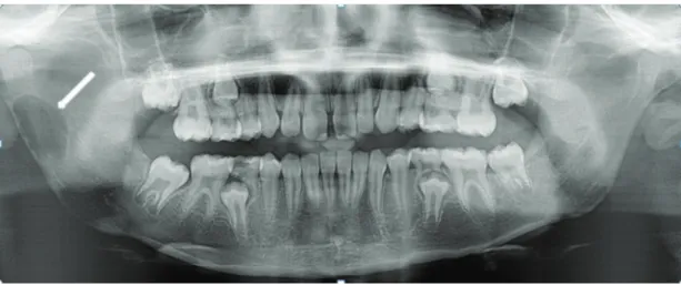
The panoramic radiograph showed unilocular radiolucent lesion with well-defined sclerotic margin just below the mandibular notch on the posterior border of the right mandibular ramus. The posterior cortical border of ramus was thin and there was no expansion of anterior and posterior border of mandibular ramus (Figure1).
Figure 1. Preoperative panoramic radiography showing well defined radiolucent lesion of the mandibular right ramus (arrow).
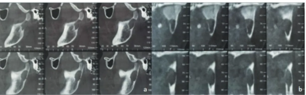
19 The cone beam computed tomography (CBCT) was performed and this examination revealed a hypodense
lesion in the right mandibular ramus which caused destruction on the posterior border of the ramus (Figure 2). The magnetic resonance imaging (MRI) was performed to determine density difference between surrounding soft tissues, contrast involvement and hemorrhagic feature of the lesion. MRI showed a high signal intensity lesion in the same location on the T2-weighted images (Figure 3 a,b). According to these images, the surgery was scheduled for the removal of the lesion.
Figure 2. CBCT shows destruction on the posterior border of ramus on sagittal view (a), coronal view (b).
Figure 3. T2 weighted MRI shows high signal intensity on sagittal (a), coronal view (b) (arrows).
For the operation, the general anesthesia was performed and the right mandibular ramus was exposed by intraoral incision. The surgical exploration has demonstrated the empty cavity and no sample could be collected for histopathological examination. The walls were curetted to induce bleeding and the fixation plate was fixed at the posterior border of mandibular ramus.
The final diagnosis is simple bone cyst of the right mandibular condyle based on the radiological and surgical findings. The patient did not have any postoperative complications in the right temporomandibular
20
joint. The postoperative panoramic radiography which was performed after 6 months showed bony formation (Figure 4).
Figure 4. Postoperative panoramic radiography after 6 months showing bony formation.
Discussion
The simple bone cyst is also known as traumatic bone cyst, solitary bone cyst, idiopathic bone cavity, and hemorrhagic bone cyst (Shigematsu et al., 1994). Although its etiology is not identified, the trauma is the most frequently considered factor.
21 It is based on the formation of an intramedullary hemorrhage following a trauma. The vascular dysfunction
causes the medullary necrosis and osteoclastic resorption due to decreased tissular pH. This theory can be applied to the maxillofacial bones due to the multiple microtraumas through the teeth and alveolar process (Harnet et al., 2008). In addition to trauma, etiologies such as calcium metabolism changes, mild infectious conditions, local bone growth changes, orthodontic treatment, and venous obstruction have been discussed (Harnet et al., 2008). Some authors have discussed this theory since there is no trauma history in most publications. Therefore, it is considered to be multifactorial etiology in most cases (Harnet et al., 2008; Suei et al., 2007).
The trauma hypothesis is supported by the occurrence of simple bone cyst in younger individuals, mostly in metaphyses of long bones and in mandibles (Olech et al., 1951). When detected early, the lesion generally contains blood or serosanguineous fluid. The aspirated fluid from the cyst cavity generally has electrolyte and protein concentrations similar to the serum. The fluid is thought to be extravasated blood. The amount of fluid decreases with the age of the lesion and it finally becomes empty (Saito et al., 1992). The radiographic characteristics of the simple bone cyst may present similar appearance with a variety of odontogenic and nonodontogenic radiolucent lesions. The differential diagnosis is difficult when detected in the body or symphysis of the mandible (Dıncer et al., 2012). In addition, since many odontogenic lesions have scalloped borders, it is not diagnostic of the simple bone cyst (Copete et al., 1998). In 2D imaging the simple bone cyst may have both unilocular appearance and multilocular appearance. The bone septa within the lesion is responsible for multilocular view (Mitchell and Ward-Booth, 1984). The true intralesional septa entity should be carefully considered because the fin-like extensions of a wall can lead to misdiagnosis by causing false septa and multilocular appearance (Mathew et al., 2012). The lesion presented in this case report had unilocular appearance with well-defined sclerotic margins.
Suei et al. suggested a relationship between prognosis and radiographic features of the simple bone cyst. The radiographic features such as absent lamina dura, scalloped margins, bone expansion, internal radiopaque masses may point out possible recurrence following treatment (Suei et al., 2010).
Diagnosis of simple bone cyst before surgical intervention is very difficult in most cases. Surgical exploration does not only lead to confirmation the diagnosis, but also curettage as treatment stimulates to bleeding and bone regeneration (Surej et al., 2015). The 6 month of follow-up panoramic radiography also showed bony formation in our case.
In conclusion, the simple bone cyst is a rare asymptomatic lesion with an unclear etiology, mostly detected incidentally on radiographic examination. It is generally associated with a good prognosis and a low rate of recurrence. Trauma history must be questioned. Advanced imaging techniques such as CBCT and MRI have an important diagnostic value for initial diagnosis and prognosis.
Conflict of Interests
22
References
An SY, Lee JS, Benavides E, Aminlari A, McDonald NJ, Edwards PC, et al. (2014). Multiple simple bone cysts of the jaws: review of the literature and report of three cases. Oral Surgery, Oral Medicine, Oral pathology and Oral Radiology, 117, e458-469.
Copete MA, Kawamata A, Langlais RP. (1998). Solitary bone cyst of the jaws. Radiographic review of 44 cases. Oral Surgery, Oral Medicine, Oral Pathology, Oral Radiology, and Endodontics, 85,221-225. Dincer O, Kose TE, Cankaya AB, Aybar B. (2012). Learning from errors: Traumatic bone cyst mimicking radicular cyst. BMJ Case Reports, 2012: bcr2012007316.
Harnet JC, Lombardi T, Klewansky P, Rieger J, Tempe MH, Cla 6. vert JM. (2008). Solitary bone cyst of the jaws: a review of the etiopatho genic hypotheses. Journal of Oral and Maxillofacial Surgery, 66, 2345-2348. Kim HK, Lim JH, Jeon KJ, Huh JK. (2016). Bony window approach for a traumatic bone cyst on the mandibular condyle: a case report with long-term follow-up. Journal of the Korean Association of Oral and Maxillofacial Surgeons, 42(4), 209-214.
Mathew R, Omami G, Gianoli D, Lurie A. (2012). Unusual cone-beam computerized tomography presentation of traumatic (simple) bone cyst: case report and radiographic analysis. Oral Surgery, oral Medicine, Oral Pathology, and Oral Radiology, 113(3), 410-413.
Mitchell DA, Ward-Booth RP. (1984). Atypical presentation of a solitary bone cyst. International Journal of oral Surgery, 13,256-259.
Olech E, Sieber H, Weinmann JP. (1951). Traumatic mandibular bone cysts. Oral Surgery, Oral Medicine, and Oral Pathology, 4, 1160-1172.
Saito Y, Hoshina Y, Nagamine T, Nakajima T, Suzuki M, Hayashi T (1992) Simple bone cyst. A clinical and histopathologic study of fifteen cases. Oral Surgery, Oral Medicine, and Oral Pathology, 74(4), 487–491. Shigematsu H, Fujita K, Watanabe K. (1994). Atypical simple bone cyst of the mandible. A case report. International Journal of Oral and Maxillofacial Surgery, 23, 298-299.
Suei Y, Taguchi A, Tanimoto K. (2007). Simple bone cyst of the jaws: eva luation of treatment outcome by review of 132 cases. Journal of Oral and Maxillofacial Surgery, 65, 918-923.
Suei Y, Taguchi A, Nagasaki T, Tanimoto K. (2010). Radiographic findings and prognosis of simple bone cysts of the jaws. Dentomaxillofacial Radiology, 39, 65-72.
Surej Kumar LK, Kurien N, Thaha KA. (2015). Traumatic Bone Cyst of Mandible. Journal of Maxillofacial and Oral Surgery, 14(2), 466–469.
Velasco I, Cifuentes J, Lobos N, San Martín F. (2012).The unusual evolution of a simple bone cyst in the mandible: A case report. Journal of Clinical and Experimental Dentistry, 4(2), e132-135
Xanthinaki AA, Choupis KI, Tosios K, Pagkalos VA, Papanikolaou SI. (2006). Traumatic bone cyst of the mandible of possible iatrogenic origin: a case report and brief review of the literature. Head and Face Medicine, 2(1), 40.