ANALYSIS OF DIFFERENTIALLY EXPRESSED GENES IN BREAST CANCER: BRCA1-INDUCED GENE EXPRESSION PROFILES AND
META-ANALYSIS GENE SIGNATURE
A THESIS SUBMITTED TO
THE DEPARTMENT OF MOLECULAR BIOLOGY AND GENETICS AND THE INSTITUTE OF ENGINEERING AND SCIENCE OF
BİLKENT UNIVERSITY
IN PARTIAL FULFILLMENT OF THE REQUIREMENTS FOR THE DEGREE OF DOCTOR OF PHILOSOPHY
By
BALA GÜR DEDEOĞLU May 2009
I certify that I have read this thesis and that in my opinion it is fully adequate, in scope and in quality, as a thesis for the degree of Doctor of Philosophy.
________________________________ Prof. Dr. Neşe Atabey
I certify that I have read this thesis and that in my opinion it is fully adequate, in scope and in quality, as a thesis for the degree of Doctor of Philosophy.
________________________________
Assoc. Prof. Dr. Işık G. Yuluğ
I certify that I have read this thesis and that in my opinion it is fully adequate, in scope and in quality, as a thesis for the degree of Doctor of Philosophy.
________________________________
Assoc. Prof. Dr. Rengül Atalay
I certify that I have read this thesis and that in my opinion it is fully adequate, in scope and in quality, as a thesis for the degree of Doctor of Philosophy.
________________________________
Asst. Prof. Dr. Özlen Konu
I certify that I have read this thesis and that in my opinion it is fully adequate, in scope and in quality, as a thesis for the degree of Doctor of Philosophy.
________________________________
Asst. Prof. Dr. Can Akçalı
Approved for the Institute of Engineering and Science
_______________________________
Prof. Dr. Mehmet Baray
ABSTRACT
ANALYSIS OF DIFFERENTIALLY EXPRESSED GENES IN BREAST CANCER: BRCA1-INDUCED GENE EXPRESSION PROFILES AND
META-ANALYSIS GENE SIGNATURE BALA GÜR DEDEOĞLU PhD in Molecular Biology and Genetics
Supervisor: Assoc. Prof. Işık G. Yuluğ May 2009, 225 Pages
The aim of the first part of this study was to find out the expression profiles of the genes, which were selected from the former BRCA1-induced gene list (OVCA1, OVCA2, ERBIN, RAD21, XRN2, RENT2, SMG1 and MAC30) in normal-matched primary breast tumors and to correlate the gene expression profiles of selected candidate genes with BRCA1 and various pathology parameters. Among the target genes, the expression of ERBIN, SMG1 and RAD21 were found to be highly
correlated with that of BRCA1 both in BRCA1 up- and down-regulated cells and this result was validated with qRT-PCR expression profiling of the eight genes in 32 normal-matched primary breast tumor samples. These genes were found to be discriminative between ER(-) and ER(+) tumors as well as grade 1 and grade 3 tumors. Target genes were also analyzed in independent microarray datasets to assess their predictive power for breast tumor grade, subtype and patient survival. ERBIN, SMG1 and RAD21 were found to have predictive roles in these datasets.
The aim of the second part of the study was to found appropriate reference genes (RGs) for accurate quantification of target gene expressions in breast tumor tissues. The expression patterns of fifteen widely-used endogenous RGs and three candidate genes that were selected through analysis of two independent microarray datasets were determined in 23 primary breast tumors and their matched normal tissues using qRT-PCR. Additionally, 18S rRNA, ACTB, and SDHA were tested using randomly primed cDNAs from 13 breast tumor pairs to assess the rRNA/mRNA ratio. The tumors exhibited significantly lower rRNA/mRNA ratio when compared to their
normals. Among the eighteen tested endogenous reference genes, ACTB and SDHA were identified as the most suitable reference genes for the normalization of qRT-PCR data in the analysis of normal-matched tumor breast tissue pairs.
The aim of the third part of this study was to develop a resampling-based meta-analysis strategy. Two independent microarray datasets that contain normal breast, invasive ductal carcinoma (IDC), and invasive lobular carcinoma (ILC) samples were used for the meta-analysis. The resampling-based meta-analysis has led to the identification of a highly stable set of genes for classification of normal breast samples and breast tumors encompassing both the ILC and IDC subtypes. A subset of this meta-gene list was shown to predict well-established molecular tumor
subtypes, e.g., basal vs luminal or ER+/ER-, with high accuracy and sensitivity based on class prediction analysis of existing breast cancer microarray datasets. Expression of selected genes, tested on 10 independent primary IDC samples and matched non-tumor controls by real-time qRT-PCR, supported the meta-analysis results.
ÖZET
MEME KANSERİNDE FARKLILAŞMIŞ İFADE GÖSTEREN GENLERİN ANALİZİ: BRCA1 TARAFINDAN İNDÜKLENEN GEN İFADE PROFİLLERİ
VE META-ANALİZ GEN İMZASI BALA GÜR DEDEOĞLU
Doktora Tezi, Moleküler Biyoloji ve Genetik Bölümü Supervisor: Doç. Dr. Işık G. Yuluğ
Mayıs 2009, 225 Sayfa
Bu çalışmanın ilk bölümünün amacı normal-eşleştirilmiş primer meme tümörlerinde daha önceki BRCA1 tarafından indüklenen gen listesinden (OVCA1, OVCA2, ERBIN, RAD21, XRN2, RENT2, SMG1 ve MAC30) seçilen genlerin ifade profillerini bulmak ve bu seçilen aday genlerin gen ifade profillerinin BRCA1 ve çeşitli patolojik parametrelerle korelasyonunu araştırmaktır. Hedef genler arasında ERBIN, SMG1 ve RAD21 ifadelerinin BRCA1'in ifadesinin artmış veya azalmış olduğu hücrelerde BRCA1 ile yüksek derecede korelasyon gösterdiği bulundu. Bu korelasyon 32 normal-eşleştirilmiş primer meme tümörü örneğinde sekiz genin qRT-PCR ile ifade profilinin çıkartılmasıyla doğrulanmıştır. Bu genlerin aynı zamanda ER(-) ve ER(+) tümörlerle evre 1 ve evre 3 tümörleri ayırmayı sağladıkları
bulunmuştur. Hedef genler ayrıca bağımsız mikrodizin veri setleri kullanılarak meme tümörü evresi, alt tipi ve hasta sağkalımı açısından öngörme güçlerini
değerlendirmek üzere analiz edilmişlerdir. Bu veri setleri için ERBIN, SMG1 ve RAD21 öngörme açısından önemli bulunmuştur.
Çalışmanın bu kısmının amacı meme tümörü dokularında hedef gen ifade miktarının hassas bir şekilde belirlenmesi için uygun referans genler (RG'ler) bulmaktı. Sık kullanılan 15 RG'nin ve iki bağımsız mikrodizin veri setinin analizi sonucunda seçilen üç aday genin ifade durumları 23 primer meme tümöründe ve eşleştirilmiş normal dokularında qRT-PCR kullanılarak belirlenmiştir. Ayrıca, 18S rRNA, ACTB, ve SDHA rRNA/mRNA oranını değerlendirmek üzere 13 meme tümörü çiftinden seçkisiz-prime edilmiş cDNA'lar kullanılarak test edildi. Tümörlerde normallerine göre önemli ölçüde düşük rRNA/mRNA oranına sahipti. Test edilen 18 endojen
referans geni arasında normal-eşleştirilmiş tümör meme dokusu çiftlerinin analizinde qRT-PCR verilerinin normalizasyonu için en uygun referans genleri olarak ACTB ve
SDHA seçildi.
Çalışmanın üçüncü kısmının amacı örnek sınıfları arasında farklılaşmış ifadenin önemini değerlendirmek için tekrar örnekleme tabanlı bir meta-analiz stratejisi geliştirmekti. Meta-analiz için normal meme, invaziv duktal karsinom (IDC) ve invaziv lobuler karsinom (ILC) örnekleri içeren iki bağımsız mikrodizin veri seti kullanıldı. Tekrar örnekleme tabanlı meta-analiz hem ILC hem IDC alt tiplerini içeren meme tümörleri ve normal meme örneklerinin sınıflandırılması için yüksek düzeyde sabit bir gen seti tanımlanmasını sağlamıştır. Bu meta-gen listesinin bir alt setinin iyi belirlenmiş moleküler tümör alt tiplerini (örn., bazal ve luminal veya ER+/ER-) mevcut meme kanseri mikrodizin veri setlerinin sınıf öngörme analizine dayanılarak yüksek doğruluk derecesiyle ve hassasiyetle öngördüğü gösterilmiştir. Seçilen genlerin 10 bağımsız primer IDC örneği ve eşleştirilmiş tümör olmayan kontrollerinde gerçek zamanlı qRT-PCR ile test edilen gen ifadeleri meta-analiz sonuçlarını desteklemiştir.
ACKNOWLEDGEMENTS
I would like to express my gratitude to Assoc. Prof. Işık G. Yuluğ for her
supervision, support and valuable suggestions throughout the course of my studies. It has always been a privilege to work with such a special person and to have her friendship. She was always more than an adviser to me. Her guiding light always helped me find my way in the science and in my life.
I would like to attend my very special thanks to Assist. Prof. Özlen Konu for sharing her excellent experiences on bioinformatics and for her endless support at every stage of my study. I always felt her friendship throughout the years in Bilkent. I would like to express my special thanks to Prof. Dr. Mehmet Öztürk for sharing his excellent scientific logic, for his support and instructive comments.
I would like to thank Assoc. Prof. Betül Bozkurt who provided us the surgical materials. In particular, I would like to thank the pathologist, Prof. Dr. Selda Seçkin and Dr. Gülüşan Ergül for their technical support.
I would like to thank to Assoc.Prof. Rengül Atalay and her student Murat İskar for helping me with the promoter analysis.
I am indebted to all my friends for providing a stimulating and fun environment in the lab. I would like to thak them on their friendship and for their endless support at every stage of my thesis work and my life. Many thanks to MBG family, past and present, who were with me during my studies. Thank you for sharing my life. I would like to thank deeply my husband Rıfat for his endless support in every step of my life and during this thesis study.
Last but not least, I wish to thank my parents and my brother. They gave birth to me, raised me, supported me, taught me, and loved me. To them and to my husband I dedicate this thesis.
TABLE OF CONTENTS
SIGNATURE PAGE Ii
ABSTRACT Iii
ÖZET v ACKNOWLEDGEMENTS vii
TABLE OF CONTENTS viii
LIST OF FIGURES xv
LIST OF TABLES xviii
ABBREVIATIONS xx
CHAPTER 1. INTRODUCTION 1
1.1 Breast cancer 1
1.1.1 Pathological subtypes of breast cancer 3 1.1.1.1 Ductal Carcinoma in Situ (DCIS) 3 1.1.1.2 Lobular Carcinoma in Situ (LCIS) 3 1.1.1.3 Infiltrating Ductal Carcinoma (IDC) 4 1.1.1.4 Infiltrating Lobular Carcinoma (ILC) 5 1.1.2 Molecular classification of breast cancer 6 1.1.2.1 Distinguishing tumors on the basis of their gene
expression profiles
6
1.1.2.2 Basal-like tumors 8 1.1.2.3 Luminal tumors: Luminal A and Luminal B 8 1.1.2.4 ErbB2 expressing tumors 9 1.1.2.5 Normal-like tumors 9 1.1.3 Genes implicated in breast cancer 9
1.2 BRCA1 11
1.2.1 Functions of BRCA1 12
1.2.1.1 BRCA1 in damage signaling and DNA repair 12 1.2.1.2 BRCA1 in regulation of transcription 14
1.2.1.3 BRCA1 modulation of sequence-specific DNA binding transcription factors
15
1.2.2 BRCA1 regulated targets 16 1.3 Genes affected by BRCA1 expression 17 1.4 Joint analysis of microarray data sets: meta-analysis 22 1.5 Measurements of gene expression with quantitative real time
RT-PCR 25 1.5.1 Detection chemistries 28 1.5.2 Quantification strategies 29 1.5.3 Normalization 31 1.6 Strategy 34
CHAPTER 2. MATERIALS & METHODS 39
2.1 Materials 39
2.1.1 General reagents 39
2.1.2 Nucleic acids and proteins 39
2.1.3 Oligonucleotides 39
2.1.4 Enzymes 39
2.1.5 Bacterial strains 40
2.1.6 Plasmids 40
2.1.7 Protein transfer materials 40 2.1.8 Photography and autoradiography 40 2.1.9 Tissue culture reagents and cell lines 40 2.1.10 Transfection reagents 41
2.1.11 Kits 41
2.1.12 Antibodies 41
2.2 Solutions and media 42
2.2.1 General solutions 42
2.2.2 RNA solutions 42
2.2.4 Protein extraction and western blotting solutions 43
2.3 General methods 44
2.3.1 Transformation of E. coli 44 2.3.2 Plasmid DNA preparation 45 2.3.3 DNA Extraction from tumor and normal tissues 45
2.3.4 RNA Extraction 45
2.3.4.1 Extraction of total RNA from tissue samples. 45 2.3.4.2 Extraction of total RNA from cultured cells 46 2.3.5 Quantification of nucleic acids 47 2.3.6 Agarose gel electrophoresis 47 2.3.6.1 Agarose gel electrophoresis of DNA 47 2.3.6.2 Agarose gel electrophoresis of RNA 48
2.4 Tissue culture techniques 49
2.4.1 Growth conditions of cell lines 49 2.4.2 Cryopreservation of cell lines 51 2.4.3 Transfection of cell lines 51 2.4.3.1 Transfection of tissue culture cells with BRCA1
containing vector.
51
2.4.3.2 shRNA and siRNA mediated knockdown of BRCA1 expression
51
2.5 Patient samples 53
2.5.1 Collection of tissue samples 53 2.5.2 Tissue sectioning and staining for pathological examination 54
2.6 cDNA synthesis 56
2.7 Amplification of DNA by polymerase chain reaction (PCR) 57
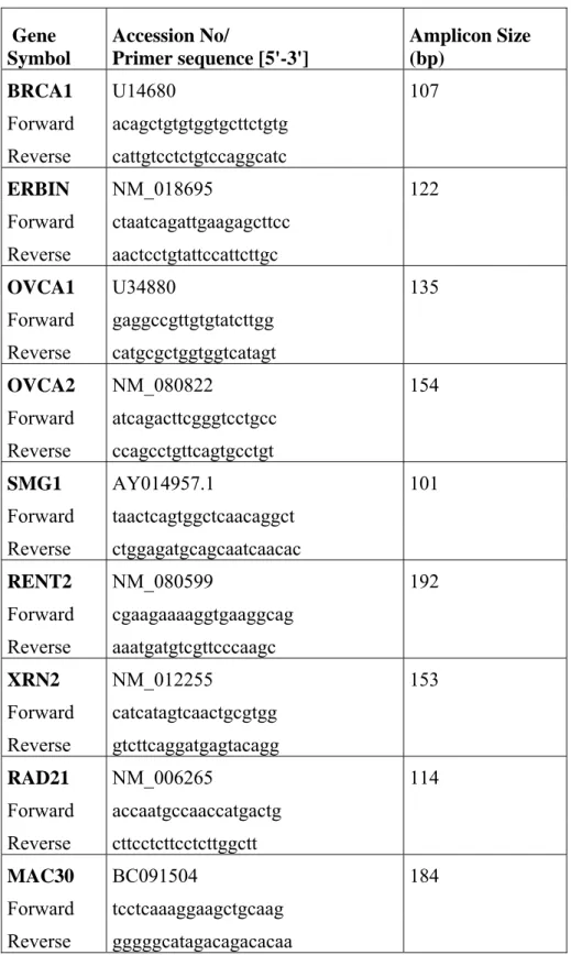
2.7.1 Primers 57
2.7.2 Reverse transcription polymerase chain reaction (RT-PCR) 62 2.7.3 Quantitative real time RT-PCR 63 2.7.3.1 Amplification efficiency calculations 64
2.8 Bisulfite sequencing 65
2.9 Selection of reference genes for normalization of real time RT-PCR data
65
2.9.1 Data retrieval and selection of candidate reference genes from microarray studies.
66
2.10 Protein extraction 67
2.10.1 Protein extraction from tissue culture cells 67 2.10.2 Protein extraction from tissue samples 67 2.10.3 Quantification of proteins 67
2.11 Western blotting 68
2.11.1 SDS polyacrylamide gel electrophoresis 68 2.11.2 Protein transfer to solid support 69 2.11.3 Immunological detection of immobilized proteins 70
2.12 Bioinformatic analysis 70
2.12.1 Data retrieval for meta-anlaysis 70 2.12.1.1 Data retrieval from Stanford Microarray Database
(SMD)
71
2.12.2 Data sets used in resampling-based meta-analysis study 72 2.12.2.1 Sorlie et al. dataset 72 2.12.2.2 Zhao et al. dataset 72
2.12.3 Data filtering 73
2.12.4 Resampling technique 74
2.12.4.1 Resampling and statistical analysis 74 2.12.4.2 Application of resampling to the breast cancer
datasets
76
2.12.5 Data retrieval and prediction analyses via BRB-Arraytools 77 2.12.6 Promoter region analysis 78
2.13 Statistical analysis 79
2.13.1 Cluster analysis 79
2.13.2 GeNorm 80
2.13.3 Normfinder 80
2.13.5 Discriminant Function Analysis 81
2.13.6 Mann-Whitney test 81
2.13.7 Student’s t-test 81
2.13.8 Kolmogorov-Smirnov test 82 2.13.9 Wilcoxon Rank-Sum Test 82
CHAPTER 3. RESULTS 83
3.1 Ectopic expression of BRCA1 in tissue culture cells 83 3.1.1 Ectopic expression of BRCA1 in MCF7 cells 83 3.1.2 Induction of BRCA1 Expression in UBR60-bcl2 cells 84 3.1.3 Control for upregulated BRCA1 activity 85
3.2 Quantitative Real-Time RT-PCR 86
3.2.1 Melt curve analysis 87
3.2.2 Standard curves and amplification efficiencies 88 3.3 Expression profiles of target genes in BRCA1 over expressed cells 91 3.4 Expression profiles of target genes in breast cancer cell lines 93 3.5 Identification of endogenous reference genes for real time qRT-PCR
analysis in normal matched breast tumor tissues
96
3.5.1 Expression patterns of candidate RGs 97 3.5.2 Expression stability of candidate RGs 99 3.5.3 Assessment of suitable RGs for normalization 103 3.5.4 Evaluation of 18S rRNA to mRNA ratio 105 3.5.5 Bisulfite sequencing of DNA samples for methylation
analysis
3.6 Human breast and paired normal tissue samples
107
110 3.6.1 Expression profiles of target genes in tumor and
paired-normal breast tissues determined by qRT-PCR
111
3.6.1.1 Correlation of target gene expression profiles 113 3.6.1.2 Evaluation of target gene expressions with clinical
information
3.7 Expression profiles and pathological contributions of BRCA1 and candidate BRCA1-target genes in independent microarray datasets
116
3.7.1 Tumor ER Status prediction in independent microarray datasets
116
3.7.2 Tumor grade status prediction in independent microarray datasets
116
3.7.3 Basal and non-basal breast tumor subtype prediction in an independent microarray dataset.
117
3.7.4 Relapse-free survival analysis for BRCA1 and BRCA1-target genes
118
3.7.5 Expression profiles of BRCA1 and candidate BRCA1-target genes in tumor and normal samples in independent
microarray datasets
120
3.8 Depletion of BRCA1 gene expression with sh- and si-RNA knockdown
121
3.8.1 shRNA mediated knock down of BRCA1 gene expression 121 3.8.1.2 Analysis of the target gene expression in
shRNA-BRCA1 depleted cells
123
3.8.2 siRNA mediated knock down of BRCA1 gene expression 124 3.8.2.1 Analysis of the target gene expression in
siRNA-BRCA1 depleted cells
124
3.9 The effect of BRCA1 on ERBIN expression 127 3.10 Promoter region analysis of BRCA1-target genes 129 3.11 The data retrieved from Stanford Microarray Database (SMD) 132 3.12 Correlation of Sorlie and Zhao Datasets
3.13 The effect of different normalization strategies on the data distribution
132 133
3.14 Distribution statistics for generation of meta-lists 134 3.15 Effects of resampling on estimates of expression and differentially
expressed gene number
3.16 Characteristics of differentially expressed meta-gene lists 138
3.17 Validation of tumor vs normal meta-gene lists by independent microarray datasets
138
3.18 Prediction of tumor-subtypes 139
3.19 Validation of ductal vs lobular meta-gene list 140 3.20 Validation of meta-analysis by real time qRT-PCR 142
CHAPTER 4. DISCUSSION 144
PART I: BRCA1 INDUCED GENE EXPRESSION PROFILES 144 PART II: A RESAMPLING BASED META-ANALYSIS FOR
DETECTION OF DIFFERENTIAL GENE EXPRESSION IN BREAST CANCER
159
CHAPTER 5. FUTURE PERSPECTIVES 166
REFERENCES 171
APENDICES 199
Appendix A Main script for “Resampling” analysis by using Matlab program
200
Appendix B Gene lists obtained through meta-analysis of Sorlie and Zhao datasets
208
Appendix C Validation of meta-analysis gene lists by three independent microarray datasets.
222
Appendix D Comparison of DL list with other published data sets 224
LIST OF FIGURES
Figure 1.1 Anatomy of the breast. 1
Figure 1.2 Functions of BRCA1 in response to DNA damage. 13 Figure 1.3 BRCA1 interacting transcription factors and their roles in
the cell.
15
Figure 1.4 Amplification curve. 27
Figure 2.1 Puc Mix Marker 8 and 1kb DNA ladder. 49 Figure 2.2 Map of pSUPER.retro.puro vector (OligoEngine). 52 Figure 2.3 General meta-analysis scheme. 76 Figure 3.1 Western blot analysis of ectopic expression of BRCA1 in
MCF7 breast carcinoma cells.
84
Figure 3.2 Western blot analysis of BRCA1 expression in tetracycline inducible UBR60-bcl2 cells.
85
Figure 3.3 Expression profile of GADD45 in BRCA1 induced cells. 86
Figure 3.4 Melt curve graph. 88
Figure 3.5 Standard curves of ACTB, SDHA and TBP for efficiency calculations.
89
Figure 3.6 Expression profiles of target genes in BRCA1 induced cells.
92
Figure 3.7 Hierarchical clustering of breast cancer cells (BCC). 94 Figure 3.8 Expression range of differences between the Ct values of
breast tumor and normal samples for each candidate reference genes.
98
Figure 3.9 Selection of reference genes for normalization in breast tumor samples using geNorm analysis.
100
Figure 3.10 The normalization of GSN gene expression with
combinations of candidate reference genes in tumor and matched normal breast samples.
Figure 3.11 The expression levels of 18S rRNA, ACTB and SDHA genes in tumor samples compared to their normal pairs.
106
Figure 3.12 The promoter region and the bisulfite sequencing result of the 45S rDNA.
108
Figure 3.13 Hematoxylin staining of the breast tissue sections. 110 Figure 3.14 Hierarchical clustering of tumor samples. 112 Figure 3.15 Discriminant Function Analysis of primary breast tumor
tissues based on their ER status.
114
Figure 3.16 Discriminant Function Analysis of primary breast tumor tissues based on their grade status.
115
Figure 3.17 Discriminant Function Analysis of primary breast tumor tissues based on their grade 1 and 3 status.
115
Figure 3.18 Relapse free survival analysis for GSE2034 and GSE2990 datasets.
119
Figure 3.19 Expression level of BRCA1 in shRNA and scrambled control RNA transfected MCF7 cells.
122
Figure 3.20 Immunoblot analysis of shRNA-BRCA1 stable MCF7 clones.
123
Figure 3.21 Expression levels of BRCA1, ERBIN and SMG1 genes in shRNA-BRCA1stable MCF7 clones.
124
Figure 3.22 Expression profiles of target genes in siRNA-BRCA1 depleted MCF7 cells.
125
Figure 3.23 GADD45 gene expression was affected by BRCA1 depletion.
126
Figure 3.24 The expression patterns of ACTB, TBP, GAPDH, PGK1 and CST6 genes in BRCA1 downregulated MCF7 cells.
127
Figure 3.25 Expression pattern of BRCA1 and ERBIN in breast cancer cell line panel.
128
Figure 3.26 BRCA1 expression changes ERBIN expression. 129 Figure 3.27 Promoter region analysis of the genes. 131 Figure 3.28 Promoter region analysis of ERBIN. 131
Figure 3.29 Pearson correlation coefficients (r) between Sorlie and Zhao datasets.
133
Figure 3.30 Correlation of global median and quantile normalized data.
134
Figure 3.31 Examples for probability distributions of Wilcoxon rank sum tests.
135
Figure 3.32 Effect of change in sample size and number of iterations on mean expression values and number of significant IMAGE clones.
137
Figure 3.33 Immunohistochemistry results of CDH1, RBP4 and TFAP2A in breast cancer tissues.
142
Figure 3.34 Validation of meta-analysis results by real-time qRT-PCR.
LIST OF TABLES
Table 1.1 List of known high- and moderate to low-risk breast cancer susceptibility genes.
10
Table 2.1 Breast carcinoma cell line information. 50 Table 2.2 Pathological features of the primary breast tumor
samples.
55
Table 2.3 The information of the primary breast tumor samples used in meta-analysis study.
56
Table 2.4 List of gene-specific primers used for expression analysis, accession numbers and amplicon sizes.
58
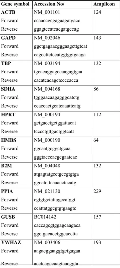
Table 2.5 List of gene-specific primers used for reference gene analysis, accession numbers and amplicon sizes.
59
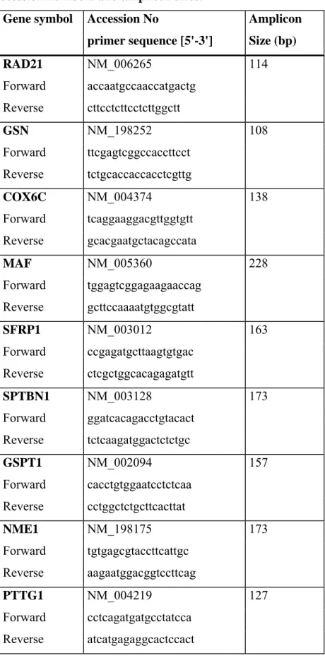
Table 2.6 List of gene-specific primers used for resampling based meta-analysis, accession numbers and amplicon sizes.
61
Table 2.7 Primers specific to 45SrRNA promoter (methylation specific primers).
65
Table 2.8 Protein sample preparation for Bradford assay. 68 Table 2.9 A standard curve preparation with BSA dilution. 68 Table 2.10 Pathology information of tumor samples used in Sorlie et
al., 2003 and Zhao et al., 2004 studies.
71
Table 3.1 Amplification efficiencies of the gene-specific primer pairs.
91
Table 3.2 Correlation coefficients and corresponding p values of eight target genes and BRCA1 expression values.
95
Table 3.3 Correlation coefficients and corresponding p values of eight target genes and BRCA1 expression values.
96
Table 3.4 Rank of candidate reference genes according to the expression stability calculated by NormFinder.
Table 3.5 Correlation coefficients and corresponding p values of GSN expression values normalized with one or
combinations of reference genes.
105
Table 3.6 Tumor cell percentages of the breast tumor samples used in this study.
111
Table 3.7 Correlation coefficients and corresponding p values of eight target genes and BRCA1 expression values.
113
Table 3.8 Validation of the prediction power of BRCA1 and BRCA1 target genes on ER status of the tumors by two independent microarray datasets.
116
Table 3.9 Validation of the prediction power of BRCA1 and BRCA1 target genes on grade status of the tumors by two independent microarray datasets.
117
Table 3.10 Prediction power of BRCA1 and BRCA1 target genes on basal and non-basal like breast cancer subtypes.
118
Table 3.11 The survival prediction analysis of BRCA1 and BRCA1-target genes.
119
Table 3.12 The results of the expression profiles of 8 genes in GEO breast cancer microarray datasets.
120
Table 3.13 Summary of GEO breast cancer microarray datasets and results of class prediction analysis for the meta-gene lists, DN (Ductal/Normal) and LN (Lobular/Normal).
ABBREVIATIONS
APS Ammonium persulphate
bp Base Pairs
BCC Basal Cell Carcinoma BCC Breast cancer cells
BRCT BRCA1 Carboxyl terminus
BSA Bovine serum albumin
cDNA Complementary DNA
CHEK CHK checkpoint homolog (S. pompe)
CP Crossing Point
Ct Cycle Threshold ddH2O Double distilled water
DCIS Ductal Carcinoma In Situ
DEPC Diethylpyrocarbonate
DFA Discriminant Function Analysis DMEM Dulbecco’s Modified Eagle’s Medium
DMSO Dimethyl Sulfoxide
DNA Deoxyribonucleic Acid
DSB Double strand break
dNTP Deoxyribonucleotide triphosphate
ds Double strand
dsDNA double-stranded DNA
EGF Epidermal Growth Factor
ERBB2 v-erb-b2 Erythroblastic Leukemia Viral Oncogene homolog 2, neuro/glioblastoma derived oncogene homolog (avian)
ER Estrogen Receptor
EtBr Ethidium Bromide
FBS Fetal Bovine Serum
HER2 ERBB2
IDC Infiltrating Ductal Carcinoma ILC Infiltrating Lobular Carcinoma
LCIS Lobular Carcinoma In Situ μg Microgram mg Miligram min Minute μl Microliter ml Mililiter μm Micrometer μM Micromolar mM Milimolar
mRNA Messenger RNA
NHEJ Nonhomologous end joining
NOS Not otherwise specified
Oligo(dT) Oligodeoxythymidylic acid
PBS Phosphate Buffered Saline
PCR Polymerase Chain Reaction
pmol Picomole
PR Progesterone Receptor
PTEN Phosphatase and Tensin homolog qRT-PCR Quantitative real time RT-PCR
Rpm Revolutions Per Minute
RT PCR Reverse Transcription PCR
Sec Second
TAE Tris-Acetate-EDTA buffer
TBP TATA Box Binding Protein
TDLU Terminal Duct Lobular Unit
Tm Melting Temperature
Tris Tris (Hydroxymethyl)- Methylamine
UV Ultraviolet
VEGF Vascular Endothelial Growth Factor
v/v volume/volume
CHAPTER 1. INTRODUCTION
1.1 Breast cancer
Breast tumors have been noted since antiquity and were probably first described in the Edwin Smith surgical papyrus originating from Egypt at around 2500 B.C. In this document tumors were described to be “cold and hard to the touch” whereas
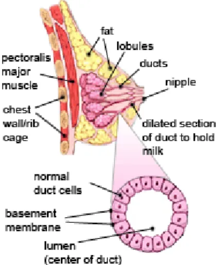
abscesses were “hot” (cross ref. from Oldenburg et al., 2007). Today breast cancer is the most common cancer among women accounting for 22% of all female cancers. A woman's breast is made up of glands that make breast milk (lobules) and ducts (small tubes that connect lobules to the nipple) and breast cancer begins in these breast tissues. The remainder of the breast is made up of fatty and connective tissue, blood vessels, and lymph vessels (Figure1.1).
Figure 1.1 Anatomy of the breast. The female breast is formed by ducts, lobules, fatty and connective tissues (http://www.cancer.org).
Breast cancer is one of the major cancer types women suffer from in the United States and Western Europe. After lung cancer, it is the second leading cause of
cancer death in women. Nearly 212,920 women in the United States were found to have invasive breast cancer in 2006 and in 2008, nearly 182,460 new cases of invasive breast cancer are expected to be diagnosed among women. The estimated number of death is about 40,460 in 2007 due to breast cancer. The chance of a woman having invasive breast cancer some time during her life is about 1 in 8. The chance of dying from breast cancer is about 1 in 33. Due to earlier diagnosis and improved treatment of breast cancer the death rates because of this disease are going down.
Many risk factors have been identified to date that contributes to the formation of breast cancer. Ethnicity, gender and the age are among the factors that were found to correlate with the incidence rates. Accordingly compared to female breast cancer incidence rate, that of male is much less and the rate of incidence increases up to 10-fold with increasing age (Medina, 2005; http://www.cancer.org). The extent and duration of exposure to sex hormones has also been consistently identified as a risk factor. Early age at menarche, delayed menopause, usage of exogenous hormones and late age of first pregnancy are expected to increase the risk of getting breast cancer (Medina, 2005, Oldenburg et al., 2007).
It is thought that breast cancer is a heterogeneous group of diseases with each
subtype having its own stable phenotype maintained during tumor progression rather than a single disease with a single tumorigenesis pathway (Mallon et al., 2000; Polyak, 2006). The most important determinants of these subtypes found are estrogen receptor (ER) and progesterone receptor (PR) status of tumor cells and the
amplification and overexpression of the HER2 oncogene. Considering these features, breast tumors are divided into pathological and molecular subtypes.
1.1.1 Pathological subtypes of breast cancer
Most breast lumps, areas of thickening, are benign; that is, they are not cancer. Benign breast tumors are abnormal growths, but they do not spread outside of the breast and they are not life-threatening. Some benign breast lumps can increase a woman's risk of getting breast cancer. Lumps are formed by fibrocystic changes in most cases. These changes include stromal fibrosis, cyst formation, and adenosis. Adenomas are also common benign lesions characterized by well-circumscribed benign epithelial elements with a variable amount of stroma.
Epithelial hyperplasia may be one of the initiating steps of breast carcinoma. Atypical hyperplasia is an epithelial proliferation in which some features of ductal carcinoma are seen in epithelial tissues (Beckmann et al., 1997; Mallon et al., 2000).
1.1.1.1 Ductal Carcinoma in Situ (DCIS)
It is the most common type of noninvasive breast cancer. Nearly all women with cancer at this stage can be cured. DCIS is a morphologically identifiable, preinvasive malignant proliferation of the breast epithelial cells (Mallon et al., 2000;
http://www.cancer.org). The abnormal cells are contained within the mammary epithelial structures. No invasion of the basement membrane and no infiltration of the breast stroma are apparent. With a true in situ carcinoma, malignant epithelial cells do not have access to the lymphatic or vascular channels present within the breast stroma. Classifications are performed according to the degree of nuclear pleomorphism (often graded on a scale of 1–3), the presence or absence of necrosis, and the mitotic activity. The most characteristic feature of DCIS is that the cells composing the intraluminal proliferation are morphologically similar to each other, but have nuclear abnormalities associated with malignancy.
1.1.1.2 Lobular Carcinoma in Situ (LCIS)
LCIS is a neoplastic proliferation of epithelial cells in the terminal duct lobular unit with specific morphological features and therapeutic implications (Beckmann et al.,
1997; Mallon et al., 2000). LCIS is a proliferation of neoplastic, epithelial cells which expand the individual acini of the lobular units involving more than 50% of the acini in a lobular unit. Both LCIS and DCIS are observed more in premenopausal women, suggesting that these lesions regress after menopause and that they are hormone dependent. This idea is supported by the ER positivity of these lesions.
1.1.1.3 Infiltrating Ductal Carcinoma (IDC)
IDC is the most common type of breast cancer. It accounts for about 80% of invasive breast cancers (http://www.cancer.org). If a tumor does not show the morphological features of a special type of invasive carcinoma or the characteristics of invasive lobular carcinoma it is called IDC if not otherwise specified (NOS) (Mallon et al., 2000, Weigelt et al., 2008). This group of tumors is morphologically heterogeneous. IDC tumors have very variable growth patterns and stromal responses. They are often hard and fibrous.
The stage is determined by spread of the tumor to the body. However, grade is determined by how the tumor cells appear under the microscope, growth rate of the tumor cells, and the tendency of tumor to spread other parts of the body. There are four stages of breast cancer (http://www.cancer.gov). If the tumor size is less than 2 centimeters and there is no metastasis, it is a stage I tumor. As it progresses to stage IV, tumor size and metastasis levels increase. In stage IIIB and IV, the metastasis spreads to other parts of the body rather than lymph nodes. As the stage increases, the severity of the disease increases, as well. It is possible to separate IDC into three grades based on the degree of tubule formation, nuclear pleomorphism, and mitotic activity. Each of the three parameters is given a score of 1–3 and the individual scores are then added together. A score of 3–5 indicates Grade 1, 6–7 indicates Grade 2, and 8–9 indicates Grade 3. The first parameter, tubule formation, is
assessed on the basis of percentage of the tumor showing distinct tubules: a score of 1 is assigned if 75% or more, a score of 2 if 10–75%, and a score of 3 if less than 10%. Nuclear pleomorphism is the second component. If the nuclei are small, with regular outlines, uniform chromatin, and little variation in size, they are assigned a score of 1. The cytoplasm of the tumor cells may also show considerable variation,
with some cells having little cytoplasm and others having abundant cytoplasm that can be eosinophilic and granular, or foamy and basophilic, or midway between the two. The third parameter is an assessment of the proliferation rate determined by counting the number of mitoses in 10 high-power fields at the periphery of the tumor. The method is standardized for each microscope objective and the tumor scored on a scale of 1–3.
1.1.1.4 Infiltrating Lobular Carcinoma (ILC)
ILC is the second most common type of invasive breast carcinoma and makes about 10-15% of all breast tumors and it is histologically characterized by uniform tumor cells arranged in single-files or concentrically localized around ducts (Yolder et al., 2007; Mallon et al., 2000; http://www.cancer.org). The cellular morphology of the tumor and the pattern of infiltration are very important in diagnosis. The tumor cells of lobular carcinoma are found in association with foci of typical LCIS and infiltrate in a very characteristic way with one cell behind the other in a defined pattern called the Indian filing pattern. They often form concentric rings around blood vessels and lobules producing a targetoid pattern. In classical lobular carcinoma, the tumor cells are relatively small. They have regular rounded nuclei with dense, evenly staining chromatin. Nucleoli are not prominent. A high grade aggressive form of ILC is known as pleomorphic lobular carcinoma (PLC) (Simpson et al., 2008). Bertucci et
al. (Bertucci et al., 2008) described that IDC and ILC were histologically and
genomically distinguishable from each other among the ER(+) grade II invasive breast tumors. Furthermore, ILC molecular subtypes were reported to include the typical and IDC-like ILCs, yet the CDH1 mutation and/or underexpression was common but not universal to ILCs in general (Yolder et al., 2007). ILC tumors mostly metastasize to gastrointestinal, gynecologic and peritoneal tissues and particularly to endocrine related sites (Zhao et al, 2004) ILC has a higher incidence of multicentricity and bilaterality than IDC and a slightly better overall survival rate than tumors in the NOS category.
There are also several other less common types of breast cancer including tubular, mucinous, medullary, papillary, invasive cribriform, and secretory carcinoma.
1.1.2 Molecular classification of breast cancer
The classifications of the breast cancer are mostly based on clinical and pathological factors, which unfortunately fail to reflect the heterogeneity of the tumors. There are some histological markers available to decide on the prognosis and treatment of breast cancer. Estrogen receptor (ER) status, as ER-positive or ER-negative, helps to categorize breast cancers into two major classes. ERBB2 (Her-2/Neu) is also
routinely used to classify breast cancer into HER-2 amplified or nonamplified categories. There are other single gene markers such as TP53, and cell proliferation markers such as Ki-67, and cyclin D1 that have emerged from detailed molecular analysis (Nielsen et al, 2004). While conventional methods were restricted to studying a single locus, current highthroughput techniques have allowed monitoring gene expression or copy number levels of almost all known genes in a single
experiment. Molecular profiling has been shown to be well-suited to phenotypic characterization of breast cancer and potentially to discover new molecular classes among cancers with similar histopathological appearance (Sorlie et al, 2001; van’t Veer et al, 2002; van de Vijver et al, 2002; Ahr et al, 2002; Sotiriou et al, 2003; Huang et al, 2003)
Several landmark microarray studies have demonstrated that one can build a
molecular taxonomy of breast tumors using this technology and can provide a more sophisticated molecular picture together with individualized recurrence risks.
1.1.2.1 Distinguishing tumors on the basis of their gene expression profiles
Gene expression profiling using DNA microarrays has provided an opportunity to perform more detailed and individualized breast tumor characterization leading to classification of breast cancer into distinct new molecular subgroups (Cleator and Ashworth, 2004). The potential advantages of improving tumor classification by expression profiling has been central to several large-scale breast cancer studies over
the past few years that have reported identification of signature gene lists with potential for prediction of clinical outcome (Sorlie et al, 2001; van’t Veer et al, 2002; Huang et al, 2003; Gruvberger et al, 2001; West et al, 2001). One of the first comprehensive studies classifying sporadic breast tumors into subtypes distinguished by differences in their expression profiles was performed by Perou et al. (Perou et al, 2000). Using 40 tumors and 20 matched pairs of samples they identified an “intrinsic geneset’ of 476 cDNAs and then used this to cluster and segregate the tumors into four major subgroups: a “luminal-like cells” group expressing estrogen receptor (ER); a “basal-like cells” group; an “ERBB2-positive” group, and a “normal like” epithelial group (Perou et al., 2000). As new samples became available they were re-evaluated the data on 85 new tumors (Sorlie et al., 2001). Not only were the breast cancer subtype definitions modified, clinical outcomes (metastasis, death and survival) were found to be significantly different between the subtypes. Gene sets corresponding to each of these groups were made on one dataset and used to classify the samples of another dataset. All groups were repeatedly found in the second dataset and differences in overall survival between the five groups were significant. Sorlie et al. (Sorlie et al., 2003) rexamined 84 of the 85 arrays used in 2001 study and added 38 new breast cancer tumor tissue arrays to this study. Once again they found the same subtypes with significant differences in overall survival between the groups and confirmed their data in independent datasets. Consequently it is now recognized that all breast cancers are not the same in molecular point of view. Subsequent studies confirmed that there are large-scale gene expression differences between ER-positive (mostly luminal-like) and ER-negative (mostly basal-like) cancers and suggested that further molecular subsets also exist (Sorlie et al, 2003; Pusztai et al, 2003; Sotiriou et al, 2003).
The prognosis and chemotherapy sensitivity of the different subgroups are different. The luminal type cancers tend to have the most favorable long-term survival,
whereas basal-like and ERBB2-positive tumors are more sensitive to chemotherapy (Sorlie et al, 2001; Rouzier et al, 2005).
In another molecular classification of breast tumor study, Van’t Veer et al. have used DNA microarray analysis on the primary breast tumors of 78 lymph node-negative young patients and compared the expression profiles of 34 patients who developed
distant metastasis within 5 years and 44 patients who remained diseasefree for at least 5 years (van’t Veer et al, 2002). Their analysis led to the identification of a 70-gene expression signature that was developed to classify tumors into the good and poor prognosis groups. The results were later confirmed in a larger set of tumors (van de Vijver et al, 2002; Buyse et al, 2006)
1.1.2.2 Basal-like tumors
Basal-like breast carcinomas are so named because in terms of gene expression, these tumors are generally characterized by high expression of some basal epithelial
markers such as KRT5, KRT6, KRT17, KRT23, c-KIT, FOXC1, P-Cadherin and LAMC2 (laminin). In particular, overexpression of LBR, DSC2, MRAS, CDCA7, FABP7, CXCL1, TRIM29, MSN, CCNE1, CCNA2, CCNB1, MYBL2, CDH3, CRYAB, MKI67, MET, AURKB, LYN, FOXM1 have been often observed (Sorlie
et al., 2001; Nielsen et al., 2004).
Basal-like breast carcinomas, as defined by gene expression microarray analysis, are the most undifferentiated breast cancers, frequently lack the expression of hormone receptors (ER) and HER2 (Perou et al., 2000; Sorlie et al., 2001; Nielsen et al., 2004), show p53 immunohistochemical expression and TP53 gene mutations (Sorlie
et al., 2001). Morphologically, basal-like breast carcinomas are characterized by high
histological grade, high mitotic indices and the presence of central necrotic zones (Turner and Reis-Filho, 2006). Finally patients with basal-like tumors experience a much shorter overall-and disease-free survival period. It is the most severe case among the other subtypes of breast cancer.
1.1.2.3 Luminal tumors: Luminal A and Luminal B
Luminal breast tumors were firstly classified according to their hormone receptor status and fell into the group of ER positive tumors. On the other hand none of the tumors in this group found to be expressed Erb-B2 at high levels (Perou et al., 2000). They are given the name “luminal tumors” since both luminal A and luminal B subtypes are positive tumors for the expression of luminal cell markers. They show a
discriminative expression of certain proteins such as TOPO II, proliferating cell nuclear antigen (PCNA) and cell cycle proteins and they have differential clinical outcomes (Melchor and Benitez, 2008).
1.1.2.4 ErbB2 expressing tumors
ErbB2 tumors show an overexpression of ErbB2 and multiple genes from the 17q11 amplicon. Overexpression of the ErbB2 oncogene was associated with the high expression of a specific subset of genes and these tumors were partially characterized by the high level of expression of this subset of genes. ErbB2 tumors also showed low levels of expression of ER and of almost all of the other genes associated with ER expression, which is a trait they share with the basal-like tumors. (Perou et al., 2000)
1.1.2.5 Normal-like tumors
Normal like tumors were the ones clustered with a group of samples that also
contained the normal breast specimens (Perou et al., 2000). The “normal-like tumor” gene expression pattern was classified by the high expression of genes characteristic of basal epithelial cells and adipose cells, and the low expression of genes
characteristic of luminal epithelial cells.
1.1.3 Genes implicated in breast cancer
The existence of a strong predisposition to breast cancer is a well known
phenomenon. To date, up to 5-10% of all breast cancers are caused by germline mutations in well-identified breast cancer susceptibility genes. These genes have been divided into high-risk and low-to moderate risk susceptibility genes. The high risk breast cancer susceptibility genes include BRCA1, BRCA2, PTEN, TP53, LKB1/STK11 and CDH1 while CHEK2, TGFβ1, CASP8 and ATM genes belong to the low-to moderate risk susceptibility genes. In one third of the hereditary breast cancers, the germline mutations of BRCA1 and BRCA2 are found to be responsible.
The other genes like p53, PTEN, CHEK2 and ATM account for a small proportion of hereditary breast cancers (Table 1.1) (Palacios et al., 2008).
Table 1.1: List of known high- and moderate to low-risk breast cancer susceptibility genes.
Gene Location Frequency Breast cancer risk
BRCA1 17q21 Rarea 46-85% lifetime risk
BRCA2 13q12 Rarea 43-84% lifetime risk
TP53 17p13.1 Rare 28-56% by age 45
PTEN 10q23.3 Rare 25-50% lifetime risk
LKB1/STK11 19p13.3 Rare 29-54% lifetime risk
CDH1 16q22.1 Rare 20-40% lifetime risk
ATM 11q22-23 Moderate RR: 2.2
TGFβ1 19q13.1 Frequent OR: 1.25 (p=0.009)
CASP8 2q33-34 Frequent OR: 0.83
CASP10 2q33-34 Frequent OR: 0.62 (p=0.0076)
CASP8/CASP10 Moderate OR: 0.37 (p=0.013)
CHEK2 22q12.1 Moderate RR: 2
Rare: <1% population frequency, moderate 1-5%, frequent >5%, OR: odds ratio and RR: relative risk a In, for example the Ashkenazi Jewish population some mutations have a moderate population frequency.(Palacios et al., 2008)
The TP53 gene encodes a protein involved in many overlapping cellular pathways that control cell proliferation and homeostasis, like cell cycle, apoptosis and DNA repair. The expression of TP53 is activated in response to stress signals including DNA damage (Oldenburg et al., 2007). While the mutations in the p53 gene were found to be responsible for Li-Fraumeni syndrome in nearly 70% of families
fulfilling the classical criteria for the disease, they were found to be less common in breast cancer (Frebourg et al., 1995). Somatic mutations are reported in 20-60% of human breast cancers (de Jong et al., 2002) and hypermethylation of the p53 gene
seems not to play a major role in breast cancer.
The tumor suppressor gene PTEN is found to be responsible for Cowden syndrome (CS) and found to be mutated in sporadic brain, breast, and prostate cancers (Liaw et
al., 1997). Women carrying a PTEN mutation have a 25-50% lifetime risk of
developing breast cancer. To date no mutations in the PTEN gene have been detected in breast cancer families without features of CS. Also in sporadic breast cancers germline and somatic mutations are rare. In addition, although LOH at the PTEN locus is found in 11-41% of sporadic breast cancers, no somatic mutations have been observed in the remaining allele (Freihoff et al., 1999; Feilotter et al., 1999).
One of the known low to moderate-risk breast cancer susceptibility gene ATM plays a central role in sensing and signaling the presence of DNA double-strand breaks. The irradiation initiates kinase activity of ATM and phosphorylates the protein products of TP53, CHEK2 and BRCA1 (Bakkenist et al., 2003). Carriers of the ATM gene mutations suffer from a recessive disorder ataxia-telangiectasia (AT). The role for the ATM gene in breast cancer is plausible but the exact association remains unclear, and most probably comprises only a modest role in familial breast cancer susceptibility (Hall, 2005).
Among the well identified breast cancer susceptibility genes BRCA1 is the one to be responsible for 45% of the all hereditary breast cancers.
1.2 BRCA1
Tumor suppressor gene BRCA1 (Breast cancer susceptibility gene 1) plays a central role in the development of breast and ovarian cancers. While inherited mutations of BRCA1 are responsible for 40-45% of the hereditary breast cancers, 71% of the BRCA1 mutation carriers have the risk of developing breast cancer (Rosen et al, 2003). Somatic BRCA1 mutations are rare in sporadic breast cancers; however both mRNA and protein expression are downregulated in 30% of sporadic cases (James et
al, 2007). It is reported that reduced expression or absence of BRCA1 protein in
sporadic cases are due to non-mutational mechanisms such as hypermethylation of BRCA1 promoter or incorrect subcellular localization of the BRCA1 protein (Birgisdottir et al, 2006, Esteller et al, 2000, Rakha et al, 2008).
1.2.1 Functions of BRCA1
BRCA1 regulates multiple cellular events including cell cycle regulation and growth control, DNA damage response and repair processes and regulation of transcription (reviewed in Rosen et al, 2003, Venkitaraman, 2002). BRCA1 is a component of BASC, a BRCA1-associated genome surveillance complex that includes proteins involved in the recognition and repair of DNA-damage (Wang et al, 2000). In addition, the carboxyl terminal of BRCA1 acts as a strong transcriptional activator when fused to a heterologous DNA binding domain (Monterio et al., 1996). BRCA1 co-purifies with RNA polymerase II holoenzyme complex, suggesting that it is a component of core transcription machinery (Scully et al., 1997a). BRCA1 also interacts with several transcription factors such as p53, CtIP, c-myc, ZBRK1, ATF, E2F, and signal transducer STAT1 (Zheng et al., 2000) and modulates their activity. These findings, together with the interaction of BRCA1 with histone deacetylases (HDACs) and the SWI/SNF-related chromatin remodeling complex, imply that transcriptional regulation is one of the main functions of BRCA1 (Bochar et al, 2000). In addition, nearly all germ-line BRCA1 mutations involve truncation or loss of the C-terminal BRCT transcriptional activation domain, supporting the
transcriptional regulation function of the BRCA1 gene.
1.2.1.1 BRCA1 in damage signaling and DNA repair
Initial evidence suggesting a role of BRCA1 in the repair of damaged DNA was derived from the observation that BRCA1 is hyperphosphorylated in response to DNA damage and relocated to sites of replication forks (Scully et al., 1997a; Thomas
et al., 1997). Additionally BRCA1 has been identified as a target for several
upstream nuclear phosphoinositide (PI) like kinases, which are implicated in DNA damage signaling through their protein kinase activities, ataxia-telangiectasia
mutated (ATM) and ATM-related (ATR) kinase (Yan et al., 2008). The studies show that ATM mediates its phosphorylation role in response to ionizing radiation, while ATR mediates the phosphorylation in response to UV irradiation (Gatei et al., 2001). The major target for ATM phosphorylation after ionizing radiation is Ser1387 of
BRCA1. In response to ultraviolet irradiation, Ser1457 residue of BRCA1 is primarily phosphorylated, mainly by ATR (Gatei et al., 2001). The G2/M control kinase, CHK2, has also been shown to phosphorylate BRCA1 at Ser988 on exposure to ionizing radiation (Chaturvedi et al., 1999) (Figure 1.2).
Figure 1.2 Functions of BRCA1 in response to DNA damage. Arrows show the phosphorylations while the dash lines show the interactions through proteins (Chaturvedi et al., 1999).
There are two mechanisms for double strand break (DSB) repair; a process known as homologous recombination (HR), which uses homologous sequences on the sister chromatid for repair and non-homologous end joining (NHEJ), which ligates either contiguous or non-contiguous sequences in the genome (Greenberg, 2008). BRCA1 was shown to be involved in the complexes that activate the repair of DSBs and initiate HR. BRCA1 and BRCA2 were found to be localized with Rad51, which is a protein required for recombination during mitosis and meiosis as well as HR repair of DSBs (Scully et al., 1997b; Shinohara et al., 1992). Co-localization of BRCA proteins with Rad51 shows that BRCA1 and BRCA2 takes role both in the detection and repair of DSBs. However the studies show that BRCA1 may not directly regulate Rad51 since they do not interact with each other.
In mammalian cells NHEJ proceeds in stepwise manner beginning with the end processing by the MRE11/RAD50/NBS1 (MRN) complex and then end joining by Ku70 and Ku80 proteins (Wu et al., 2000; Chan et al., 1999). It is not clear how BRCA1 promotes NHEJ but studies have shown that BRCA1 co-localizes with MRN complex and negatively regulates end-processing by MRE11 endo- and exonucleases (Zhong et al., 1999a). Suppression of MRN-mediated end-processing by BRCA1may enhances NHEJ accuracy (Shrivastav et al., 2008).
1.2.1.2 BRCA1 in regulation of transcription
A role for BRCA1 in transcriptional regulation was first suggested by the finding that BRCA1 has a conserved acidic COOH-terminal transcriptional activation domain, which is a globular domain, found in proteins involved in repair and cell-cycle control (Monteiro et al., 1996). Results showing that the BRCA1 C terminus (aa 1560-1863) has the ability to activate transcription when fused to GAL4 DNA-binding domain (DBD) provided the initial experimental evidence of the
involvement of BRCT domain of BRCA1 in transcription (Monteiro et al., 1996). Series of experiments have demonstrated that the C-terminus of BRCA1 can be used to recruit RNA polymerase II (RNAPII) to synthetic reporters showing that BRCA1 plays some role in transcriptional activation (Monteiro et al., 2000). However, direct evidence that BRCA1 binds to promoter regions of genes is lacking. Although BRCA1 is not known to bind to specific DNA sequences, it may regulate
transcription through protein:protein interactions. BRCA1 physically associates with many proteins (transcription factors) involved in transcription and is paradoxically involved in both transcriptional activation and repression (Mullan et al., 2006). ER-alpha, p53, STAT1, CtIP, c-Myc and ZBRK1 are the known transcription factors that have interaction with BRCA1 (Mullan et al., 2006) (Figure 1.3).
Figure 1.3 BRCA1 interacting transcription factors and their roles in the cell. (Mullan et al., 2006)
1.2.1.3 BRCA1 modulation of sequence-specific DNA binding transcription factors
Still no specific DNA binding sequence of BRCA1 was stated but it has been established that it can bind to various sequence specific DNA binding transcription factors to stimulate or inhibit transcription. BRCA1 interacts with tumor suppressor protein p53 and act by both stabilizing and stimulating its transcriptional activity (Chai et al., 1999; Zhang et al., 1998). This stabilization appears to induce a subset of p53-regulated genes involved in DNA repair and cell cycle arrest other than apoptosis which may show that BRCA1:p53 interaction may influence the cell fate decision during the DNA damage (Ongusaha et al., 2003; MacLachlan et al., 2002).
It was found that a physical interaction between BRCA1 and STAT1, which is a transcription factor that transduces the cellular response to interferon-γ (INF- γ), induced a subset of INF- γ responsive genes (Ouchi et al., 2000). In addition to its role in inducing gene expression with INF- γ, BRCA1 also potentiated INF- γ mediated apoptosis (Andrews et al., 2002).
BRCA1 can also regulate the promoter activity and expression of growth inhibitory genes like p21, Gadd45α and p27. BRCA1 was found to physically interact with sequence specific transcription factors Oct-1 and NF-YA, which directly bind to the OCT-1 and CAAT motifs on GADD45 promoter thus induce the expression of Gadd45α (Fan et al., 2002). On the other hand Zheng et al. showed that BRCA1 interacts with a zing finger and KRAB domain protein ZBRK1 to bind a specific DNA sequence on the 3rd intron of GADD45 and in this context ZBRK1 appeared to repress GADD45 transcription in a BRCA1 dependent manner (Zheng et al, 2000). Addition to this direct binding of BRCA1 to some transcription factors Cable et al. published a specific DNA sequence that can be bound by BRCA1 protein complexes (BRCA1:USF2) to control gene expression (Cable et al, 2003). These known
sequences have paramount importance to find out the new downstream targets of BRCA1 to explain or to clarify the exact role of BRCA1 in transcriptional regulation.
1.2.2 BRCA1 regulated targets
BRCA1 is known to be a multifunctional protein and it locates at different sites and takes role in many functional events in the cell. Although many of these functions are clear and the mechanism of its action is known some of the functions of BRCA1 and how it controls these functions is still a gap. Hence it is important to find out its exact role in these cellular events to clarify its role in breast cancer development, transcriptional regulation and in other cellular events. Finding out transcriptional targets of BRCA1 is one of the strategies at least to identify the subgroups of genes from different molecular and cellular functions.
In order to identify a series of downstream targets of BRCA1, Harkin et al.
established an osteosarcoma cell line with tightly regulated BRCA1 expression with the tetracycline inducible system (Harkin et al., 1999). High density oligonucleotide
arrays were used to analyze the gene expression profiles at various times following BRCA1 induction. They found 20 BRCA1 target genes. Among those genes, they identified GADD45 as one of the major targets of BRCA1 and showed that this activation was p53 independent. Concordant with the previous study Mullan et al. used the same tetracycline inducible system with a breast cancer cell line and found GADD45 as the main target of BRCA1 (Mullan et al., 2001). Later on MacLachlan
et al. have overexpressed BRCA1 by using an adenovirus vector and have identified
45 major targets in the SW480 colorectal cancer cell line. Most of the genes they found were DNA damage response genes and the ones involved in cell cycle control (MacLachlan et al., 2000). Furthermore Welch et al. identified 62 genes that are targets of BRCA1 by using ecdysone inducible expression of BRCA1 in human embryonal kidney epithelial cells (Welch et al., 2002). Finally Atalay et al. used suppression subtractive hybridization (SSH) technology to generate a library of partial-length cDNAs representing mRNAs in BRCA1-overexpressing MCF7 cells. By this approach 60 genes have been identified that are upregulated as a result of BRCA1 overexpression in breast cancer cells (Atalay et al., 2002).
1.3 Genes affected by BRCA1 expression
ERBB2 interacting protein, ERBIN
ERBIN was initially found to interact specifically with ErbB2 by its PDZ domain and acts in the localization of ErbB2 to the basolateral domain in epithelia which is important for its activation and signaling of ERBB2/HER2 in epithelia (Borg et al., 2000). It has 16 LLRs and a single PDZ domain in its C-terminus which interacts directly with the C-terminal aminoacids of unphosphorylated ERBB2. It is
constitutively associated with ErbB2 in living cells (Borg et al., 2000, Huang et al., 2001;). They reported that the Erbin PDZ domain binds preferentially to the C terminus of ErbB2, which is non-Tyr1248-phosphorylated (Borg et al., 2000). Importantly phosphorylation of this residue following ErbB2 activation is a critical event for the mitogenic signaling and oncogeneity of this receptor (Dittmar et al., 2002). Overexpression of ErbB2 correlates with poor prognosis and resistant
chemotherapy in breast and ovarian cancers (Klapper et al., 2000). Despite the close relation of ERBIN and ErbB2 the functional role of ERBIN has not been studied extensively in breast cancer yet. Recently Liu et al. studied the expression and the regulation of ERBIN and its binding partner ErbB2 in the MCF7 breast cancer cell line. One of their finding was that the affinity of Erbin-ErbB2 interaction was reduced by ErbB2 posphorylation (Liu et al., 2008).
Furthermore Erbin was found to be a novel suppressor of the Ras signaling (Huang et
al., 2003). It can inhibit the activation of Ras pathway by disrupting the interaction of
Sur-8, which is a positive regulator of the Ras pathway, with Ras and Raf (Dai et al., 2006). The requirement of LRRs for this process rather than PDZ domain shows that ERBIN has dual function (functions as a signaling molecule in addition to being a scaffold protein) in cells with its LRR and PDZ domains (Huang et. al., 2003, Dai et
al., 2006). On the other hand a new role of ERBIN was described in inflammatory
responses by McDonald et al. They found the inhibitory effect of ERBIN by its carboxyl terminus on Nod2-dependent activation of NF-κB and cytokine secretion (McDonald et al., 2005). In addition a very recent novel negative regulatory role was added to the functions of ERBIN and expanded the physiological role of it to the regulation of TGFβ signaling through its direct interaction with Smad2/Smad3 (Dai
et al., 2007).
Two tumor suppressor genes, OVCA1 and OVCA2
OVCA1 and OVCA2 are the two tumor suppressor genes mapped to chromosome 17p13.3, that is most commonly lost in ovarian and breast tumors. The two OVCA genes are expressed from the same genetic locus using two different promoters; however since OVCA2 transcript contains a unique exon and only the 3’ UTR of the OVCA1 transcript, they share no coding sequence (Chen and Behringer, 2004). Northern blot analysis reveled that they are both expressed in normal surface
epithelial cells of the ovary but reduced or undetectable in ovarian tumors and tumor cell lines (Schultz et al., 1996). In an independent study OVCA1 was shown to be reduced in protein level in breast and ovarian tumors and shown to inhibit growth of ovarian cancer cells (Bruening et al., 1999). To address the role of OVCA1 and
OVCA2 in development and proliferation Chen and Behringer (2004) generated OVCA1 disrupted and OVCA1 and OVCA2 disrupted mouse models. The identical results obtained from the two strains showed that OVCA1 was more important than OVCA2 with respect to development and proliferation. They finally concluded that OVCA1 acted as a positive regulator for cell cycle progression and it was a tumor suppressor (Jensen and Helin, 2004; Chen and Behringer, 2004).
Diphthamide is a unique post-translationally modified histidine residue found only on translational elongation factor 2 (EF-2). The biosynthesis of the diphthamide is one of the most complex post-translational modifications. Addition to its tumor suppressor activity Nobokini et al. (2005) and Chen et al. (2005) showed that OVCA1 was a component of the biosynthetic pathway of diphthamide on EF-2.
Nonsense mediated decay genes, RENT2 and SMG1
Nonsense-mediated mRNA decay (NMD) is a quality control mechanism that selectively degrades mRNAs harboring premature termination (nonsense) codons. The core NMD machinery comprises three trans-acting factors, called up-frameshift (UPF) proteins, which were initially discovered in S. cerevisiae and later identified in higher eukaryotes. UPF1, UPF2 also known as RENT2 and UPF3 proteins comprise the core NMD machinery. The SMG-1, SMG-5, SMG-6 and SMG-7 proteins mediate the phosphorylation and dephosphorylation cycle of UPF1 (Chang et al., 2007).
RENT2 (UPF2) is an adapter molecule that brings together UPF1 and UPF3 to elicit NMD. It is a part of large complex of proteins that is deposited on mRNAs at exon-exon junctions during RNA siplicing in the nucleus. With UPF3, RENT2 is a part of the exon-junction complex (EJC), a large dynamic protein complex deposited just upstream of exon-exon junctions during RNA splicing. Many EJC components, including RENT2 and UPF3, remain bound to the mRNA after its export to the cytoplasm, where they function as a second signal to elicit NMD when the mRNA is proofread during translation (Chang et al., 2007).
SMG-1 is a kinase that phosphorylates serine residues. It is a member of the phosphoinositide 3-kinase related kinase (PIKK) family, whose other members function in DNA damage and growth responses. SMG-1 mediated phosphorylation of UPF-1 is likely to be crucial for NMD. SMG-1 forms the SMG-1-Upf1-eRF1-eRF3 complex (SURF) prior to phosphorylation of Upf1. The SURF recognizes downstream Upf2-EJC and they associate to induce Upf-1 phosphorylation. SMG-1 also phosphorylates the p53 protein upon genotoxic stress (Yamashita et al., 2005, Chang et al., 2007). Furthermore, siRNA-mediated depletion of SMG-1 results in accumulation of spontaneous DNA damage and increases cellular sensitivity to ionizing radiation (Brumbaugh et al., 2004). Recently it was found that loss of SMG-1 function dramatically increased the rate and extent of apoptotic cell death induced by tumor necrosis factor-alpha (TNFα). Thus it protects human cells from TNFα induced apotosis through a mechanism unrelated to its role in NMD (Oliveira et al., 2008).
5’ to 3’ exonuclease, XRN2
The XRN2 gene is the human homologue of the Saccharomyces cerevisiae RAT1 gene, which encodes a nuclear 5' to 3' exoribonuclease, and is essential for RNA metabolism and cell viability. Xrn2 /Rat1, product of XRN2/RAT1 gene, functions in the mRNA degradation and processing of rRNAs and small nucleolar RNAs (snoRNAs) in the nucleus (Li et al., 2005).
Polymerase II (Pol II) transcriptional termination depends on two independent genetic elements: poly (A) signals and downstream terminator sequences. The latter may either promote cotranscriptional RNA cleavage or pause the elongating of Pol II. It was found that the previously characterized MAZ(4) pause element promotes Pol II termination downstream of a poly(A) signal, dependent on both the proximity of the pause site and poly(A) signal and the strength of the poly(A) signal (Gromak
et al., 2006). The 5' to 3' exonuclease Xrn2 facilitates this pause-dependent
termination by degrading the 3' product of poly (A) site cleavage. The human beta-actin gene also possesses poly(A) site proximal pause sequences. Xrn2 depletion causes an increase in both steady-state RNA and Pol II levels downstream of the
beta-actin poly (A) site. All these data provided new insights into the mechanism of pause site-mediated termination and establish a general role for the 5' to 3'
exonuclease Xrn2 in Pol II termination (West et al., 2004; Gromak et al., 2006).
The meningioma associated protein, MAC30
The meningioma associated protein, MAC30 gene is located on 17q11.2, and the protein has a small segment which is similar to an apical gut membrane polyprotein of Haemonchus contortus, to olfactory receptor 30 of Mus musculus, and to
cytochrome b in several organisms. MAC30 mRNA has been found to express as a non-erythropoietic gene in the fatal liver during the early stages of the development but not in the adult liver. In the light of this evidence it was suggested that MAC30 may play a role in growth and differentiation of the liver. MAC30 expression is seen in many types of normal organs including brain, lung, heart, skeletal muscles, testis and ovary but it is changed during tumor development. The decrease in its expression was shown in pancreatic and renal cancers while in meningiomas, ovarian, gastric and colarectal cancers its expression was shown to be increased (Murphy et al., 1993; Moparthi et al., 2007).
In a recent study performed with rectal cancers, MAC30 expression in radiated-primary tumors was related to more aggressive morphological and biological factors that were involved in cell proliferation, invasion and metastasis of the tumors. Stronger MAC30 expression was shown to be an indicator of poor prognosis (Zhang
et al., 2007). In another study dealing with the colorectal cancers, MAC30
expression was found to be much stronger in cytoplasm in lymph node metastasis compared to primary tumor and normal mucosa. Addition to overexpression of MAC30 at the invasion margins they suggested that the protein may play an
important role in the development and aggressiveness of colorectal cancer (Moparthi