Case Report
Cholangiocarcinoma Presenting with
Hypercalcemia and Thrombocytopenia
Muharrem Battal,
1Bünyamin Gürbulak,
2Ozgür Bostanci,
1Müveddet Banu Y
Jlmaz,
3Yasar Ozdenkaya,
4and Oguzhan Karatepe
51General Surgery Department, Sisli Etfal Teaching and Research Hospital, Sisli, 34173 Istanbul, Turkey 2Arnavutkoy Government Hospital General Surgery Department, Arnavutkoy, 34555 Istanbul, Turkey 3Pathology Department, Sisli Etfal Teaching and Research Hospital, Sisli, 34173 Istanbul, Turkey 4Medipol University Hospital General Surgery Department, Bagcılar, 34214 Istanbul, Turkey 5Bezmi Alem University Hospital General Surgery Department, Fatih, 34093 Istanbul, Turkey Correspondence should be addressed to Muharrem Battal; [email protected]
Received 29 January 2014; Revised 25 May 2014; Accepted 26 May 2014; Published 15 June 2014 Academic Editor: Thomas J. Vogl
Copyright © 2014 Muharrem Battal et al. This is an open access article distributed under the Creative Commons Attribution License, which permits unrestricted use, distribution, and reproduction in any medium, provided the original work is properly cited.
Malignant hypercalcemia and thrombocytopenia may result from bone metastasis of cholangiocarcinoma (CC). Our case was 53-year-old man admitted to emergency department with symptoms of anorexia, weight loss, nausea, vomiting, and general fatigue in February 2012. His laboratory findings showed hypercalcemia and thrombocytopenia. CT showed a large multinodular mass in the right lobe and, extending through left lobe of the liver. We considered the diagnosis of hypercalcemia of malignancy with elevated calcium levels and suppressed PTH level with the existence of skeletal bone metastasis and the absence of parathyroid gland pathology. Treatment of hypercalcemia with IV saline, furosemide, and calcitonin improved the patient symptoms. After the 8th day of admission, calcium level, thrombocytopenia, and other symptoms were normalized. Patient was sustained surgically inoperable and transferred to medical oncology department for the purpose of palliative chemotherapy and intended radiotherapy for bone metastasis. Hypercalcemia relapsed 4 weeks after discharge and patient died at the 5th month after admission due to disseminated metastasis. We should be aware of CC with symptomatic hypercalcemia and rarely low platelet count. The correction of hypercalcemia provides symptomatic relief and stability of patients.
1. Introduction
CC is a well-known malignancy which has a poor prognosis. Its prevalence has been estimated to be 5.3% in patients with hepatocellular carcinoma (HCC) and 17.5% in patient with
CC [1]. Generally, hypercalcemia with an advanced disease
and the interval between the discovery of hypercalcemia and
the patient death is typically less than one year [2]. Although
chemotherapy for CC is administered to inoperable patients,
the results are largely disappointing [3].
2. Case Presentation
53-year-old man was admitted to our clinic at February 2012 with symptoms of general fatigue, anorexia, weight loss,
nausea, and vomiting. His past history was unremarkable and he only complained of 20 kilos’ weight loss extending over 4 months. Physical examination was unremarkable other than an enlarged liver extending to 5 cm below the right costal margin. Laboratory tests showed WBC of 9430 K/𝜇L, hbg of 12.4 g/dL, hct of 36.2%, plt of 2000 K/𝜇L, calcium of 14.9 mg/dL (8.6–10.2), phosphate of 1.8 mg/dL (2.6–4.5), albumin of 3.4 g/dL, alkaline phosphatase (ALP) of 295 (40– 130), and gamma-glutamyl transferase (GGT) of 363 U/L (0– 60). PTH was suppressed to 5 pg/dL (15–65), and CA 19-9 was as high as 2384 U/mL (<27). Patient consulted endocrinology and hematology departments and medical treatment was applied. Peripheral blood smear staining showed throm-bocytopenia. Thorax and abdominal CT, abdominal MRI, and FDG-PET studies were performed. Abdominal USG
Hindawi Publishing Corporation Case Reports in Medicine
Volume 2014, Article ID 246817, 4 pages http://dx.doi.org/10.1155/2014/246817
2 Case Reports in Medicine
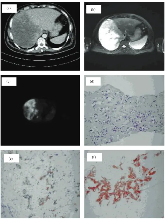
(a) (b)
(c) (d)
(e) (f)
Figure 1: (a) CT scan,20×14 cm size hypoattenuating multinodular tumor mainly filling the right lobe of the liver with irregular boundaries. (b) MR, T2-weighted images as a slightly hyperintense area with contrast enhancement of peripheral zone being liable to confluent multiple lesions in the right lobe of liver. (c) Fluorodeoxyglucose-positron emission tomography (FDG-PET) showed 10.7 of a standardized uptake value for FDG in CC of liver. (d) Tumor cells diffusely formed ductal structures in fibrotic stroma and have a big hyperchromatic nucleus in narrow cytoplasm, H&E,×200. (e) Tumor cells diffusely replaced the liver parenchyma and formed ductal structures in fibrotic stroma which were positively stained for cytokeratin 19 immunoreaction,×200. (f) Tumor cells diffusely replaced the liver parenchyma and formed ductal structures in fibrotic stroma which were positively stained for cytokeratin 7 immunoreaction×200.
showed a large tumor with hypoechogen pattern in right lobe
of the liver. CT revealed 20 × 14 cm size hypoattenuating
multinodular tumor mainly filling the right lobe of the
liver with irregular boundaries (Figure 1(a)). Thorax and
brain CT were normal. T1-weighted MR images showed the tumor as a hypointense area and T2-weighted images showed it as a slightly hyperintense area with contrast
enhancement of peripheral zone being liable to confluent multiple lesions in right lobe of liver with retrocaval and aor-tocaval multiple metastatic lymphadenopathy and metastatic
lesions on right iliac and right femoral bones (Figure 1(b)).
Fluorodeoxyglucose-positron emission tomography (FDG-PET) showed pathological signal intensity of 10.7 of a stan-dardized uptake value in CC of liver, in renal hilum, in
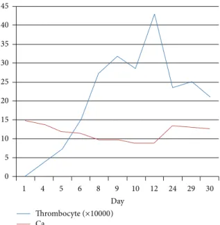
Case Reports in Medicine 3 45 40 35 30 25 20 15 10 5 0 Day 1 4 5 6 8 9 10 12 24 29 30 Ca Thrombocyte (×10000)
Figure 2: After treatment, relationship with calcium and thrombo-cyte level.
bilateral aortocaval lymph nodes, and in six places of bones as C-5, T-11, and S-1 vertebral corpus, right iliac bone, and
left and right ischium (Figure 1(c)).
The patient was treated with IV saline, furosemide, and calcitonin. After the 8th day of treatment, calcium level, thrombocytopenia, and other clinical symptoms were
nor-malized (Figure 2). After the patient stability and normal
platelet count were obtained, we performed USG guided trucut liver biopsy. USG guided trucut liver biopsy confirmed the diagnosis of CC. Neoplastic cells stained positively by cytokeratins 7 and 19. Patient was evaluated surgically as inoperable with evidence of radiological and pathological findings and was directed to medical oncology department for purpose of palliative chemotherapy with intended
radio-therapy for bone metastasis (Figures1(d),1(e), and1(f)).
At the end of first month of the oncologic treatment, patient was readmitted to hospital with worsening of con-sciousness state and relapsed hypercalcemia (11.5 mg/dL), while thrombocyte count was normal. Calcium level was higher than normal levels but not as high as first admittance to lead to thrombocytopenia again. Medical treatment of hypercalcemia rapidly stabilized the patient consciousness state. In spite of the chemoradiation treatment, disseminated metastasis developed and patient died in the 5th month of diagnosis.
3. Discussion
CC is a rare neoplasm which accounts for approximately
3% of the gastrointestinal cancers worldwide [4]. Although
surgical resection remains the only curative treatment, most
patients with CC are not operable candidates [3].
Hypercalcemia is a serious and frequent complication and a sign of advanced stage of malignant disease, occurring in
10% to 20% of patients with malignancies [5]. It has been
estimated to be of 5.3% in patients with HCC and 17.5% in
patients with CC [6]. Approximately 85% of patients with
cancer and hypercalcemia have metastatic bone disease. The remaining 15% have some other etiology for hypercalcemia
as paraneoplastic syndrome [7]. Humoral hypercalcaemia
of malignancy (HHM) is characterised by elevated calcium, low serum phosphorus, low PTH, and low vitamin D levels
[8]. The type of hypercalcemia has been associated with the
abnormal secretion of various proteins, including PTH-rP by
the tumor cells [7,8]. HHM is usually manifested as certain
type of malignancies and is generally associated with squ-amous-cell carcinomas of esophagus, lung, head, and neck.
HHM is rarely seen with CC [9]. The present case had
mult-iple metastatic bone lesions. So, predominantly, these lesions were thought of as the reason for hypercalcemia. And the mechanism of hypercalcemia had no effect on treatment of our case, so we did not need to differentiate etiology of hyper-calcemia.
Medical treatment of hypercalcemia is composed of IV hydration with saline, loop diuretics, and antiresorptive
agents such as calcitonin and bisphosphonates [10,11].
Thrombocytopenia in these cases may occur for many reasons and can be seen in some liver tumors like angiosarco-mas and hemangioangiosarco-mas. The relationship between hypercal-cemia and thrombocytopenia is not clear but in literature it is stated that there is correlation between hypercalcemia and hypophosphatemia. Hypophosphatemia also impairs granu-locyte function by interfering with ATP synthesis, increas-ing platelet diameter, and shortenincreas-ing platelet survival so it triggers the marked platelet disappearence. And these lead
to thrombocytopenia and reactive megakaryocytosis [12]. In
this case thrombocyte level was as low as 2000 K/𝜇L. But there was not any spontaneous bleeding. So we did not need transfusion. The patient was treated with IV saline, diuretics, and calcitonin.
4. Conclusion
We presented a case of metastatic CC with symptomatic hypercalcemia, hypophosphatemia, and low platelet count. With increased calcium levels and suppressed PTH with exis-tence of metastatic bone disease clinician should recognize this rare presentation of CC. The correction of hypercalcemia and hypophosphatemia provides correction of thrombocy-topenia.
Consent
Written informed consent was obtained from the patient’s and his first degree relatives for publication of this case and involving images.
Conflict of Interests
The authors declare that there is no conflict of interests regarding the publication of this paper.
Authors’ Contribution
Muharrem Battal and Ozgur Bostanci were involved in man-agement and treatment of this patient, Bayindir G¨urbulak and
4 Case Reports in Medicine Oguzhan Karatepe drafted the paper, and Yasar Ozdenkaya
reviewed the literature. All authors have read and approved the paper.
References
[1] W. A. Oldenburg, J. A. van Heerden, G. W. Sizemore, C. F. Abboud, and P. F. Sheedy II, “Hypercalcemia and primary hepatic tumors,” Archives of Surgery, vol. 117, no. 10, pp. 1363– 1366, 1982.
[2] J. M. Davis, R. Sadasivan, T. Dwyer, and P. van Veldhuizen, “Case report: cholangiocarcinoma and hypercalcemia,” The
American Journal of the Medical Sciences, vol. 307, no. 5, pp. 350–
352, 1994.
[3] M. J. Olnes and R. Erlich, “A review and update on cholangio-carcinoma,” Oncology, vol. 66, no. 3, pp. 167–179, 2004. [4] J.-N. Vauthey and L. H. Blumgart, “Recent advances in the
man-agement of cholangiocarcinomas,” Seminars in Liver Disease, vol. 14, no. 2, pp. 109–114, 1994.
[5] A. F. Stewart, “Humoral hypercalcemia of malignancy,” in
Primer on the Metabolic Bone Diseases and Disorders of Mineral Metabolism, M. J. Favus, S. Christakos, S. R. Goldring et al.,
Eds., pp. 198–203, Lippincott-Raven, Philadelphia, Pa, USA, 3rd edition, 1996.
[6] I. D. Xynos, S. Sougioultzis, A. Zilos, K. Evangelou, and G. S. Hatzis, “Hypercalcemia in a patient with cholangiocarcinoma: a case report,” International Archives of Medicine, vol. 2, no. 1, article 35, 2009.
[7] K. Sato, E. Onuma, R. C. Yocum, and E. Ogata, “Treatment of malignancy-associated hypercalcemia and cachexia with humanized parathyroid hormone-related protein anti-body,” Seminars in Oncology, vol. 30, no. 5, supplement 16, pp. 167–173, 2003.
[8] W. M. Philbrick, J. J. Wysolmerski, S. Galbraith et al., “Defining the roles of parathyroid hormone-related protein in normal physiology,” Physiological Reviews, vol. 76, no. 1, pp. 127–173, 1996.
[9] A. F. Stewart, R. Horst, L. J. Deftos, E. C. Cadman, R. Lang, and A. E. Broadus, “Biochemical evaluation of patients with can-cer-associated hypercalcemia. Evidence for humoral and non-humoral groups,” The New England Journal of Medicine, vol. 303, no. 24, pp. 1377–1383, 1980.
[10] G. A. Clines and T. A. Guise, “Hypercalcaemia of malignancy and basic research on mechanisms responsible for osteolytic and osteoblastic metastasis to bone,” Endocrine-Related Cancer, vol. 12, no. 3, pp. 549–583, 2005.
[11] C. S. Kovacs, S. M. MacDonald, C. L. Chik, and E. Bruera, “Hypercalcemia of malignancy in the palliative care patient: a treatment strategy,” Journal of Pain and Symptom Management, vol. 10, no. 3, pp. 224–232, 1995.
[12] K. A. Hruska, Hyperphosphatemia and Hypophosphatemia, Chapter 41, American Society for Bone and Mineral Research, 2006.
View publication stats View publication stats