Hearing Thresholds and Serum Apelin Levels in Patients
with Vertebrobasilar Insufficiency
Hande Ezerarslan1, Tuba Çandar2, Sedat Özdemir2, Gökçe Kaan Ataç3, Sinan Kocatürk1, Selda Demirtaş2 1Department of Otolaryngology, Ufuk University Faculty of Medicine, Ankara, Turkey
2Department of Biochemistry, Ufuk University Faculty of Medicine, Ankara, Turkey 3Department of Radiology, Ufuk University Faculty of Medicine, Ankara, Turkey Original Investigation
Objective: Vertebrobasilar insufficiency (VBI) is a clinical entity that results from the decrease in flow volumes due to hemodynamic changes in the vertebral artery and its branches. Herein, we aimed to investi-gate the hearing thresholds and serum apelin levels in VBI patients who were admitted to our clinics with vertigo.
Methods: Patients who were admitted to our clinics with vertigo were included in our study. They under-went bilateral carotid and vertebral artery color Dop-pler ultrasound investigation to determine VBI. Then, they were grouped into two groups: having VBI or not. Pure tone audiometry tests between 2500-8000 Hertz (Hz) were applied, and plasma levels of apelin, routine biochemistry parameters, and levels of C-reactive pro-tein were studied in these patients.
Results: Thirty-eight (47% women, mean age 57.6±9.3) patients with VBI and 24 controls (50% women, mean
age 57.0±10.1) were included. In patients with VBI, hearing thresholds were higher in the left ear except at the 500 Hz frequency. Serum apelin levels were not statistically different between the 2 groups.
Conclusion: In this study, several abnormalities in hearing tests were determined in VBI patients. The hypothesis was that basilar artery dysfunction caused by asymmetry of vertebral artery flow volumes in VBI might increase apelin levels, which have functions in angiogenesis, hemostasis, and cardiovascular hemo-dynamics. However, we could not find a significant difference in apelin levels between the 2 groups. This result was thought to be due to the absence of severe hemodynamic abnormalities and atherosclerosis in the study groups.
Keywords: Vertebrobasilar insufficiency, hearing im-pairment, apelin, color doppler ultrasound
Address for Correspondence:
Hande Ezerarslan, Ufuk Üniversitesi Tıp Fakültesi, Kulak Burun Boğaz Hastalıkları Anabilim Dalı, Ankara, Turkey
Phone: +90 533 430 95 28 E-mail: [email protected] Received Date/Geliş Tarihi: 21.10.2014 Accepted Date/Kabul Tarihi: 16.11.2014
© Copyright 2014 by Offical Journal of the Turkish Society of Otorhinolaryngology and Head and Neck Surgery Available online at www.turkarchotolaryngol.net DOI:10.5152/tao.2014.727
Abstract
Introduction
The vertebral artery arises from the subclavian artery, passes through the foramina transversar-ia of the cervical vertebrae, and extends upward. It unites with the contralateral vertebral artery at the lower border of the pons and forms the basilar artery. The posterior 1/3 region of the brain is supplied with blood by these vessels called the vertebrobasilar system. Vertebrobasilar insufficiency (VBI) is a clinical entity resulting from decreased flow volume due to hemodynam-ic changes in this artery and its branches. The symptoms of VBI include ataxia, focal neurologic signs, or dysarthria according to the region where ischemia occurs. Similarly, peripheral vertigo, po-sitional nystagmus, and hearing loss can be found in patients as the reason of cocleo-vestibular sys-tem dysfunction. The diagnosis of VBI should be confirmed with imaging techniques, and doppler ultrasonography (US) is frequently used for this aim. Moreover, angiography, magnetic resonance
imaging, magnetic resonance angiography, and computed tomography angiography are used in addition to US (1).
Apelin is an adipokine released from the adipose tissue that is accepted to be an endocrine tissue (2, 3). Apelin has been detected in the endothelium of the vessel wall as well as the heart and peripheral organs in the humans (4). Apelin affects angiogen-esis and central nervous system and has a role in providing fluid hemostasis and cardiovascular he-modynamics (5).
The aim of this study was to determine the hear-ing thresholds and serum apelin levels of patients with VBI. Considering the possible occurrence of endothelial dysfunction in the basilar artery due to asymmetry of vertebral artery flow volumes while planning the study, it was decided to measure the apelin levels in the serum samples for VBI diagno-sis. Thus, the attainability of objective data rather
than subjective data, such as dizziness and dysarthria, were in-vestigated while VBI was suspected in the differential diagnosis of patients having peripheral vertigo.
Methods
This clinical and prospective cross-sectional study was conduct-ed with patients having appliconduct-ed to the outpatient clinic of Oto-rhinolaryngology with the complaint of vertigo.
The study included patients with vertigo, whose etiology did not involve otologic reasons, such as Meniere’s disease, otosclero-sis, or benign paroxysmal positional vertigo, and who did not have ischemic encephalopathy. Moreover, patients with a known chronic disease, with kidney, liver or heart failure, with a history of continuous use of antihypertensive or lipid lowering drugs or a history of alcohol abuse, and with thyroid dysfunction and obesity were excluded from the study.
The patients were divided into two groups as the ones with VBI (Total volume value for both vertebral arteries <200 mL/min) and without VBI (Total volume value for both vertebral arteries >200 mL/min) in the color Doppler US analysis of the carotid– vertebral artery (6).
Following detailed neurootologic examinations of patients, they underwent pure tone audiometry (Interacoustic AC - 33, As-sens, Denmark) at the frequencies between 250 and 8000 Hertz (Hz). Hearing threshold values and the mean pure tone (MPT) were calculated for each frequency. Furthermore, speech dis-crimination levels (SDL) were evaluated. On the other hand, tympanometry (Interacoustic AZ-26, Assens, Denmark) was performed to eliminate the pathology of the middle ear. Color Doppler US examination was performed by a radiolo-gist having 20-year experience in this area. Color Doppler US evaluation of the carotid and vertebral arteries was performed with high-frequency broadband linear probe (Logiq 7, General Electric, WI, USA). Bilateral carotid arteries were first exam-ined in grayscale and intima media thickness (IMT), existence of plaque, and stenosis were detected. The diameters of vertebral arteries (VD) were similarly measured. Then, peak systolic flow velocity (PSV), end-diastolic flow velocity (EDV), and vertebral artery flow volumes (VF) were calculated.
Blood serum samples were collected between 09:00 a.m. and 10:00 a.m. after at least 8-hour fasting. Routine biochemis-try parameters, complete blood count, and C-reactive protein (CRP) measurements were performed simultaneously. Apelin was evaluated with ELISA method using the unit of ng/ml (Phoenix Pharmaceuticals Inc., Belmont, USA).
Body mass indices (BMI) of patients were calculated with the weight (kg) / height (m2) formula. Patients with BMI value of
18–25 kg/m2 were accepted to be normal and they were includ-ed in the study.
Statistical Analysis
All data obtained were analyzed with PASW (Predictive An-alytics Software) Statistics 21.0 software (SPSS Inc., Chicago, USA). In statistical evaluation, Kolmogorov Smirnov test was applied for the test of normality. For variables with normal dis-tribution, t-test was used. Mann–Whitney U test was employed for variables without normal distribution. Chi-square analysis was performed for categorical variables. Partial Spearman cor-relation test was also performed.
The ethical approval for this study was received from the Ethics Committee of Ufuk University Medical School (Date-Number: 31.10.2013-6). Furthermore, patients were informed about the study in detail and their written informed consents were ob-tained.
Results
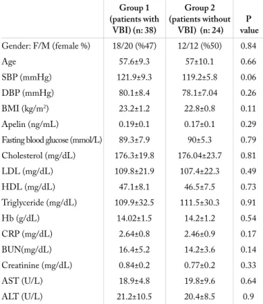
The study included 38 patients with VBI (47% female) and 24 patients without VBI (50% female). The mean age of patients with VBI was 57.6±9.3 years, whereas the mean age of the con-trol group was 57.0±10.1 years. No statistically significant dif-ference was found between two groups with regard to the mean ages and gender distribution (p: 0.66 and p: 0.84, respectively). The distribution of the demographic features and laboratory findings of the participants are summarized in Table 1 accord-ing to the groups.
The MPT values (right ear: 40.2±15.2; left ear: 37.4±16.9) and SDL values (right ear: 88±8.3; left ear: 88.9±8.4) of pa-tients with VBI were found to be higher than those of the control group (MPT; right ear: 22.2±4.16, left ear: 22.2±3.9 and SDL; right ear: 96.7±4.03, left ear: 96.7±4.03) (p<0.001). It was observed that hearing thresholds in the group with VBI increased at all frequencies except the frequency of 500 Hz in the left ear (Figure 1, 2). Moreover, it was detected that VF affected the occurrence of hearing loss (Odds ratio: 0.97; CI 95%: 0.94–0.99; p: 0.04). In patients having VBI, no statisti-cally significant difference was found between the right and left ears in terms of MPT and SDL values (p: 0.06 and p: 0.05, respectively). Moreover, power analysis conducted for deter-mining the adequacy of the sampling size revealed the power of the study as 99%.
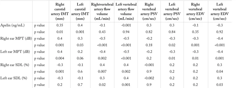
The parameters obtained through color Doppler US are pre-sented in Table 2. There was no statistically significant difference between two groups with regard to apelin levels (p: 0.29). A positive correlation was found between apelin and carotid artery IMT [Rho (p): 0.4; p: 0.001]. Table 3 presents the correlation values between apelin value, carotid artery IMT, vertebral artery
PSV, EDV, and VF values and MPT and SDL after correction of the effects of some factors such as age, systolic and diastolic blood pressures, and BMI.
Discussion
This is the first study investigating the relationship between the serum apelin value and the existence of VBI and its effects on hearing thresholds. In our study, no difference was observed be-tween patients with and without VBI, whose ages, genders and BMI averages were similar, in terms of serum apelin values. Fur-thermore, it was revealed that the hearing thresholds of patients with VBI elevated in all bilateral frequencies (except 500 Hz in the left ear).
Of today’s radiological imaging techniques, angiography is considered to be the gold standard for the diagnosis of verte-brobasilar insufficiency (7). On the other hand, Doppler US is a method with high specificity and sensitivity for hemodynamic examination of the carotid and vertebral arteries (97% and 93%, respectively) (8, 9). Owing to its common usage and being a non-invasive method, US was performed for imaging the carot-id and vertebral artery by using both grayscale and color Dop-pler features in our study.
Vertebral arteries unite at the pontomedullary junction and form the basilar artery. The 85% supply of the inner ear is carried out by the anterior inferior cerebellar artery arising from approxi-mately 1 cm over the basilar artery proximally. The labyrinthine artery supplies the cochlea by one or two cochlear artery branch-es. Unequal mechanical force between two vertebral arteries can increase the stress in the vessel endothelium of the basilar artery and lead to atherosclerosis by allowing the production of free oxygen radicals. The low amount of blood flow in the verte-brobasilar system causes ischemia in the cochlea and can lead to Table 1. Demographic features and laboratory findings of groups
Group 1 Group 2
(patients with (patients without P VBI) (n: 38) VBI) (n: 24) value
Gender: F/M (female %) 18/20 (%47) 12/12 (%50) 0.84 Age 57.6±9.3 57±10.1 0.66 SBP (mmHg) 121.9±9.3 119.2±5.8 0.06 DBP (mmHg) 80.1±8.4 78.1±7.04 0.26 BMI (kg/m2) 23.2±1.2 22.8±0.8 0.11 Apelin (ng/mL) 0.19±0.1 0.17±0.1 0.29
Fasting blood glucose (mmol/L) 89.3±7.9 90±5.3 0.79
Cholesterol (mg/dL) 176.3±19.8 176.04±23.7 0.81 LDL (mg/dL) 109.8±21.9 107.4±22.3 0.49 HDL (mg/dL) 47.1±8.1 46.5±7.5 0.73 Triglyceride (mg/dL) 109.9±32.5 111.5±30.3 0.91 Hb (g/dL) 14.02±1.5 14.2±1.2 0.54 CRP (mg/dL) 2.64±0.8 2.46±0.9 0.17 BUN(mg/dL) 16.4±5.2 14.2±3.6 0.14 Creatinine (mg/dL) 0.84±0.2 0.77±0.2 0.33 AST (U/L) 18.9±4.8 19.8±9.6 0.64 ALT (U/L) 21.2±10.5 20.4±8.5 0.9
(ALT: Alanine aminotransferase; AST: Aspartate aminotransferase; BUN: Blood urea nitrogen; CRP: C-reactive protein; DBP: Diastolic blood pressure; Hb: hemoglobin; HDL: High density lipoprotein; LDL: Low density lipoprotein; SBP: Systolic blood pressure; BMI: Body mass index.)
Table 2. Distribution of color Doppler ultrasonography parameters between the groups
Group 1 Group 2 (patients with (patients without P
VBI) (n: 38) VBI) (n: 24) value
Right carotid artery IMT (mm) 0.7±0.3 0.4±0.1 <0.001 Left carotid artery IMT (mm) 0.6±0.2 0.5±0.1 0.01 Right vertebral artery diameter (mm) 3.3±0.6 3.5±0.4 0.08 Left vertebral artery diameter (mm) 3.3 ±0.8 3.6±0.6 0.07 Right vertebral artery flow 83.9±24.2 165±42.4 <0.001 volume (mL/min)
Left vertebral artery flow 81.6±8.2 204.3±63.6 <0.001 volume (mL/min)
Right vertebral artery PSV (cm/sec) 43.9±18.1 49.04±12.9 0.07 Left vertebral artery PSV (cm/sec) 42.4±14.1 54.6±19.4 0.01 Right vertebral artery EDV (cm/sec) 11.2±4.7 14.4±4.4 0.02 Left vertebral artery EDV (cm/sec) 11.5±5.3 17.5±5.6 <0.001 (EDV: End-diastolic flow velocity; IMT: Intima media thickness, PSV: Peak systolic flow velocity; VBI: Vertebrobasilar insufficiency)
Figure 1. The mean hearing thresholds for the right ear Frequency (Hz)
dB H
L
Patients with VBI
250 500 1000 2000 4000 6000 8000 60 50 40 30 20 10 0 Control group
Figure 2. The mean hearing thresholds for the left ear Frequency (Hz)
dB H
L
Patients with VBI
250 500 1000 2000 4000 6000 8000 60 50 40 30 20 10 0 Control group
endothelial dysfunction of the vessel wall with free radicals (10). In our study, elevated IMT in patients with VBI supports the increased risk for atherosclerosis. Considering these conditions, it is more likely that patients with VBI have sensorineural hear-ing loss at all frequencies, apparently at bilateral high frequen-cies, as in our study, rather than hearing loss pattern at unilateral high frequencies as stated in literature (11, 12). However, this study should be supported with further studies that will include higher number of patients and be conducted employing tests such as autoacoustic emission or auditory brain stem response audiometry for finding the causes of hearing loss.
In vertebral artery stenosis, PSV decreases in the distal of lesion, whereas EDV increases (6). In our study, the lower averages of vertebral artery flow volume accompanied by low averages of PSV and EDV than that in the control group were associated to the fact that the cause of lesion in our patients was low level of flow volumes rather than a segmental stenosis in the verte-bral artery. Moreover, negative correlation was detected between PSV and EDV and MPT in our study, which shows that the end organ cochlea had ischemic damage. Absence of an increase in hearing thresholds at only 500 Hz in the left ear may have resulted from relatively narrow sampling size.
Apelin receptor and ligand have been detected at high rate in the vessel endothelial cells (13, 14). While apelin creates vasodi-latation in the vessels through nitric oxide, it has positive inotro-pic effect on the heart (15). Owing to these features, apelin has cardio-protective effects. The presence of negative correlation is known between apelin and IMT, one of the indicators of athero-sclerosis increasing the risk for cardiovascular diseases (16-18). On the other hand, a positive correlation was found between apelin and IMT in this study, but no difference was revealed between two groups with regard to serum apelin values. This
indicates that there was no IMT at pathologic level in VBI pa-tients included in the study and they were not at advanced ages. Moreover, the patients in the study were included in the low risk group for cardiovascular diseases.
The limitations of this study include the low number of patients and its being a cross-sectional study. Furthermore, for confirm-ing the diagnosis of VBI and obtainconfirm-ing more detailed hemody-namic measures, invasive imaging techniques should have been used instead of Doppler US.
Conclusion
In this study, abnormalities detected in the hearing test in pa-tients with VBI were demonstrated objectively. The absence of correlation between plasma apelin values and hearing test results can be explained by lack of severe atherosclerosis in patients in-cluded in the study.
Ethics Committee Approval: Ethics committee approval was received for this study from the ethics committee of Ufuk University Medical School, 31.10.2013-6.
Informed Consent: Written informed consent was obstained from the patients who participated in this study.
Peer-review: Externally peer-reviewed.
Author Contributions: Concept - H.E., S.D., S.K.; Design - H.E., T.Ç.; Supervision - S.D., S.K.; Funding - S.D., G.K.A., S.Ö.; Materi-als - H.E., T.Ç., S.D.; Data Collection and/or Processing - H.E., T.Ç., S.Ö.; Analysis and/or Interpretation - T.Ç., H.E., G.K.A.; Literature Review - H.E., S.Ö., G.K.A.; Writing - H.E.; Critical Review - S.K., S.D.
Conflict of Interest: No conflict of interest was declared by the authors. Table 3. The mean pure tone, speech discrimination levels, and factors affecting apelin
Right Left Rightvertebral Left vertebral Right Left Right Left carotid carotid artery flow artery flow vertebral vertebral vertebral vertebral artery IMT artery IMT volume volume artery PSV artery PSV artery EDV artery EDV
(mm) (mm) (mL/min) (mL/min) (cm/sec) (cm/sec) (cm/sec) (cm/sec)
Apelin (ng/mL) p value 0.35 0.4 -0.1 -0.001 0.3 0.3 -0.1 -0.3
p value 0.01 0.001 0.43 0.94 0.82 0.84 0.35 0.92
Right ear MPT (dB) p value 0.4 0.3 -0.5 -0.5 -0.2 -0.3 -0.3 -0.4
p value 0.001 0.03 <0.001 <0.001 0.18 0.02 0.001 <0.001
Left ear MPT (dB) p value 0.4 0.2 -0.4 -0.5 -0.2 -0.3 -0.3 -0.4
p value 0.004 0.06 0.002 <0.001 0.2 0.01 0.01 0.001
Right ear SDL (%) p value -0.3 -0.1 0.4 0.4 -0.001 0.2 0.2 0.3
p value 0.001 0.6 0.007 0.002 0.9 0.2 0.2 0.04
Left ear SDL (%) p value -0.3 -0.1 0.3 0.4 -0.002 0.2 0.2 0.3
p value 0.2 0.7 0.02 0.001 0.9 0.2 0.2 0.03
Financial Disclosure: The authors declared that this study has re-ceived no financial support.
References
1. Nakagawa T, Yamane H, Nakai Y, Shigeta T, Takashima T. Evalua-tion of the vertebrobasilar arterial system by magnetic resonance an-giography in the diagnosis of the vertebrobasilar insufficiency. Acta
Otolaryngol 1998; 538: 54-7. [CrossRef]
2. Liu Y, Song CY, Wu SS, Liang QH, Yuan LQ, Liao EY. Novel adipokines and bone metabolism. Int J Endocrinol 2013; 2013:
895045. [CrossRef]
3. Wozniak S, Gee L, Wachtel M, Frezza E. Adipose Tissue: The New Endocrin Organ A Review Article. Digestive Diseases and Sciences
2009; 54: 1847-56. [CrossRef]
4. Kleinz MJ, Davenport AP. Emerging roles of apelin in biology and medicine Pharmacology & Therapeutics 2005; 107: 198-211.
[CrossRef]
5. Lee DK, George, S.R., O’Dowd, B.F. Unravelling the roles of the apelin system: prospective therapeutic applications in heart failure
and obesity. Trends Pharmacol Sci 2006; 27: 190-4. [CrossRef]
6. Bendick PJ, Glover JL. Hemodynamic evaluation of vertebral arteries by duplex ultrasound. Surg Clin North Am 1990; 70: 235-44.
7. Wardlaw JM, Chappell FM, Best JJ, Wartolowska K, Berry E; NHS Research and Development Health Technology Assessment Carot-id Stenosis Imaging Group. Noninvasive imaging compared with intra-arterial angiography in the diagnosis of symptomatic carotid
stenosis: a meta-analysis. Lancet 2006; 367: 1503-12. [CrossRef]
8. Hong JM, Chung CS, Bang OY, Yong SW, Joo IS, Huh K. Ver-tebral artery dominance contributes to basilar artery curvature and peri-vertebrobasilar junctional infarcts. J Neurol Neurosurg
Psychi-atry 2009; 80: 1087-92. [CrossRef]
9. Bartels E, Knauth M, Liebetanz D, Paulus W. Traumatic dissection of the vertebral artery: value of sonographic diagnostics.
Cerebro-vasc Dis 2006; 22: 209-13. [CrossRef]
10. Society of Atherosclerosis Imaging and Prevention Developed in collaboration with the International Atherosclerosis Society. Ap-propriate use criteria for carotid intima media thickness testing.
Atherosclerosis 2011; 214: 43-6. [CrossRef]
11. Pirodda A, Ferri GG, Modugno GC, Borghi C. Systemic Hypoten-sion and the Development of Acute Sensorineural Hearing Loss in Young Healthy Subjects. Arch Otolaryngol Head Neck Surg 2001;
127: 1049-52. [CrossRef]
12. Suckfull M, Zacharias S, Mees K. Exploratory analysis of risk factors of acute inner ear dysfunction. Laryngorhinootologie 1999; 78: 4-8. 13. Medhurst AD, Jennings CA, Robbins MJ, Davis RP, Ellis C,
Win-born KY, et al. Pharmacological and immunohistochemical charac-terization of the APJ receptor and its endogenous ligand apelin. J
Neurochem 2003; 84: 1162-72. [CrossRef]
14. Lee DK, Cheng R, Nguyen T, Fan T, Kariyawasam AP, Liu Y, et al. Characterization of apelin, the ligand for the APJ receptor. J
Neuro-chem 2000; 74: 34-41. [CrossRef]
15. Tatemoto K, Takayama K, Zou MX, Kumaki I, Zhang W, Kumano K, et al. The novel peptide apelin lowers blod presure via a nitric oxide-dependent mechanism. Regulatory Peptides 2001; 99: 87-92.
[CrossRef]
16. Grobbee DE, Bots ML. Carotid artery intima-media thickness as an indicator of generalized atherosclerosis. J Intern Med 1994;
236: 567-73. [CrossRef]
17. Nambi V, Chambless L, Folsom AR, He M, Hu Y, Mosley T, et al. Carotid intima-media thickness and presence or absence of plaque improves prediction of coronary heart disease risk: the ARIC (Ath-erosclerosis Risk In Communities) study. J Am Coll Cardiol 2010;
55: 1600-7. [CrossRef]
18. Kadoglou NP, Sailer N, Moumtzouoglou A, Kapelouzou A, Gera-simidis T, Kostakis A, et al. Adipokines: a novel link between adi-posity and carotid plaque vulnerability. Eur J Clin Invest 2012; 42: