Herz 2015 · 40:481–486 DOI 10.1007/s00059-013-4020-y Received: 23 July 2013 Revised: 26 October 2013 Accepted: 2 November 2013 Published online: 21 December 2013 © Urban & Vogel 2013
A.F. Erkan1 · G.K. Beriat2 · B. Ekici1 · C. Doğan2 · S. Kocatürk2 · H.F. Töre1 1 Department of Cardiology, School of Medicine, Ufuk University, Dr. Rıdvan Ege Hospital, Ankara 2 Department of Ear, Nose and Throat, School of Medicine, Ufuk University, Dr. Ridvan Ege Hospital, Ankara
Link between angiographic
extent and severity of coronary
artery disease and degree of
sensorineural hearing loss
Atherosclerotic coronary artery disease (CAD) is the leading cause of morbidi-ty and mortalimorbidi-ty around the world. Ath-erosclerosis is a systemic, chronic, pro-gressive disease that mainly involves me-dium-sized arteries. Clinically, it can be-come apparent as ischemic heart disease, cerebrovascular disease, or peripheral ar-terial disease [1]. Presbycusis, or age-re-lated hearing loss, is the cumulative ef-fect of aging on hearing. Presbycusis is defined as progressive bilateral symmetri-cal age-related sensorineural hearing loss, with onset during the 3rd or 4th decade of life [2, 3, 4]. The etiology of presbycusis is not fully understood, yet it is thought to be multifactorial. Many authors have as-serted that presbycusis has a genetic basis, and there is a general presumption based on clinical observation that presbycusis is an inherited disorder and that genet-ic factors may influence both the rate and severity of hearing loss [5, 6].
On the other hand, atherosclerosis may diminish the vascularity of the co-chlea, thereby reducing its oxygen supply [7]. Hypoperfusion of the cochlea may al-so lead to senal-sorineural hearing loss. In a long-term follow-up study that examined elderly individuals, low-frequency hear-ing loss (low pure-tone average, 0.25– 1 kHz) was associated with cardiovascular disease events including CAD and stroke in both genders but more prominently in women [16].
In this study, we aimed to investigate the relationship between the angiograph-ic severity and extent of CAD, whangiograph-ich is a surrogate of atherosclerotic burden, and
the overall degree of sensorineural hear-ing impairment, includhear-ing both hearhear-ing loss and speech discrimination.
Patients and methods
Patient selection
In this study, 381 consecutive patients who underwent coronary angiography for symptoms suggesting ischemic heart disease and who had ischemia detected by a noninvasive stress test were screened. Seventy-nine patients were excluded be-cause they met one of the exclusion cri-teria defined below, and 37 patients were excluded because they did not meet the inclusion criteria. The remaining 265 pa-tients [mean age, 61.5±13.0 years; medi-an age (25th–75th percentile), 59 years (50.5–67)] including 146 male (55.1%) and 119 female subjects (44.9%) constitut-ed the study population. This was a sam-ple of adult patients who may represent
the general population. The study proto-col was approved by the local ethics com-mittee.
The patients were included in the study if they were 18 years of age or old-er, if their coronary angiogram was clear enough to enable efficient calculation of the Gensini score, and if they gave con-sent to participate in the study.
The patients with acute coronary syn-drome, cardiomyopathy, congenital heart disease, severe valvular heart disease, a history of previous revascularization (percutaneous transluminal coronary an-gioplasty or coronary artery by-pass graft surgery) were excluded from the study. From an audiological point of view, pa-tients with a history of exposure to oto-toxic drugs or excessive noise, a history of Meniere’s disease, acoustic neuroma or otological surgery, and patients with an air-bone gap of more than 20 dB hear-ing level units (HL) or non-type-A tym-panogram or nonstapedial reflex were
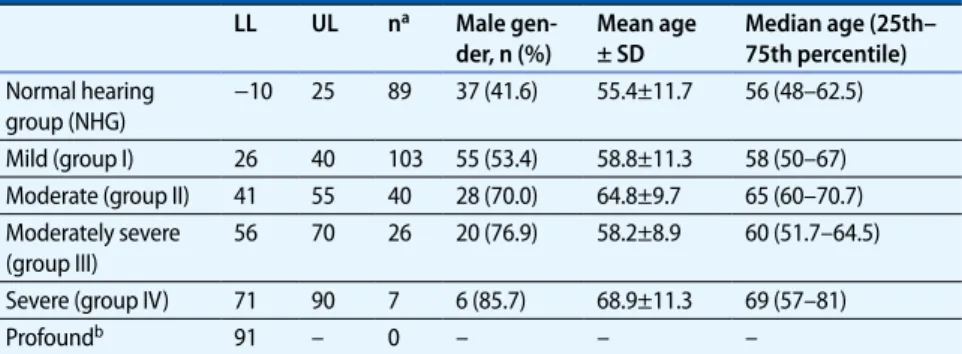
ex-Tab. 1 Classification of hearing loss and distribution according to age and sex LL UL na Male gen-der, n (%) Mean age ± SD Median age (25th– 75th percentile) Normal hearing group (NHG) −10 25 89 37 (41.6) 55.4±11.7 56 (48–62.5) Mild (group I) 26 40 103 55 (53.4) 58.8±11.3 58 (50–67) Moderate (group II) 41 55 40 28 (70.0) 64.8±9.7 65 (60–70.7) Moderately severe (group III) 56 70 26 20 (76.9) 58.2±8.9 60 (51.7–64.5) Severe (group IV) 71 90 7 6 (85.7) 68.9±11.3 69 (57–81) Profoundb 91 – 0 – – –
LL and UL denote lower and upper limits of hearing loss, respectively, in dB(HL) defining each group, SD stan-dard deviation. a Denotes the number of patients in each group, mean and median ages are expressed as years. b As none of the patients in our study had profound hearing loss, no such group was assigned
cluded. Patients with asymmetrical hear-ing loss (a difference in terms of hearhear-ing loss of more than 20 dB(HL) between the two ears) were also excluded.
Coronary angiography and
assessment of CAD
Selective coronary angiography was per-formed using the femoral approach em-ploying the Judkins technique and utiliz-ing an Innova angiographic system (Gen-eral Electric, USA). Multiple views were obtained for all patients, with visualiza-tion of the left anterior descending and left circumflex coronaryrtery in at least
four projections, and the right coronary artery in at least two projections. Coro-nary angiograms were recorded in DI-COM format. The extent and severity of the CAD were evaluated by using the Gensini score [8]. In this scoring system, a severity score is derived for each coro-nary stenosis based on the degree of lu-minal narrowing and its topographic im-portance. Reductions in luminal diameter of 1–25%, 26–50%, 51–75%, 76–90%, 91– 99%, and total occlusion are scored as 1, 2, 4, 8, 16, and 32, respectively. Each princi-pal vascular segment is assigned a multi-plier that represents its functional impor-tance in maintaining myocardial blood
supply. The multipliers are: 5 for the left main coronary artery; 2.5 for the proxi-mal segment of the left anterior descend-ing coronary artery (LAD) and circum-flex artery; 1.5 for the mid-segment of the LAD; 1 for the right coronary artery, the distal segment of the LAD, the postero-lateral artery, and the obtuse marginal ar-tery; and 0.5 for other segments (. Fig. 1). Angiographic scoring was performed by two interventional cardiologists who were blinded to audiological measure-ments and clinical data. The Gensini score for each angiogram was the average of the scores assigned by these two ob-servers. In case of discrepancy between the two observers, the angiogram was re-scored. The study population was divided into three groups according to the Gensi-ni score as follows: GensiGensi-ni score 0, nor-mal coronary arteries; Gensini score be-tween 0 and 20, mild CAD; Gensini score >20, significant CAD [9].
Audiological assessment
All patients who underwent coronary angiography also underwent an audio-metric evaluation before the procedure per the study protocol. Hearing impair-ment was not the presenting symptom in any of the patients. All audiological tests were performed in the Ear, Nose, and Throat Department, Division of Hear-ing and Speech, by the same audiologist to avoid variation in technique. The au-diologist was also blinded to angiograph-ic and clinangiograph-ical data. Detailed hearing as-sessment included ear, nose, and throat examination, tympanograms, stapedi-al acoustic reflexes, pure-tone hearing levels, and speech discrimination scores (SDS). An interacoustics AC-40 audi-ometry device, TDH-49 earphones, and B71 bone vibrator for bone conduction were used in the audiological testing. All subjects underwent air-conduction and bone-conduction pure-tone threshold measurement at 125–8,000-Hz frequen-cies, and degree of hearing loss was not-ed in dB(HL). The frequency of 8,000 Hz was excluded from the analysis because hearing loss at this relatively high fre-quency could be more likely to be age-re-lated, and thus confounding. The degree of hearing loss was classified as
previous-Fig. 1 8 Schematic explanation for the calculation of the Gensini score. (With permission from [8]) 100 80 60 40 20 0
Hearing loss in decibel
s 0 50 100 150 200 250 Gensini score Fig. 2 9 Scatterplot depicting the positive correlation between the degree of hearing loss at 2,000 Hz and the Gensini score (for the right ear)
e-Herz: Original article
ly described by Clark et al. [10]. This clas-sification and the distribution of patients according to the degree of hearing loss are summarized in . Tab. 1.
Speech discrimination is described as a person’s ability to not only hear words but to identify them. The procedure in-volved presentation of 50 selected mono-syllabic words at an easily detectable in-tensity level, which was defined as 40 dB above the pure-tone average for each
in-dividual patient. The SDS was defined as the percentage of words correctly identi-fied.
Statistical analysis
Statistical analysis was performed by us-ing SPSS 15.0 for Windows (SPSS, Chica-go, Ill., USA). The normal distribution of variables was verified with the Kolmogo-rov–Smirnov test. The Gensini score and
the degree of hearing loss (in decibels) did not display a normal distribution. To demonstrate the relationship between the Gensini score and the audiological measurements, Spearman’s rho correla-tion analysis was performed. Compari-sons between the groups were made with Kruskal–Wallis test or the Mann–Whit-ney U test where appropriate. The χ2 test was used to investigate whether distribu-tions of categorical variables differ
with-Herz 2015 · 40:481–486 DOI 10.1007/s00059-013-4020-y © Urban & Vogel 2013
A.F. Erkan · G.K. Beriat · B. Ekici · C. Doğan · S. Kocatürk · H.F. Töre
Link between angiographic extent and severity of coronary artery
disease and degree of sensorineural hearing loss
Abstract Aims. Atherosclerosis is a systemic disease that can affect the whole arterial tree. An im-portant cause of neuronal degeneration is atherosclerosis, which may lead to sensori-neural hearing loss. We aimed to investigate the relationship between the angiograph- ic severity and extent of coronary artery dis- ease, which is a surrogate of atherosclerot-ic burden, and the degree of sensorineural hearing loss. Patients and methods. Out of 381 consec- utive patients who underwent coronary an- giography for symptoms suggesting isch- emic heart disease and who had ischemia de- tected by a noninvasive stress test, 265 pa-tients [mean age, 61.5±13.0 years; median age (25th–75th percentile), 59 years (50.5– 67)], including 146 male (55.1%) subjects met the eligibility criteria and were enrolled. Au-diological measurements (hearing levels and discrimination scores) were performed before the coronary angiography. The Gensini score was calculated for each angiogram. Results. There was a statistically significant positive correlation between the degree of hearing loss at all frequencies analyzed (250, 500, 1,000, 2,000, 4,000 Hz) and the Gensi- ni score (p<0.05 for all frequencies), which re- mained significant after adjustment accord-ing to age and other risk factors. A statistically significant negative correlation was observed between the Gensini score and the speech discrimination score (p<0.05). Conclusion. The findings of this study sug- gest that the angiographic severity and ex- tent of coronary artery disease are signifi-cantly and independently correlated with the degree of hearing loss. Sensorineural hear-ing loss was more prominent in patients with higher Gensini scores. We propose that the findings of this study warrant further re-search and should be verified in large-scale studies. Keywords Atherosclerosis · Coronary artery disease · Sensorineural hearing loss · Neuronal degeneration · Angiography
Beziehung zwischen Ausmaß und Schweregrad der koronaren Herzkrankheit
in der Angiographie und Grad der sensorineuralen Schwerhörigkeit
Zusammenfassung Ziel. Die Atherosklerose ist eine systemi-sche Erkrankung, die alle Arterien erfassen kann. Eine bedeutende Ursache der neuro-nalen Degeneration ist die Atherosklerose, die zur sensorineuralen Schwerhörigkeit füh-ren kann. Ziel der vorliegenden Studie war es, den Zusammenhang zwischen dem Schwe- re grad und dem Ausmaß der koronaren Herz- krankheit (KHK), die einen Surrogatparame-ter der atherosklerotischen Last darstellt, und dem Grad der sensorineuralen Schwerhörig-keit zu untersuchen. Methoden. Von 381 konsekutiven Patien-ten, bei denen eine Koronarangiographie wegen Symptomen erfolgte, die den Ver-dacht auf eine ischämische Herzerkrankung nahelegten, oder wegen einer Ischämie, die mit einem nichtinvasiven Belastungs- test festgestellt wurde, erfüllten 265 Patien- ten [Durchschnittsalter: 61,5±13,0 Jahre, Al-tersmedian (25.–75. Perzentile): 59 (50,5– 67) Jahre], darunter 146 Männer (55,1%), die Auswahlkriterien und wurden in die Studie aufgenommen. Vor der Koronarangiographie wurden audiologische Messungen durchge- führt (Hörschwelle und Wert für Sprachver-ständlichkeit). Für jede Angiographie wurde der Gensini-Score berechnet. Ergebnisse. Es bestand eine statistisch sig-nifikante positive Korrelation zwischen dem Grad der Schwerhörigkeit bei allen ausgewer-teten Frequenzen (250, 500, 1000, 2000, 4000 Hz) und dem Gensini-Score (p<0,05 für alle Frequenzen), die auch nach Berück- sichtigung des Alters und anderer Risikofak-toren signifikant blieb. Andererseits wurde eine statistisch signifikante negative Korrela-tion zwischen dem Gensini-Score und dem Wert für Sprachverständlichkeit festgestellt (p<0,05). Schlussfolgerung. Den Ergebnissen der vor- liegenden Studie zufolge sind der Schwere-grad und das Ausmaß der KHK signifikant und unabhängig mit dem Grad der Schwer-hörigkeit korreliert. Die sensorineurale Schwer hörigkeit war bei Patienten mit ei- nem höheren Gensini-Score deutlicher aus- geprägt. Nach Ansicht der Autoren rechtfer- tigen die Ergebnisse der Studie weitere Un-tersuchungen und sollten in großangelegten Studien verifiziert werden. Schlüsselwörter Atherosklerose · Koronare Herzkrankheit · Sensorineurale Schwerhörigkeit · Neuronale Degeneration · Angiographie
in groups. Data are shown as mean ± SD and/or median (25th–75th percentile) for continuous variables and absolute num-bers (%) for dichotomous variables. A p value less than 0.05 was considered sta-tistically significant.
Results
Baseline characteristics
and biochemical and
audiological findings
The study population consisted of 265 patients, the mean age of whom was 61.5±13.0 years, and 146 (55.1%) of whom were male.
When the patients were classified ac-cording to the degree of hearing loss, it was seen that 89 subjects had normal hearing (normal hearing group, NHG), 103 subjects had mild hearing loss (group I), 40 subjects had moderate hearing loss
(group II), 26 subjects had moderately se-vere hearing loss (group III), and 7 sub-jects had severe hearing loss (group IV). The audiological definitions of hearing loss and the age and sex distributions of these groups are summarized in . Tab. 1. The differences between the groups in terms of age and sex were significant (p<0.001 and p=0.001, for age and sex, respectively, see . Tab. 1).
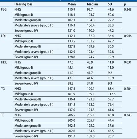
There was no significant difference between the groups with regard to tra-ditional risk factors for atherosclerot-ic CAD, namely, diabetes mellitus, hy-perlipidemia, hypertension, tobacco use, and family history of premature CAD. As for the biochemical findings, the mean HDL cholesterol level of group IV was significantly lower than that of the NHG and group I (p=0.031). Other biochemi-cal parameters did not significantly differ across the groups (. Tab. 2).
Angiographic findings and
degree of hearing loss
According to the Gensini score, 32.8% of the subjects had normal coronary ar-teries, 24.5% of the subjects had mild CAD, and 42.7% of them had significant CAD. Comparison of the groups with re-spect to the Gensini score revealed that CAD became progressively more sig-nificant as the hearing status worsened; the NHG had the lowest average Gen-sini score, and group IV had the high-est (mean Gensini scores, 3.63±8.69, 16.75±18.86, 55.76±32.93, 82.79±32.68, and 121.57±44.26 for the NHG, group I, group II, group III, and group IV, respec-tively, p<0.001).
There was a statistically significant positive correlation between the degree of hearing loss (in decibels), which was cal-culated according to the pure-tone hear-ing thresholds for 125, 250, 500, 1,000, 2,000, and 4,000 Hz and the Gensini score (. Tab. 3). This correlation main-tained its significance after adjustment for age and other risk factors. As an ex-ample, . Fig. 2 depicts the positive corre-lation between the degree of hearing loss and the Gensini score at 2,000 Hz for the right ear.
A traditional three-frequency (500, 1,000, and 2,000 Hz) pure-tone average was also obtained in order to assess over-all hearing levels. The pure-tone average for 500, 1,000, and 2,000 Hz was posi-tively and significantly correlated with the Gensini score (r=0.713, p<0.001 and r=0.668, p<0.001 for the left and the right ear, respectively).
A low-frequency pure-tone average (125, 500, 1,000 Hz) was also calculated, and it was found that the average hear-ing loss at these low frequencies was al-so positively and significantly correlated with the Gensini score (r=0.594, p<0.001 and r=0.358, p<0.001 for the left and the right ear, respectively).
There was a statistically significant negative correlation between the SDS and the Gensini score (r= −0.841, p<0.01 and r= −0.813, p<0.01 for the left and the right ear, respectively). This correlation also maintained its significance after adjust-ment for age and other risk factors.
Tab. 2 Biochemical parameters with regard to degree of hearing lossa
Hearing loss Mean Median SD p
FBG NHG 110.9 98.7 41.6 0.248 Mild (group I) 118.4 103.7 50.6 Moderate (group II) 107.3 104.3 22.2 Moderately severe (group III) 116.3 106.4 35.3 Severe (group IV) 131.0 110.9 47.2 LDL NHG 132.1 132.0 36.4 0.946 Mild (group I) 133.5 132.2 42.2 Moderate (group II) 127.8 129.9 30.5 Moderately severe (group III) 132.9 123.4 39.8 Severe (group IV) 128.8 124.7 19.9 HDL NHG 47.5 45.9 11.8 0.031 Mild (group I) 45.4 43.6 11.0 Moderate (group II) 41.0 41.7 9.2 Moderately severe (group III) 42.8 41.6 10.9 Severe (group IV) 38.2 34.8 9.3 TG NHG 147.5 129.1 83.4 0.204 Mild (group I) 161.9 139.1 112.6 Moderate (group II) 136.4 123.8 59.7 Moderately severe (group III) 181.5 153.2 79.4 Severe (group IV) 137.0 124.3 61.6 TC NHG 206.5 205.1 43.8 0.343 Mild (group I) 205.0 205.7 44.4 Moderate (group II) 192.5 192.2 37.6 Moderately severe (group III) 202.6 184.6 43.5 Severe (group IV) 191.7 189.0 20.7
a There is no significant difference between the groups except for HDL cholesterol levels. All biochemical param-eters are expressed as mean ± SD, in mg/dlNHG normal hearing group, FBG fasting blood glucose, LDL low-density lipoprotein cholesterol, HDL high-low-density lipoprotein cholesterol, TG triglycerides, TC total cholesterol, SD standard deviation
Discussion
Atherosclerosis and
sensorineural hearing loss
Atherosclerotic cardiovascular disease is a major health problem around the world. Atherosclerosis is an inflammato-ry process that may affect the entire arte-rial tree. Normal blood supply to the co-chlea is crucial to auditory transduction, the mechanism by which sounds are con-verted to nerve impulses that travel along the auditory pathways to the gyri tempo-rales transversi (Heschl’s gyri). Thus, co-chlear ischemia is followed almost im-mediately by hearing loss [11]. Accord-ing to Fang [12], there is a negative cor-relation between the diameter of the ar-terial vessels of the internal auditory me-atus and the degree of hearing loss. In ad-dition, the lumen of the vessels reduced more in patients with atherosclerosis than those without this condition, and fibroid thickness was seen in the tunica adven-titia [12]. Pertinent to these findings, the data obtained from our study demon-strated that the severity of CAD, which reflects the atherosclerotic burden, is sig-nificantly correlated with audiological pa-rameters: Sensorineural hearing impair-ment was more prominent in patients with higher Gensini scores and vice ver-sa. These findings suggest that there is a relationship between sensorineural hear-ing impairment and the severity of ath-erosclerotic CAD, a surrogate of systemic atherosclerosis. One explanation may be that the progress of the vessel changes in
patients with presbycusis might be accel-erated by atherosclerosis. Atherosclero-sis is a systemic disease that may dimin-ish blood supply of the cochlear-neural structures, and this might be a plausible explanation for this relationship. It can be hypothesized that audiological assess-ments may be predictive of the severity of CAD, or systemic atherosclerosis in gen-eral. This hypothesis must be tested fur-ther in large-scale studies.
In a recent, population-based, retro-spective cohort study [13], diabetic pa-tients were significantly more likely to experience sudden sensorineural hearing loss when compared with nondiabetic age- and sex-matched controls. Of note, comorbidities such as retinopathy and CAD increased the likelihood of sudden sensorineural hearing loss [13]. Ciccone et al. [14] reported an association between vascular endothelial dysfunction and sen-sorineural hearing loss, suggesting a vas-cular etiology for this condition.
It has previously been reported in the literature that the audiometric pat-tern may be an indicator of cardiovascu-lar health status [15, 16, 17]. Of note, in a long-term follow-up study by Gates et al. [16], low-frequency hearing loss (low pure-tone average, 0.25–1 kHz) was asso-ciated with cardiovascular disease events including CAD and stroke. In addition to the findings of Gates et al., we found significant correlations of the severity of CAD not only with low-frequency hear-ing loss, but with overall and relatively high-frequency (i.e., 4,000 Hz) hearing loss, and with speech discrimination as
fied with large-scale studies.
Study limitations
This study has certain limitations. A ma-jor limitation was that the cochlear blood flow measurements using laser Doppler velocimetry were not performed because of cost restrictions. Genetic evaluation for hereditary hearing loss syndromes was al-so not performed.
Conclusion
In conclusion, the results of this study suggest that the degree of sensorineu-ral hearing loss may be correlated with the severity and extent of atherosclerot-ic CAD. This may be explained by the fact that atherosclerosis is a diffuse and sys- temic process that affects the entire ar- terial tree. Increased atherosclerotic bur-den, as reflected by a higher Gensini score, may explain the diminished blood supply to the cochlea, eventually lead-ing to sensorineural hearing loss. These findings need to be verified in further re-search, involving large-scale studies.Corresponding address
Assist. Prof. Dr. A.F. Erkan
Department of Cardiology, School of Medicine, Ufuk University, Dr. Rıdvan Ege Hospital Mevlana Bulvarı 86–88, Balgat, 06520 Ankara Turkey [email protected] Acknowledgments. We would like to thank audiologist Ms. Figen Bağcı for performing the audiometric measurements.
for left) Gensini score (Hz, for right) score
125 r 0.141 125 r 0.153 p 0.023 p 0.013 250 r 0.159 250 r 0.102 p 0.010 p 0.101 500 r 0.408 500 r 0.321 p 0.000 p 0.000 1,000 r 0.750 1000 r 0.205 p 0.000 p 0.001 2,000 r 0.828 2000 r 0.853 p 0.000 p 0.000 4,000 r 0.806 4000 r 0.868 p 0.000 p 0.000
Compliance with ethical
guidelines
Conflict of interest. A.F. Erkan, G.K. Beriat, B. Ekici, C. Doğan, S. Kocatürk, and H.F. Töre state that are no con-flicts of interest. All studies on humans described in the present man- uscript were carried out with the approval of the re-sponsible ethics committee and in accordance with national law and the Helsinki Declaration of 1975 (in its current, revised form). Informed consent was ob-tained from all patients included in studies.