1 Kayseri Eğitim Araştırma Hastanesi, Anestezi Bölümü Kayseri, Turkey
2 Medipol university Medical Faculty Department of Anaestesiology and Reanimation, İstanbul, Turkey 3 Erciyes University Medical Faculty Department of Anaestesiology and Reanimation, Kayseri, Turkey
Yazışma Adresi /Correspondence: Günhan Gökahmetoğlu,
Kayseri Eğitim Araştırma Hastanesi, Anestezi Bölümü Kayseri Email: [email protected] Geliş Tarihi / Received: 16.02.2013, Kabul Tarihi / Accepted: 27.05.2013
Copyright © Dicle Tıp Dergisi 2013, Her hakkı saklıdır / All rights reserved
ORIGINAL ARTICLE / ÖZGÜN ARAŞTIRMA
Comparison of the depth of anesthesia in sevoflurane and halothane anesthesia with
bispectral index and 95% spectral edge frequency
Bispektral indeks ve %95 spektral edge frekans ile halotan ve sevofluran anestezi
derinliklerinin karşılaştırılması
Günhan Gökahmetoglu1, Elvan Tercan2, Cihangir Bicer3, Recep Aksu3, Adem Boyaci3 ÖZET
Amaç: Bu çalışmanın amacı, bispektral indeks (BIS) ve
% 95 spektral edge frekans (SEF) monitorizasyon sistem-leri kullanarak, halotan ve sevofluran anestezi derinliksistem-leri- derinlikleri-nin değerlendirilmesidir.
Yöntemler: 2001-2002 yılları arasında, yaşları 20 ile 60
arasında değişen, genel anestezi altında jinekolojik ope-rasyon geçiren 40 hasta çalışmaya dahil edildi. Aneste-zi indüksiyonu Grup I hastalarda (n=20) halotan, Grup II hastalarda sevofluran ile konvansiyonel spontan inhalas-yon indüksiinhalas-yon metodu ile sağlandı. SEF ve BIS indeks değerleri kaydedildi.
Bulgular: Her iki grupta entübasyon öncesi ve sonrası,
cilt insizyon öncesi ve sonrası SEF değerleri, bazal de-ğerlerine göre anlamlı derecede düşüktü (p<0,05). Aynı zamanda, her iki grupta anestezi indüksiyonu sonrası kaydedilen SEF değerleri anlamlı olarak bazal değerler-den yüksekti (p<0,05). Ekstübasyon sonrası kaydedilen SEF değerleri kıyaslandığında, iki grup arasında istatis-tiksel olarak anlamlı fark bulundu (p<0.05). Her iki grupta da, anestezi indüksiyonu sonrası, entübasyon öncesi, en-tübasyon sonrası, cilt insizyon öncesi ve sonrası len BIS değerleri, bazal ve ekstübasyon sonrası kaydedi-len değerlere kıyasla anlamlı derecede düşüktü (p<0.05). Entübasyon öncesi ve entübasyondan bir dakika sonra kaydedilen BIS değerlerine bakıldığında, her iki grup ara-sında istatistiksel olarak anlamlı fark bulundu (p<0,05).
Sonuç: Sonuç olarak BIS ve SEF değerleri anestezinin
hipnotik komponentini değerlendirmede yardımcı olabilir ve bunlar peroperatif sempatik ve cerrahi uyaranlara karşı otonomik cevapların gösterilmesinde yeterli değildir.
Anahtar kelimeler: Bispectral indeks, halotan,
sevoflu-ran, anestezi derinliği, hemodinamik değişiklikler
ABSTRACT
Objective: The aim of the present study was to evaluate
the depth of anesthesia provided by halothane or sevoflu-rane by using the bispectral index (BIS) and 95% spectral edge frequency (SEF) monitoring systems.
Methods: Forty patients, between the ages of 20-60
years, scheduled for gynecological surgery under general anesthesia were studied between the years 2001-2002. Anesthesia induction was maintained by conventional spontaneous inhalation induction method with sevoflu-rane in the group I patients (n=20), with halothane in the group II patients (n=20). SEF and BIS index values were recorded.
Results: In both groups, SEF values recorded before
intubation, after intubation, before and after skin incision were significantly lower compared with values recorded at baseline (p<0.05). Also, SEF values in both groups re-corded after anesthesia induction were significantly high-er compared with baseline values (p<0.05). Regarding the recorded SEF values after extubation, there were sta-tistically significant differences between the two groups (p<0.05). In both groups, BIS values recorded after anes-thesia induction, before intubation, after intubation, before and after skin incision were significantly lower compared with values recorded at baseline and after extubation (p<0.05). Regarding the recorded BIS values before intu-bation and 1 min after intuintu-bation, there were statistically significant differences between the two groups (p<0.05).
Conclusion: We conclude that BIS index and SEF
val-ues will only be helpful in the evaluation of the hypnosis component of anesthesia and it is not sufficient in show-ing the autonomous response, which is formed against the perioperative sympathetic and surgical stimulations.
Key words: Bispectral index, halothane, sevoflurane,
INTRODUCTION
The purposes of general anesthesia are to provide hypnosis, analgesia and appropriate surgical area (immobility, muscle relaxation). Unfortunately, the difference between hypnosis and analgesia is delet-ed in the operation room and patient replies (such as hypertension or movement) cannot be said to dem-onstrate the analgesic or hypnotic demands. The cli-nicians using anesthetic and sedatives need to direct the hypnotic status of their patients. For many years, monitoring the effects of the central nervous system (CNS) in an objective and reliable way has been a purpose for anesthesiology [1]. By discovering that the brain produces electrical activity, the fact was regained regarding that the anesthetic agents change electroencephalography (EEG). Analgesic medi-cines have effects on both the cerebral physiology and the EEG patterns. Insufficient anesthesia gener-ally causes EEG activation. EEG may be considered as a criterion of the anesthetic depth due to many reasons [2]. Despite the dramatic developments in anesthesia safety, it is not accurately clear whether these benefits are due to more intense monitoring or due to new monitoring standards. Bispectral index (BIS) is an EEG parameter, which has been specifi-cally developed to measure the patient responses during anesthetics and sedatives administration. The opportunity to arrange and develop better hypnotic drug administration doses are provided for the anes-thesia operators. The results show that BIS monitor-ing durmonitor-ing anesthetic administration provides vari-ous benefits, both clinically and economically [1].
In this study, we aimed to evaluate the anes-thesia depth provided by halothane and sevoflurane by using the BIS and 95% spectral edge frequency (SEF) monitoring systems during the general anes-thesia induction, maintenance and wakeup periods.
METHODS
After Erciyes University Medical Faculty Ethics Committee approval, 40 patients of ASA I-II group, between the ages of 20-60 years, scheduled for gy-necological surgery under general anesthesia were studied between the years 2001-2002. All patients gave their informed consent. Surgical interventions which may possibly take more than two hours, pa-tients with intelligence problem which may cause cooperation difficulty or who had a history of
neu-rologic disease, patients who had taken inhalation anesthesia until 6 months ago, age below 20 or over 60 years and the patients who did not accept this technique when explained were excluded from the study.
Patients did not receive any sedative, hypnotic or analgesic premedication. Forty patients were ran-domly divided into two groups. Twenty were as-signed to the sevoflurane group (group I) and 20 to the halothane group (group II). After the patients in both groups were taken into the operation room, iv serum physiologic infusion was started before induction. Anesthesia was given to all patients by the same team. All patients were monitored in or-der to monitor their continuous heart rates (HR), noninvasive artery blood pressure, oxygen satura-tion (SpO2) and the gas ratios in the inspiration and expiration mixture (Hewlett Packard Monitor and Anaesthesia Viridiazuc). After skin preparation, disposable BIS sensor (Aspect Medical Systems, Natick, MA, USA) was attached to the patient’s forehead according to a standard montage and BIS and 95% SEF were monitored continuously using A-2000 BIS monitor (Aspect Medical Systems In-ternational B.V., Leiden, Netherlands) for display of the Bispectral Index processed data and real-time EEG waveforms.
The mixtures of each anesthetic agent together with oxygen were delivered by an Ohmeda Vapor vaporizer into the circle system of an Ohmeda an-esthesia machine. The inspiratory and expiratory limbs of the circle were attached to a Y connector. Respiratory gases were sampled into the multi-gas monitor (Datex), between the Y connector and the elbow connector with the mask attached to it. The patients were told to breath with the conventional spontane inhalation induction technique and anes-thesia induction was provided by volatile anesthetic inhalation. In Group I (n=20) patients, the anesthe-sia induction was provided by sevoflurane starting from 1% within 6L/min O2 and by increasing 1% in every 3 breathes and lastly at 7% concentration. The period from the beginning of the induction un-til the perishment of the eye lash reflex which we controlled at every 10 seconds was recorded as the duration of induction. 0.1 mg/kg iv vecuronium bromide was given in order to facilitate the tracheal intubation. Then, volatile anesthetic agent concen-tration was decreased with 1% at every 3 breathes to 2% concentration and endotracheal intubation
was performed. The anesthesia induction in Group II patients was started with the same conditions with Group I. However, the halothane concentration was increased to 5%. Once again, the period from the beginning of the induction until the perishment of the eye lash reflex which we controlled at every 10 seconds was recorded as the duration of induc-tion. Neuromuscular blocking agent was given at the same doses. In induction; complications such as coughing, bucking, laryngospasm, bronchospasm, nausea vomiting, extremity movements, secretion increase, excitation, agitation, and arrhythmia were recorded.
Laryngoscopy quality was assessed by tracheal intubation scoring system as: excellent (easy pas-sage of endotracheal tube without coughing; vo-cal cords relaxed and abducted), good (passage of endotracheal tube with slight coughing or bucking; vocal cords relaxed and abducted), poor (passage of endotracheal tube with moderate coughing or buck-ing; vocal cords moderately abducted) and not pos-sible (unable to intubate) [3]. Excellent and good tracheal intubation conditions were accepted as clinically sufficient.
After the tracheal intubation, all patients were mechanically ventilated with 10-12 ml/kg of tidal volume to maintain end-tidal carbon dioxide pres-sure (ETCO2) 30-35 mmHg. Systolic arterial pres-sure (SAP), diastolic arterial prespres-sure (DAP), heart rate (HR), BIS index, SEF value, SpO2 and ETCO2 values were recorded before anesthesia induction; after the anesthesia induction; before intubation; at minutes 1, 2 and 3 after intubation; before skin in-cision, after skin incision and at the end of the tra-cheal extubation.
In order to have a BIS index value in the range of 55 - 60 within the last 10-15 minutes of the sur-gery, the volatile anesthetic concentrations were re-duced by titration. After skin closure at the end of the surgery, residual neuromuscular blockade was reversed and the anesthetics discontinued. The peri-od from this moment till extubation was recorded as the duration of extubation. The response time to the command ‘open your eyes’ and the time at which the patient was oriented to time and place were re-corded as duration of recovery.
Data are presented as mean ± SD. Data between the treatment groups were analyzed using Student’s t-test or multiple analysis of variance (ANOVA).
Nonparametric data were analyzed by Chi square test. Pearson correlation test was applied to assess the recorded anesthesia depth parameters and he-modynamic parameters and p values <0.05 consid-ered statistically significant.
RESULTS
There was no statistically significant difference be-tween the groups, in terms of age, weight, type of surgery and duration of surgery (p>0.05). The mean times for induction of anesthesia, extubation and re-covery were significantly shorter with sevoflurane than with halothane (p<0.05) (Table 1). There were statistically no significant differences between the groups regarding to the recorded complications ob-served during the anesthesia induction (p>0.05).
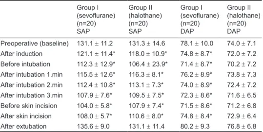
There were statistically no significant differ-ences in the SAP and DAP values between the two groups (p>0.05), however, SAP values in groups I and II and DAP values in group I were significantly lower at all recorded times except after extubation period compared with baseline values (p<0.05) (Ta-ble 2). Regarding the recorded HR and SpO2 values, there were statistically no significant differences be-tween the two groups (p>0.05).
In both groups, SEF values recorded before in-tubation, at 1., 2. and 3. minutes after inin-tubation, be-fore and after skin incision were significantly lower compared with values recorded at baseline, after anesthesia induction and after extubation (p<0.05) (Table 3). Also SEF values in both groups recorded after anesthesia induction were significantly higher compared with baseline values (p<0.05) (Table 3). Regarding the recorded SEF values after extuba-tion, there were statistically significant differences between the two groups (p<0.05) (Table 3).
In both groups, BIS values recorded after anes-thesia induction, before intubation, at 1., 2. and 3. minutes after intubation, before and after skin inci-sion were significantly lower compared with values recorded at baseline and after extubation (p<0.05) (Table 3). Regarding the recorded BIS values before intubation and 1 min after intubation, there were statistically significant differences between the two groups (p<0.05) (Table 3).
There was no statistically significant correla-tion observed between the SEF and BIS index val-ues recorded in both Groups and the cardiovascular parameters (p>0.05).
Table 1. Patients and
surgical characteristics Group I (sevoflurane)
(n=20)
Group II (halothane)
(n=20) t p
Age (yr)
Type of surgery (AH/VH/M) 48.2 ± 7.69/7/4 48.2 ± 10.411/6/3 0.017 >0.05
Weight (kg)*
Duration of induction (sec)* 67.6 ± 10.5119.8 ± 7.3 67.6 ± 11.3152.1 ± 9.3 0.01412.09 >0.05<0.05 Duration of surgery (min)*
Duration of extubation (min)* Duration of recovery (min)*
103.1 ± 12.3 8.1 ± 0.5 6.8 ± 1.0 102.9 ± 14.1 9.6 ± 0.8 9.8 ± 1.0 0.03 6.86 8.8 >0.05 <0.05 <0.05 *Data are presented as mean ± SD. AH/VH/M=abdominal hysterectomy, vaginal hysterec-tomy, myomectomy
Table 2. Systolic (SAP)
and diastolic (DAP) ar-terial pressures. Group I (sevoflurane) (n=20) SAP Group II (halothane) (n=20) SAP Group I (sevoflurane) (n=20) DAP Group II (halothane) (n=20) DAP Preoperative (baseline) 131.1 ± 11.2 131.3 ± 14.6 78.1 ± 10.0 74.0 ± 7.1 After induction 121.1 ± 11.4* 118.0 ± 10.9* 74.8 ± 8.7* 72.0 ± 7.2 Before intubation 112.3 ± 12.9* 106.4 ± 23.9* 71.4 ± 8.7* 70.2 ± 7.2
After intubation 1.min 115.5 ± 12.6* 116.3 ± 8.1* 76.2 ± 8.9* 73.8 ± 7.3
After intubation 2.min 112.4 ± 10.8* 113.1 ± 7.3* 74.0 ± 8.9* 72.4 ± 7.2
After intubation 3.min 107.9 ± 7.6* 109.5 ± 7.5* 72.3 ± 8.6* 71.6 ± 6.5
Before skin incision 104.0 ± 5.8* 107.9 ± 7.4* 71.5 ± 8.6* 71.2 ± 6.8
After skin incision 108.0 ± 5.7* 110.6 ± 8.0* 74.8 ± 8.4* 72.9 ± 6.4
After extubation 135.6 ± 9.0 131.1 ± 11.4 80.2 ± 9.3 76.8 ± 6.8
Data are presented as mean ± SD, *p < 0.05 vs. baseline
Table 3. Bispectral
index (BIS) and 95% Spectral Edge Fre-quency (SEF) values
Group I (sevoflurane) (n=20) SEF Group II (halothane) (n=20) SEF Group I (sevoflurane) (n=20) BIS Group II (halothane) (n=20) BIS Preoperative (baseline) 17.8 ± 1.7 17.1 ± 1.1 96.7 ± 1.3 96.4 ± 1.1 After induction 19.7 ± 1.5# 19.0 ± 1.5# 53.6 ± 5.5+ 52.4 ± 7.5+ Before intubation 15.1 ± 1.2* 14.5 ± 1.4* 45.1 ± 5.0+ $ 42.1 ± 4.2+
After intubation 1.min 14.1 ± 1.7* 13.4 ± 2.0* 44.5 ± 5.3+ $ 39.1 ± 5.6+
After intubation 2.min 14.2 ± 1.8* 14.2 ± 1.8* 42.9 ± 5.7+ 40.6 ± 6.2+
After intubation 3.min 14.4 ± 1.4* 14.5 ± 1.5* 42.4 ± 5.8+ 40.4 ± 6.3+
Before skin incision 14.5 ± 1.4* 14.6 ± 1.2* 43.0 ± 5.7+ 40.6 ± 4.9+
After skin incision 15.6 ± 1.9* 14.7 ± 1.6* 43.2 ± 5.6+ 41.4 ± 5.5+
After extubation 18.7 ± 1.7$ 17.6 ± 1.0 86.5 ± 4.5 85.4 ± 4.3
*p < 0.05 vs. baseline and after induction, # p < 0.05 vs. baseline $ p < 0.05 vs. group II, + p < 0.05 vs. baseline and after extubation
DISCUSSION
In the modern application; anesthesia is generally induced rapidly and safely in adults through iv agents. Induction by inhalation is frequently used in children but prolonged anesthesia induction can frighten both the children and the adults [4]. Thus, generally both the patients and the anesthetists avoid from induction by volatile agents. However, in recent years, the discovery of the new volatile anesthetics with less adverse effects, having lower blood: gas solubility ratios and providing rapid anesthesia induction, easy control for anesthesia depth, popularized the usage of inhalation induc-tion method again in general anesthesia applicainduc-tion both in children and in adults. [3-8]. In our study, we have used conventional inhalation induction method for the adults. The time elapsed as the an-esthesia induction was 119.8 ± 7.3 sec. for Group I; and 152.0 ± 9.3 sec. for Group II. Smith et al. [9] have found the anesthesia induction time with sevo-flurane as 109 seconds, and they have stated that the reason of the shortness of that time was giving the inhalation induction agent from 5% concentration up to the completion of the induction. In the study of Oguz et al. [10], the period in sevoflurane induc-tion performed by the conveninduc-tional method was de-termined as 107 seconds. Here, using the N2O-O2 mixture in 2:1 ratio has shortened this period with respect to the periods which we have obtained.
In this study, we found shorter extubation and recovery times in the sevoflurane group compared with halothane group. In rats, cerebral sevoflurane concentration was shown to decrease two times faster than the halothane [11]. Also, sevoflurane is more rapidly taken and eliminated faster than halo-thane [12]. These studies demonstrate the reason of why the extubation times and recovery times are shorter in sevoflurane and longer in halothane.
All inhalation agents have irritation risk for the respiratory tract and this risk increases with longer induction periods. Coughing is accepted as a good indicator of respiratory tract irritation. In our study, we observed 5% coughing in the sevoflurane group and 10% in the halothane group. We did not see bucking, laryngospasm or bronchospasm in both groups. In a study which compares vital capac-ity rapid inhalational induction of anesthesia with sevoflurane and isoflurane, Yurino et al [13] report-ed extremity movements in 12% of the patients in
sevoflurane group, and they observed no cough, la-ryngospasm, breath holding or a secretion increase. Oguz et al [10] reported that the length of the induc-tion period leads patient to anxiety and extremity movements. We have only encountered extremity movements in 1patient in the halothane group. The movements were in the form of moderate extremity movements joined by the wrist joint. Immamura et al [14] reported that in patients having sevoflurane anesthesia, the risk of arrhythmia inducted with the adrenaline discharge under hypoxic and hypercar-bic conditions was less than in halothane. Sevoflu-rane and halothane were reported as the most suit-able agents in terms of cardiovascular parameters in anesthesia induction with mask [15]. We observed arrhythmia in 2 patients in the halothane group, while there was no arrhythmia in any case among the sevoflurane group.
In a study conducted by Hall et al [16], anes-thesia induction was performed with sevoflurane in concentrations of 3% and 8%; mean arterial pres-sure after the loss of consciousness was found to be lower than the baseline value. Tanaka et al [15] reported that sevoflurane and halothane caused minimal change in SAP and DAP values compared to other volatiles. The decrease of SAP in the halo-thane group in our study was more than the decrease in DAP but it was not statistically insignificant.
Glass et al [17] conducted a series of volunteer studies in order to show the effect of BIS monitor-ing in the measurement of hypnotic drug effects. Propofol, midazolam, isoflurane, midazolam-alfen-tanil, propofol-alfentanil or propofol-nitrous oxide combinations were administered to healthy subjects in gradual doses; the anesthetic agent doses were gradually increased and decreased and BIS index was constantly recorded. Clinic measurement for sedation, hypnosis and memory were also carried out. It was proven that BIS is a very good determi-nant of the hypnotic state.
In a study conducted by Flaishan et al [18], in which the benefits of BIS monitorization were re-searched in the return of consciousness after the an-esthesia induction with propofol and tiopental, the consciousness was regained when the BIS values exceeded 60, and the changes in blood pressure and heart rate failed to reflect those. No statistical sig-nificance was found in our study during the
correla-tion test with HR and blood pressure changes in the intraoperative assessment of BIS and SEF values.
SEF is a frequency, where the total EEG power is below 95%. It was suggested that SEF can be used in order to measure the anesthesia depth. In a study conducted by Schwander et al [19], SEF decreased during general anesthesia inducted by isoflurane or propofol compared to the conscious state, and it in-creased similarly to the conscious state values dur-ing the periods, where the effects of the isoflurane or propofol anesthesia disappear, or during intraop-erative spontaneous movements. We have observed an increase of SEF during our post-induction mea-surements in our cases. SEF values did not show a relationship with the intraoperative hemodynamic changes. In a study conducted by Sawtelle et al [20], SEF 95% values of patients were compared with BIS, and the hypnotic state of the patients were determined with BIS at least 15 minutes before SEF 95%. We have observed no such correlations in our study. The SEF value in the moment of extubation was significantly higher in sevoflurane group com-pared to halothane group. There was no such dif-ference between the groups in terms of BIS values.
The idea of finding a reliable index of the anes-thesia depth has changed toward the finding of vari-ous indexes for different anesthesia components. Therefore, a monitor can only measure one of the general anesthesia components. We especially think that the BIS index is associated with the hypnotic component of the anesthesia. In our study; the fol-lowing results were obtained: anesthesia induction is faster with sevoflurane than halothane; during the anesthesia induction and the peroperative period, the hemodynamic differences in both halothane and sevoflurane treated patients are similar; the compli-cations regarding to the usage of halothane or sevo-flurane in anesthesia induction and maintenance are similar; postoperative extubation and recovery peri-ods are shorter in the sevoflurane group than halo-thane group; BIS index and SEF values which are recorded during the surgery show a similar change in both groups; the numerical values of SEF, ob-tained after the induction and extubation are higher than the basal values and they are insufficient pa-rameters in showing the anesthesia depth; there is no correlation between the hemodynamic data and the recorded BIS index and the SEF values.
With these results, it is considered that BIS index and SEF values will only be helpful in the evaluation of the hypnose component of anesthesia and it is not sufficient in showing the autonomous response which is formed against the peroperative sympathetic and surgical stimulations.
REFERENCES
1. Rosow C, Manberg PJ. Bispectral index monitoring. Anesth Clin N Am 1998;2:89-107.
2. Stanski DR. Monitoring depth of anesthesia. In: Miller RD (ed), Anesthesia. Churchill Livingstone Inc, New York 2000, pp 1087-1116.
3. Pino RM, Ali HH, Denman WT, et al. A comparison of the intubation conditions between mivacurium and rocuronium during balanced anesthesia. Anesthesiology 1998;88:673-678.
4. Ruffle JM, Snider MT, Rosenberger JL, et al. Rapid in-duction of halothane anaesthesia in man. Br J Anaesth 1985;57:607-611.
5. Muzi M, Robinson BJ, Ebert TJ, et al. Induction of anesthe-sia and tracheal intubation with sevoflurane in adults. Anes-thesiology 1996;85:536-543.
6. Jellish WS, Lien CA, Fontenot HJ, et al. The comparative effects of sevoflurane versus propofol in the induction and maintenance of anesthesia in adult patients. Anesth Analg 1996;82:479-485.
7. Ruffle JM, Snider MT. Comparison of rapid and convention-al inhconvention-alation inductions of hconvention-alothane oxygen anesthesia in healthy men and women. Anesthesiology 1987;67:584-587. 8. Drummond GB. Rapid inhalation induction of anaesthesia.
Br J Anaesth 1988;61:373-375.
9. Smith I, Nathanson M, White PF. Sevoflurane a long awaited volatile anaesthetic. Br J Anaesth 1996;76:435-445. 10. Oğuz S, Özalp G, Özmert G, et al. Sevofluran ile
anes-tezi indüksiyonunda klasik spontan inhalasyon indüksi-yon yöntemi ile tek vital kapasite soluması yöntemlerinin karşılaştırılması. Türk Anesteziyoloji ve Reanimasyon Ce-miyeti Mecmuası 1998;26:130-133.
11. Stern R, Towler S, White P, et al. Elimination kinetics of sevoflurane and halothane from blood, brain and adipose tissue in the rat. Anesth Analg 1990;7:237-244.
12. Yasuda N, Lockhart SH, Eger II EI, et al. Comparison of kinetics of sevoflurane and isoflurane in humans. Anesth Analg 1991;72:316-324.
13. Yurino M, Kimura H. Vital capacity breathing respiration technique for rapid anaesthetic induction comparison of sevoflurane and isoflurane. Anaesthesia 1992;47:946-949. 14. Immamura S, Ikeda K. Comparison of the epinephrine
in-duced arrhytmogenic effect of sevoflurane and halothane. J Anaesth 1987;1:62-68.
15. Tanaka S, Tsuchida H, Nakabayashi K-I, et al. The effects of sevoflurane, isoflurane, halothane and enflurane on hae-modynamic responses during an inhaled induction of anes-thesia a mask in humans. Anesth Analg 1996;82:821-826.
16. Hall J, Ebert TJ, Harmer M. Induction characteristics with 3% and 8% sevoflurane in adults: An evaluation of the second stage of anaesthesia and its haemodynamic conse-quences. Anaesthesia 2000;55:545-550.
17. Glass PS, Bloom M, Kearse L, et al. Bispectral analysis measures sedation and memory effects of propofol, mid-azolam, isoflurane and alfentanil in healthy volunteers. An-esthesiology 1997;86:836-847.
18. Flaishon R, Windsor A, Sigl J, et al. Recovery of conscious-ness after thiopental or propofol: BIS and the isolated fore-arm technique. Anesthesiology 1997;86:613-619.
19. Schwender D, Daunderer M, Mulzar S, et al. Spectral edge frequency of the electroencephalogram to monitor “depth” of anaesthesia. Br J Anaesth 1996;77:179-184.
20. Sawtelle K, Rampil I. Bispectral EEG index predicts awak-ening. Anesthesiology 1994;81:A213.