• Sonuç bulunamadı

Bedside ultrasonography for the confirmation of gastric tube placement in the neonate

N/A
N/A
Protected

Academic year: 2021

Share "Bedside ultrasonography for the confirmation of gastric tube placement in the neonate"

Copied!
5
0
0

Yükleniyor.... (view fulltext now)

Tam metin

(1)

Yunus oktaY ataLaY, ahMet veYseL PoLat1, eLiF ozYaziCi ozkan2, LeMan toMak3, Canan aYGun2,

JosePh DreW toBias4

Department of Anesthesiology, Istanbul Medipol University, International School of Medicine, Istanbul, 1Department of Radiology, Ondokuz Mayis University, 2Division of Neonatology, Department of Pediatrics, 3Department of Biostatistics and Medical Informatics, Ondokuz Mayis University, Samsun, Turkey, 4Department of Anesthesiology and Pain Medicine, Nationwide Children’s Hospital, Columbus, Ohio, USA

Address for correspondence: Dr. Joseph Drew Tobias, Department of Anesthesiology and Pain Medicine, Nationwide Children’s Hospital, 700 Children’s Drive, Columbus, Ohio 43205, USA. E‑mail: joseph.tobias@nationwidechildrens.org

ABSTRACT

Background: Naso/Orogastric tube (NOGT) misplacement can lead to significant complications. Therefore, the assessment

of tube position is essential to ensure patient safety. Although radiography is considered the gold standard for determining NOGT location, new methods may be helpful in reducing repetitive radiation exposure, especially for neonates. In this study, we sought to investigate if bedside ultrasonography (BUSG) can be used to verify NOGT placement in neonatal intensive care patients.

Materials and Methods: Infants requiring NOGT placement were enrolled. After insertion of the NOGT, the location was

first identified using BUSG and then confirmed using abdominal radiography for comparison.

Results: The study cohort included 51 infants with an average gestational age of 34 ± 4.9 weeks. BUSG determined the

NOGT location correctly with a sensitivity of 92.2%. The location of the NOGT could not be determined by BUSG in four neonates (7.8%). In one infant, the NOGT was positioned in the esophagus, as determined both by BUSG and radiography.

Conclusion: BUSG is a promising diagnostic tool for determining NOGT location in neonates, thereby eliminating the need

for abdominal radiography.

Key words: Enteral nutrition; infant; intensive care units; nasogastric tube; ultrasonography

Introduction

The placement of a naso/orogastric tube (NOGT) is commonly performed in the neonatal intensive care unit (NICU) for enteral feeding, medication administration, and gastric decompression.[1] Misplacement of the NOGT may result in

serious complications related to the location or inadvertent trauma including gastric perforation, placement within the tracheobronchial tree, aspiration, pneumothorax, and

pulmonary hemorrhage.[2‑4] The risk of NOGT misplacement

is higher in various clinical scenarios, including patients with neurologic disabilities or sedated and critically ill newborns.[5]

The incidence of misplacement has been reported to be as high as 1.3–2.4% with the most serious complications related to misplacement into the tracheobronchial tree and respiratory tract.[6] Sites of incorrect placement also

include the upper esophagus and too deep into the stomach,

Bedside ultrasonography for the confirmation of gastric tube

placement in the neonate

This is an open access journal, and articles are distributed under the terms of the Creative Commons Attribution-NonCommercial-ShareAlike 4.0 License, which allows others to remix, tweak, and build upon the work non-commercially, as long as appropriate credit is given and the new creations are licensed under the identical terms.

For reprints contact: reprints@medknow.com

How to cite this article: Atalay YO, Polat AV, Ozkan EO, Tomak L,

Aygun C, Tobias JD. Bedside ultrasonography for the confirmation of gastric tube placement in the neonate. Saudi J Anaesth 2019;13:23-7.

Original

Article

Access this article online

Website: www.saudija.org

Quick Response Code

DOI:

(2)

representing 6% and 61% of cases, respectively.[7] While

pneumonia, atelectasis, lung abscess, apnea, bradycardia, desaturation, and aspiration may result from inadvertent placement into the tracheobronchial tree, incorrect placement within the GI tract may result in gastroesophageal reflux, esophageal perforation, malabsorption, diarrhea, dumping syndrome, and inadequate weight gain, especially in low‑birth weight neonates.[7‑10] Therefore, it is essential

to ensure correct placement and verify the NOGT location during placement and before use.

Verification methods such as measuring the NOGT length, auscultation, capnography, observation of gastric aspirate, gastric pH testing, and radiography have specific limitations.[7]

While standard radiography is accepted as the gold standard method, its use may be limited because of concerns related to repeated radiation exposure for multiple placement verifications.[4] Furthermore, abdominal radiography does not

define the position of the gastroesophageal junction and the pylorus, which are critical landmarks in ensuring the correct NOGT location and cannot be used in real time during tube placement.[11] Currently, there are no radiological standards

to confirm NOGT placement, especially in neonates, and therefore, new methods for bedside confirmation of NOGT placement and ongoing verification of its location in neonates are needed.[5]

Ultrasonography is being used more frequently in various clinical scenarios. Advantages of ultrasonography include its ready availability in most ICUs, lack of radiation exposure, and lower cost.[12] The efficacy of ultrasonography for the

verification of the NOGT location has been reported for adult patients in various settings including prehospital management, the emergency room, and the intensive care unit.[13‑15] We have previously shown that bedside

ultrasonography (BUSG) can confirm NOGT location in patients in the pediatric intensive care unit when performed by a radiologist.[16] The current study prospectively evaluates

the efficacy of BUSG when determining NOGT location in neonates. Since it is not always feasible to consult a radiologist to perform BUSG for the verification of the NOGT location, we also investigated whether a critical care physician could perform BUSG.

Materials and Methods

This prospective study was performed over a 4‑month period in a tertiary NICU. The local clinical research ethical committee approved the study (Ethics Committee Approval: OMU‑KAEK 2016/158), and written informed consent was obtained from parents. According to the statistical power

analysis (statistical power of 98% and an alpha of 5%), 51 neonates requiring NOGT insertion were included in the study. The decision to place the NOGT was made by the attending neonatologist. All NOGTs were inserted by NICU physicians. The depth of insertion was determined by measuring the distance from the tip of the patient’s nose or the corner of mouth to the earlobe and then from the earlobe to the distance midway between the xiphoid process and the umbilicus (Nose‑ear‑mid‑umbilicus (NEMU) method).[17]

According to our standard NICU protocol, the NOGT position was confirmed using plain abdominal radiography before its use for enteral feeding or medications.

For the purpose of this study, verification was first conducted after insertion using BUSG followed by abdominal radiography. A critical care physician who had ultrasound experience for other clinical uses (central venous catheter placement) received additional training on esophageal and gastric ultrasound from a radiologist. The gastric ultrasound was then performed by the physician and the radiologist. As part of the training, the critical care physician observed gastric ultrasound and verification of the NOGT by the radiologist in 10 neonates. Afterward this, the critical care physician performed the ultrasonography and the radiologist evaluated his performance in an additional cohort of neonates. When the technique and expertise of the NICU physician were approved by the radiologist, the critical care physician was approved to participate in the evaluation of the 51 patients that were included in this study.

Ultrasonography was performed using the Toshiba Xario SSA‑770A (Toshiba Medical Systems Corporation, Otawara, Japan) with a 12‑MHz linear‑array transducer. The ultrasound examinations included a longitudinal scan performed in the sternal region. Two parallel hyperechogenic lines in the esophageal lumen were visualized behind the heart during longitudinal scanning using a cardiac window, and the location of the NOGT was verified in the thoracic esophagus [Figure 1a]. After visualizing the NOGT in the esophagus, the NOGT was followed with a transducer through the esophagogastric (EG) junction. At the EG junction, the transducer was directed into the semi‑sagittal plane and the NOGT was visualized in the stomach using a liver window [Figure 1b]. Confirmation was verified using an abdominal radiograph [Figure 2]. The location of the NOGT was then recorded using these two different methods. Feeding and/or enteral medications were administered after verification of the NOGT tip in the stomach. The time required to perform the ultrasonography (procedure time) was also recorded for each patient.

(3)

Statistical analyses were performed using SPSS 18.0 for Windows (SPSS Inc., Chicago, IL, USA). The data are presented as the mean ± standard deviation, median (minimum–maximum), and frequency (%). The Shapiro–Wilk test was used to analyze the normal distribution assumptions of the quantitative outcomes. The data were then analyzed using a Mann–Whitney U test for non‑normal data. Sensitivity and positive predictive values were also evaluated. A P value <0.05 was considered statistically significant. Results

Fifty‑one patients were included in the study including 22 (43%) preterm neonates. The average gestational age was 34 ± 4.9 weeks and the birth weight was 1910 ± 895 g [Table 1]. During the study process, 72.5% of the neonates were breathing spontaneously while the remainder of the cohort (27.5%) was receiving positive pressure support (mechanical ventilation in 11.8% and nasal continuous positive airway pressure in 15.7%).

Ultrasonography verified the correct location of the NOGT with a sensitivity of 92.2% (84.8–99.5%). The positive predictive value was 100%. In one term neonate, the NOGT was identified in the esophagus using ultrasonography and then subsequently confirmed by direct radiography. The NOGT could not be visualized in four (7.8%) patients (confidence level: 95%). Two patients had gas bowel interposition and two had hepatomegaly that obscured visualization of the stomach during ultrasonography. Table 2 lists the verification percentage of the NOGT location using ultrasonography and radiography. The mean time to perform the ultrasound (procedure time) was 3.6 ± 2.1 min (range: 1.4–10 min). The procedure time for the first 25 patients was 3.9 ± 2.2 min and 3.2 ± 2.0 min for the last 26 patients (P = 0.17).

Discussion

The current study evaluates the efficacy of BUSG for confirmation of NOGT placement in neonates and compares these results with abdominal radiography. BUSG has a high sensitivity when determining NOGT location in this patient population. BUSG was performed quickly with an average procedure time of 3.6 min. The major obstacle with BUSG identified in our study was failure to identify the NOGT in 4 of 51 patients due to the presence of bowel gas or hepatomegaly. One other concern is that for the current study, the NOGT was placed by a physician which may not be standard practice in other countries and other clinical scenarios. As such, further studies are needed to identify whether the NOGT and use of

Figure 2: A plain radiograph showing the naso/orogastric tube (arrows)  in the stomach

Figure 1: (a) A transverse sonogram showing a naso/orogastric tube (arrows)  in  the  esophagus.  The  heart  (H)  and  liver  (L)  can  also  be  observed.  (b) A coronal oblique sonogram showing the naso/orogastric tube in the  stomach (S) entering through the esophagogastric junction. The liver  (l) can be observed

b a

Table 1: Demographic and procedure data of the study cohort

Demographic n (%); Median (range); mean±SD Gender

Male 28 (54.9)

Female 23 (45.1)

Gestational age (weeks) 34±4.932 (23‑40)

Weight (g) 1910±8951; 570 (500‑3970)

Diagnosis

Prematurity 22 (43.1)

Respiratory distress syndrome 15 (29.4)

Transient tachypnea of the

newborn 5 (9.8) Meningomyelocele 2 (3.9) Chromosomal disorders 2 (3.9) Intracranial hemorrhage 1 (2) Choanal stenosis 1 (2) Hypothyroidism 1 (2) Hydronephrosis 1 (2) Neonatal pneumonia 1 (2) Total 51 (100)

Data are presented as the frequency (%), mean±SD, or median (range). Study cohort=51 patients. SD: Standard deviation

(4)

ultrasound can be accomplished by the nursing staff or other health‑care providers.

Previous studies have demonstrated various methods for identifying the correct location of a bedside NOGT placement. While previous studies have investigated and compared the efficacy of various techniques, each method has its own limitations.[18‑25] Although auscultation is one of

the most commonly used methods given its ease of use and its noninvasiveness, it alone is not sufficient to determine the correct placement of the NOGT as sounds from air insufflation can be heard in various locations even when the tip of the NOGT is in the lung, esophagus, or stomach.[26,27]

Therefore, this method is no longer recommended without other confirmatory tests.[2,27] Calculating the approximate

proper length of the NOGT may be the first step in preventing potential insertion errors; however, various measuring methods have been proposed to predict the proper length of the NOGT.[4] A commonly used and referenced technique

is to measure the distance from the tip of the patient’s nose to the earlobe and then to the xiphoid process (Nose‑ear‑ xiphoid (NEX) method). However, esophageal placement of the NOGT has been frequently noted with the NEX method. Therefore, the NEMU method (direct measurement of the distance from the nose to the ear to the mid‑umbilicus) or age, weight, and height‑based placement techniques have become more popular.[28,29] In the present study, the NEMU

method predicted the appropriate NOGT insertion length in 98% of the patients.

Other placement techniques have relied on aspirates from the gastric tube to measure pH, pepsin, and trypsin. Among these techniques, pH testing is the most simple and widely available bedside test.[21] While a cutoff point of pH ≤4 has

been used to confirm gastric aspirate, the pH of the gastric aspirate in pediatrics is often >4.[22] In a study comparing

the sensitivity and specificity as well as the negative and positive predictive values of different pH cutoff points (<4.0, <4.5, <5.0, and <5.5), a pH <5 was a positive

predictor in 100% of infants.[30] However, difficulties in

obtaining an adequate aspirate from small‑bore tubes and similarities of the pH between the stomach and intestine limit the utility of the gastric aspirate, pH, bilirubin, and enzyme testing techniques.[22,25]

Powers et al. demonstrated the efficacy of an electromagnetic device when verifying feeding tube placement compared to abdominal radiography.[23] However, the smallest tube

diameter available for the electromagnetic device is excessive, even for term infants. For the other nonradiological methods, capnography can verify endotracheal misplacement, but differentiation of the location (i.e., esophageal, gastric, or intestinal) is not possible.[31] Among the verification methods,

radiography remains the gold standard. However, this method is not practical for the verification of every NOGT due to multiple radiation exposures, feeding delays, and cost.[9,32]

These concerns may be magnified in the vulnerable, preterm population where prolonged use of feeding tubes may result in the need for repeated X‑ray exposure.[29]

With these concerns in mind, it is necessary to find an ideal technique to verify the accurate placement of a wide range of feeding tube sizes (5 F–10 F) with reasonable cost and portability. The efficacy and sensitivity of BUSG in verifying gastric tube location has been reported in both adult and pediatric ICU patients.[14‑16,33] Despite its efficacy, the routine

use of ultrasonography requires additional equipment, which may delay the process when the equipment is located outside of the intensive care unit. However, as BUSG has seen increased use in the ICU setting for several indications, routine availability of equipment is being more common. In addition to equipment availability, the technique is dependent on the user’s skills and training.[12] However, as demonstrated

by our study and other investigations, clinicians can gain adequate ultrasound skills in a relatively short period of time through didactic and hands‑on courses.[34] Other limitations

of ultrasound when confirming nasogastric tube locations include poor visualization due to obesity, hepatomegaly, excessive patient movement, and bowel gas interposition. Conclusions

In summary, the present study demonstrates that BUSG can be used to verify NOGT location accurately with a high sensitivity rate (92.2%) in the neonatal population. BUSG may be an effective and promising tool for determining the location of NOGT in neonates, even when performed by a non‑radiologist. This method can be used for the verification of correct NOGT location following initial placement, as well as subsequent verifications, which may lead to a reduction Table 2: Verification percentages using ultrasonography and

radiography

Modality and tube location Frequency (%)

Ultrasound Gastric 46 (90.2) Esophageal 1 (2) Not identified 4 (7.8) Radiography Gastric 50 (98) Esophageal 1 (2) Not identified 0 (0) Total 51 (100)

(5)

in radiation exposure and costs. Furthermore, it is feasible to use this technique in real time to observe the passage of the NOGT into the esophagus and the stomach. Future studies with a larger cohort are needed to confirm these preliminary results.

Financial support and sponsorship Nil.

Conflicts of interest

There are no conflicts of interest. References

1. Lyman B, Kemper C, Northington L, Yaworski JA, Wilder K, Moore C,

et al. Use of temporary enteral access devices in hospitalized neonatal

and pediatric patients in the United States. JPEN J Parenter Enteral Nutr 2016;40:574‑80.

2. Metheny NA, Meert KL, Clouse RE. Complications related to feeding tube placement. Curr Opin Gastroenterol 2007;23:178‑82.

3. Creel AM, Winkler MK. Oral and nasal enteral tube placement errors and complications in a pediatric Intensive Care Unit. Pediatr Crit Care Med 2007;8:161‑4.

4. Society of Pediatric Nurses (SPN) Clinical Practice Committee, SPN Research Committee, Longo MA. Best evidence: Nasogastric tube placement verification. J Pediatr Nurs 2011;26:373‑6.

5. Irving SY, Lyman B, Northington L, Bartlett JA, Kemper C, NOVEL Project Work Group. et al. Nasogastric tube placement and verification in children: Review of the current literature. Nutr Clin Pract 2014;29:267‑76.

6. Sorokin R, Gottlieb JE. Enhancing patient safety during feeding‑tube insertion: A review of more than 2,000 insertions. JPEN J Parenter Enteral Nutr 2006;30:440‑5.

7. Quandt D, Schraner T, Ulrich Bucher H, Arlettaz Mieth R. Malposition of feeding tubes in neonates: Is it an issue? J Pediatr Gastroenterol Nutr 2009;48:608‑11.

8. Pillai JB, Vegas A, Brister S. Thoracic complications of nasogastric tube: Review of safe practice. Interact Cardiovasc Thorac Surg 2005;4:429‑33. 9. Clifford P, Heimall L, Brittingham L, Finn Davis K. Following the

evidence: Enteral tube placement and verification in neonates and young children. J Perinat Neonatal Nurs 2015;29:149‑61.

10. Maruyama K, Shiojima T, Koizumi T. Sonographic detection of a malpositioned feeding tube causing esophageal perforation in a neonate. J Clin Ultrasound 2003;31:108‑10.

11. Cohen MD, Ellett ML, Perkins SM, Lane KA. Accurate localization of the position of the tip of a naso/orogastric tube in children; where is the location of the gastro‑esophageal junction? Pediatr Radiol 2011;41:1266‑71.

12. Srinivasan S, Cornell TT. Bedside ultrasound in pediatric critical care: A review. Pediatr Crit Care Med 2011;12:667‑74.

13. Brun PM, Chenaitia H, Bessereau J, Leyral J, Barberis C, Pradel‑Thierry AL, et al. Ultrasound evaluation of the nasogastric tube position in prehospital. Ann Fr Anesth Reanim 2012;31:416‑20. 14. Kim HM, So BH, Jeong WJ, Choi SM, Park KN. The effectiveness

of ultrasonography in verifying the placement of a nasogastric tube in patients with low consciousness at an emergency center. Scand J Trauma Resusc Emerg Med 2012;20:38.

15. Vigneau C, Baudel JL, Guidet B, Offenstadt G, Maury E. Sonography as an alternative to radiography for nasogastric feeding tube location. Intensive Care Med 2005;31:1570‑2.

16. Atalay YO, Aydin R, Ertugrul O, Gul SB, Polat AV, Paksu MS, et al. Does bedside sonography effectively identify nasogastric tube placements in pediatric critical care patients? Nutr Clin Pract 2016;31:805‑9. 17. Beckstrand J, Cirgin Ellett ML, McDaniel A. Predicting internal distance

to the stomach for positioning nasogastric and orogastric feeding tubes in children. J Adv Nurs 2007;59:274‑89.

18. Freeman D, Saxton V, Holberton J. A weight‑based formula for the estimation of gastric tube insertion length in newborns. Adv Neonatal Care 2012;12:179‑82.

19. Westhus N. Methods to test feeding tube placement in children. MCN Am J Matern Child Nurs 2004;29:282‑7.

20. Ellett ML, Croffie JM, Cohen MD, Perkins SM. Gastric tube placement in young children. Clin Nurs Res 2005;14:238‑52.

21. Stock A, Gilbertson H, Babl FE. Confirming nasogastric tube position in the emergency department: PH testing is reliable. Pediatr Emerg Care 2008;24:805‑9.

22. Gilbertson HR, Rogers EJ, Ukoumunne OC. Determination of a practical pH cutoff level for reliable confirmation of nasogastric tube placement. JPEN J Parenter Enteral Nutr 2011;35:540‑4.

23. Powers J, Luebbehusen M, Spitzer T, Coddington A, Beeson T, Brown J,

et al. Verification of an electromagnetic placement device compared with

abdominal radiograph to predict accuracy of feeding tube placement. JPEN J Parenter Enteral Nutr 2011;35:535‑9.

24. Ellett ML, Cohen MD, Perkins SM, Croffie JM, Lane KA, Austin JK,

et al. Comparing methods of determining insertion length for placing

gastric tubes in children 1 month to 17 years of age. J Spec Pediatr Nurs 2012;17:19‑32.

25. Ellett ML, Cohen MD, Croffie JM, Lane KA, Austin JK, Perkins SM,

et al. Comparing bedside methods of determining placement of gastric

tubes in children. J Spec Pediatr Nurs 2014;19:68‑79.

26. Metheny N, McSweeney M, Wehrle MA, Wiersema L. Effectiveness of the auscultatory method in predicting feeding tube location. Nurs Res 1990;39:262‑7.

27. Metheny NA. Preventing respiratory complications of tube feedings: Evidence‑based practice. Am J Crit Care 2006;15:360‑9.

28. Cirgin Ellett ML, Cohen MD, Perkins SM, Smith CE, Lane KA, Austin JK, et al. Predicting the insertion length for gastric tube placement in neonates. J Obstet Gynecol Neonatal Nurs 2011;40:412‑21. 29. Nguyen S, Fang A, Saxton V, Holberton J. Accuracy of a weight‑based

formula for neonatal gastric tube insertion length. Adv Neonatal Care 2016;16:158‑61.

30. Metheny NA, Pawluszka A, Lulic M, Hinyard LJ, Meert KL. Testing placement of gastric feeding tubes in infants. Am J Crit Care 2017;26:466‑73.

31. Araujo‑Preza CE, Melhado ME, Gutierrez FJ, Maniatis T, Castellano MA. Use of capnometry to verify feeding tube placement. Crit Care Med 2002;30:2255‑9.

32. Chenaitia H, Brun PM, Querellou E, Leyral J, Bessereau J, Aimé C, et al. Ultrasound to confirm gastric tube placement in prehospital management. Resuscitation 2012;83:447‑51.

33. Gok F, Kilicaslan A, Yosunkaya A. Ultrasound‑guided nasogastric feeding tube placement in critical care patients. Nutr Clin Pract 2015;30:257‑60.

34. Chalumeau‑Lemoine L, Baudel JL, Das V, Arrivé L, Noblinski B, Guidet B, et al. Results of short‑term training of naïve physicians in focused general ultrasonography in an Intensive‑Care Unit. Intensive Care Med 2009;35:1767‑71.

Şekil

Table 1: Demographic and procedure data of the study cohort Demographic n (%); Median (range); mean±SD Gender

Referanslar

Benzer Belgeler

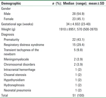
For this reason, we examined 78 preterm infants hospitalized between 2007 and 2013 in the Fatih University Neonatal Intensive Care Unit with a gestation age of 25-36 weeks

Akışkan yataklı reaktörler, çok miktarda sabit biyokütle bulundurması, düşük bekletme süresinde yüksek giderme verimi sağlaması, tıkanma probleminin olmaması

Concerning the use of octreotide in our two cases, it is es- pecially noteworthy that this drug was effective for treating chylothorax in a case where surgery had failed.. We did

[r]

[r]

Dahili yoğun bakım ünite- lerinde santral venöz kateter, üretral kateter, periferik arteryel kateter ve endotrakeal tüpler; cerrahi yoğun bakım ve postoperatif sıkı

Proteus mirabilis gibi gram olumsuz basillere bağlı beyin apsesi çocuklarda nadir görülmekte, özellikle risk gruplarında düşünülüp erken tanı konulması ve

All neonates admitted to the neonatal intensive care unit (NICU), Uni- versiti Kebangsaan Malaysia Medical Centre (UKMMC) were screened with a two-step protocol using an