Pediatric Urology
Relationship Between Procalcitonin Levels
and Presence of Vesicoureteral Reflux During
First Febrile Urinary Tract Infection in Children
Ilke Ozahi Ipek, Rabia Gonul Sezer, Evrim Senkal, and Abdulkadir Bozaykut
OBJECTIVE To investigate the association between the procalcitonin (PCT) level during the first febrile
urinary tract infection (UTI) in children and the presence of vesicoureteral reflux (VUR). VUR-associated UTI is among the primary causes of chronic renal failure in Turkey.
METHODS From March 2008 to November 2009, patients admitted with their first febrile UTI were
included in the present prospective hospital-based study. The serum concentrations of C-reactive protein, complete blood count, and PCT were measured. All patients underwent renal ultra-sonography and voiding cystourethrography.
RESULTS Of the 66 patients who were diagnosed with UTI, 18 had VUR. The geometric mean of the PCT
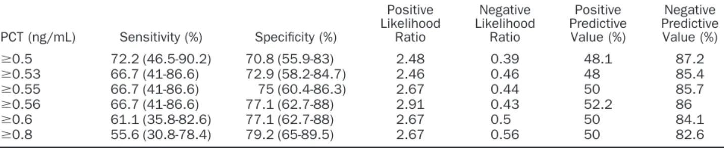
levels was significantly greater in the children with VUR than in those without (P⫽ .006). After logistic regression adjustment, the association between the PCT levels and the presence of VUR remained significant (odds ratio 5.08, 95% confidence interval [CI] 1.43-18.02). A PCT level ⬎0.56 ng/mL had 66.7% sensitivity (95% CI 41-86.6) and 77.1% specificity (95% CI 62.7-88) for diagnosing VUR. The area under the receiver operating characteristic curve for PCT was 0.715 (95% CI, 0.56-0.86, P⫽ .007), and the area under the curve for C-reactive protein was 0.723 (95% CI 0.58-0.86, P⫽ .006).
CONCLUSION A PCT-guided strategy could help in detecting patients with VUR. Large cohort studies are needed
to define an accurate cutoff value for children who are at risk of VUR, which increases the risk of renal damage and subsequent scarring. UROLOGY79: 883– 887, 2012. © 2012 Elsevier Inc.
T
he cumulative incidence of urinary tractinfec-tions (UTIs) during childhood is estimated at 7.8% for girls and 1.6% for boys.1UTIs are the most common indication necessitating radiographic eval-uation to detect the presence of vesicoureteral reflux (VUR). VUR is found in 25%-40% of children during the evaluation of their first UTI episode.2,3 With an incidence of 32.4%, VUR-associated UTIs have been reported to be the primary cause of childhood hyperten-sion and chronic renal failure in Turkey.4VUR has been diagnosed in 22% of those with a first UTI and 26% of
those with a second UTI in Turkish children.5 The
American Academy of Pediatrics recommends that ultra-sonography of the kidneys and bladder be performed after the first febrile UTI in children.6Voiding cystourethrog-raphy (VCUG) should be performed in children with a recurrence of febrile UTI, renal and bladder ultrasonog-raphy exhibiting hydronephrosis, scarring, findings
sug-gestive of high-grade VUR, or obstructive uropathy.6 Because renal ultrasonography alone is not satisfactory for predicting VUR, and VCUG has disadvantages, in-cluding exposure to radiation, a risk of iatrogenic UTI, and pain, new predictors are required to determine the infants at high risk of VUR.
Procalcitonin (PCT) is a very specific marker for acute bacterial infection, although it is unclear whether PCT is a hormone, a cytokine, or an acute-phase protein, studies have found that high PCT levels in the context of a bacterial infection suggest a rigorous acute immune re-sponse.7PCT can be detected in the blood 2 hours after endotoxin injection, and the increase in the PCT con-centration often correlates with the severity and mortal-ity of the disease.7,8Increases in PCT occur more rapidly than increases of C-reactive protein (CRP), an acute phase protein also used to differentiate between bacterial and viral infection.9 Many studies assessing the role of PCT in neonatal infections, invasive infections, infec-tions of the lower respiratory tract, UTI, fever of un-known origin, and pediatric oncology have concluded PCT is a valuable tool for the early diagnosis of bacterial disease in children.10
PCT has previously been demonstrated to be associ-ated with renal scars and VUR.11-13Because PCT values
From the Department of Pediatrics, Medipol University School of Medicine, Unkapanı, Istanbul, Turkey; and Department of Pediatrics, Zeynep Kamil Maternity and Child-rens’ Diseases Training and Research State Hospital, Uskudar, Istanbul, Turkey
Reprint requests: Rabia Gonul Sezer, M.D., Zeynep Kamil Hastanesi, Arakiyeci Haci Mehmet Mah, Op Dr. Burhanettin U¨ stünel Caddesi, Uskudar, Istanbul 34668 Turkey. E-mail:[email protected]
Submitted: October 5, 2011; received (with revisions): November 15, 2011
can be determined from venous blood samples and are generally available even in hospitals with limited facili-ties, we aimed to investigate the association between PCT and the presence of VUR.
MATERIAL AND METHODS
The present hospital-based prospective study was conducted from March 2008 to November 2009 in the Zeynep Kamil Maternity and Childrens’ Diseases Training and Research State Hospital (Istanbul, Turkey). Informed parental consent was obtained for all subjects, and the local ethics committee ap-proved the study.
Patients who were admitted with their first febrile UTI were included in the present study. The inclusion criteria were de-fined as follows: patients of either sex aged 1 month to 14 years, an axillary temperature⬎37.2°C, and a positive urine culture with a single microorganism. The patients with the following criteria were excluded from the study: newborns, those with a history of previous UTI, a history of previous urinary tract operations, previously known uropathy (eg, congenital malfor-mations, urolithiasis), antibiotic use at urinalysis and blood analysis, positive urine cultures with⬎1 organism, or negative urine cultures. Urine samples were obtained by suprapubic aspiration or transurethral catheterization in nontoilet-trained children and midstream clean-catch in toilet-trained children. Pyuria was defined as the presence ofⱖ5 leukocytes per a 40⫻ objective high power field of urine. All urine samples were cultured on blood and eosin-methylene blue agar plates using a standard loop. The plates were incubated at 37°C for 24 hours, and bacteria were identified using standard methods. The diag-nosis of UTI according to a positive urine culture was defined as any growth of a single bacterial pathogen from suprapubic aspiration,ⱖ105colony-forming units/mL for midstream urine
or ⱖ104 colony-forming units/mL of urine for transurethral
catheterization.
All eligible patients with a suspected UTI were examined, and the leukocyte (WBC) counts, CRP levels, PCT levels, and urine cultures were obtained. Urinalysis was conducted for all patients. The blood and urine were sampled at admission. Empirical antibiotic treatment after blood sampling was started for patients with pyuria. Children who were unable to retain their oral intake, presented with symptoms of toxicity, or were dehydrated were hospitalized. The children with leukocyte es-terase positivity were followed clinically until the culture results were obtained. The laboratory parameters of children with positive urine culture results were analyzed in the present study. A renal ultrasound scan was obtained within the first 48 hours after confirmation of UTI by the urine culture findings. All patients had undergone VCUG within 3 months, after UTI treatment when the urine was sterile, and VUR was graded using the International System for Radiological Grading of VUR.14
Serum CRP concentrations were measured using a nephelo-metric method (IMMAGE 800, Beckman Coulter, Brea, CA), and values⬎1 mg/dL were considered abnormal. The quanti-tative measurement of the PCT levels was performed using a chemiluminescence assay (Liaison, DiaSorin Laboratories, Sa-luggia, Italy), and values⬎0.5 ng/mL were considered abnor-mal. The PCT results were compared with the CRP values.
Statistical Analysis
Statistical analysis was performed using the Statistical Package for Social Sciences, version 17.0 (SPSS, Chicago, IL). Univar-iate analysis was conducted using the chi-square and t tests to evaluate the relationship between the high PCT levels and VUR. A logistic regression model was used to assess the inde-pendence of this relationship. Potential confounders, including age, sex, family history of uropathy, pathologic renal ultrasonog-raphy, WBC count, and CRP (factors) were adjusted for the prediction of the presence of VUR (dependent variable). These variables were dichotomized as follows: age⬍12 months versus ⬎12 months, boys versus girls, family history of uropathy versus no history, pathologic ultrasound findings versus normal find-ings, PCT ⬍0.5 ng/mL versus ⬎0.5 ng/mL, CRP ⬍1 mg/dL versus⬎1 mg/dL, WBC count ⬍15 000 mm3versus⬎15 000
mm3. Because all the children had fever, fever did not affect the
model outcome and was not included in the model. The best model was based on the Nagelkerke R2value. The
discriminat-ing power of PCT was determined by its sensitivity and speci-ficity for VUR. A cutoff value for PCT was determined using the receiver operating characteristic (ROC) curve. P⬍ .05 was considered statistically significant.
RESULTS
A total of 66 children, 47 girls (71.2%) and 19 (28.8%) boys, were enrolled in our prospective study. Their mean age was 38.62⫾ 39.21 months (range 1-144, median 19). VUR was diagnosed in 18 children.
The mean PCT level was 1.96⫾ 5.92 ng/dL (range
0.1-43, median 0.39). The geometric mean of the PCT levels was significantly greater in the children with VUR than in those without (P ⫽ .006;Table 1). Of the 66 children, 39 (59.1%) had negative serum PCT levels
(⬍0.5 ng/mL). Of the 39 patients with normal PCT
levels, 5 (12.8%) had VUR (2 with grade 1 and 1 each with grade 2, 3, and 4). Logistic regression adjustment, including age, sex, ultrasound results, family history, CRP level, and WBC count as potential confounders, was applied. The best model explaining the association be-tween the presence of VUR and the risk factors included
sex, WBC count, CRP level, and PCT level (P⫽ .02).
The association between the PCT level and the presence of VUR remained significant (odds ratio 5.08, 95% con-fidence interval [CI] 1.43-18.02;Table 2). All the chil-dren had fever; thus, fever was not included in the model.
A PCT level ⬎0.56 ng/mL gave a 66.7% sensitivity
(95% CI 41%-86.6%) and 77.1% specificity (95% CI
62.7%-88%) for diagnosing VUR (Table 3). The area
under the ROC curve for PCT was 0.715 (95% CI 0.56-0.86, P⫽ .007). The area under the ROC curve for CRP was 0.723 (95% CI 0.58-0.86, P⫽ .006;Fig. 1). The comparison of the ROC area of CRP and PCT was not significant (P⫽ .9). The mean CRP level was 7.2 ⫾ 6.83 mg/dL. A significant positive correlation was found
be-tween the CRP and PCT values (r⫽ 0.28, P ⫽ .02).
The axillary temperature of the children ranged from 38° to 40.3°C. Of the 66 children, 12 (18.2%) presented with vomiting, 8 (12.1%) with stomachache, and 16 (24.2%) with other symptoms, such as costovertebral
angle tenderness, dysuria, and enuresis, in addition to fever. Pyuria was present in 55 patients and nitrate pos-itivity in 26 patients. Leukocyte esterase pospos-itivity was observed in 29 urine samples. No significant difference was found with regard to age, sex, fever status, WBC count, isolated organism found in the urine culture, method of urine collection, or renal ultrasound results of the patients with and without VUR. No correlation was found between the age of the children and the PCT levels (r⫽ ⫺0.06, P ⫽ .58), and no significant difference was detected between the PCT level when stratified by sex (P⬎ .05).
A comparison of PCT levels according to the VUR grade did not reveal any statistically significant results, although the geometric mean of the PCT levels was relatively greater for grade 1 VUR (1.77 ng/dL in grade 1, 0.41 ng/dL in grade 2, and 0.84 ng/dL in grade 3; P ⫽ .34).
COMMENT
In the presence of a febrile, culture-documented UTI, elevations in PCT correlated with the presence of VUR and might prompt an earlier evaluation with VCUG.
Table 1. Baseline characteristics of children and comparison of parameters stratified by presence of vesicoureteral reflux
Characteristic VUR P Value No (n⫽ 48) Yes (n⫽ 18) Age (mo) 38.37⫾ 39.01 39.30⫾ 40.85 .93* Sex .18† Male 16 (33.3) 3 (16.7) Female 32 (66.7) 15 (83.3) Fever (°C) 38.80⫾ 0.37 38.84⫾ 0.67 .78* WBC (mm3 ) 14 868⫾ 7232 15 338⫾ 6264 .80* CRP (mg/dL) 5.82⫾ 5.04 10.87⫾ 9.42 .007*‡
Pathogens isolated in urine culture .55†
Escherichia coli 38 (79.2) 15 (83.3)
Proteus 6 (12.5) 1 (5.6)
Staphylococcus aureus 0 1 (5.6)
Enterobacter 1 (2.1) 1 (5.6)
Group D-hemolytic streptococci 1 (2.1) 0
Klebsiella 1 (2.1) 0
Citrobacter freundii 1 (2.1) 0
Method of urine collection .88†
Suprapubic aspiration 12 (75) 4 (25) Transurethral catheterization 13 (68.4) 6 (31.6) Clean void 23 (74.2) 8 (25.8) PCT (ng/mL) Geometric mean 0.35 1.03 .006* Mean⫾ SD 1.12⫾ 3.03 4.19⫾ 10.07 .06*‡ PCT group (n) .002†‡ ⬍0.5 ng/mL 34 (70.8) 5 (27.8) ⬎0.5 ng/mL 14 (292) 13 (72.2)
Urinary ultrasound findings .35†
Normal 35 (72.9) 13 (72.2) Pelviectasis 5 (10.4) 3 (16.7) Hydronephrosis 2 (4.2) 0 (0) Nephrolithiasis 0 (0) 1 (5.6) Other 6 (12.5) 1 (5.6) Family history .13† No 38 (79.2) 17 (94.4) Yes 10 (20.8) 1 (5.6)
WBC, leukocyte; CRP, C-reactive protein; PCT, procalcitonin.
Data presented as mean⫾ SD or numbers, with percentages in parentheses. * t Test.
†
Chi-square test.
‡
Statistically significant.
Table 2. Analysis of role of various factors in detecting vesicoureteral reflux on multivariate analysis
Variable Standard Error P Value Odds Ratio 95% Confidence Interval
CRP⬎1 mg/dL 1.17 0.628 1.76 1.17-17.51 PCT⬎0.5 ng/dL 0.64 0.012 5.08 1.43-18.02 WBC⬎15 000 mm3 0.62 0.367 1.76 0.51-6.03 Female sex 0.78 0.451 1.80 0.38-8.37 Abbreviations as inTable 1.
The early diagnosis of VUR is essential owing to the high mortality and morbidity associated with the condition. Be-cause of the inconvenience of VCUG and the inaccuracy of ultrasonography for the detection of VUR, new diagnostic methods are continuously being sought. A test that distin-guishes VUR without requiring the use of an imaging tech-nique would help in treatment planning.
PCT, a precursor of calcitonin, was first discovered in 1993 to be an infection marker by Assicot et al.9PCT levels increase 3-4 hours after bacterial lipopolysaccharide expo-sure, peak at approximately 6 hours, and then plateau for ⱕ24 hours.8 PCT has a half-life of 25-30 hours. Studies have confirmed PCT to be an indicator of systemic bacterial infection. In healthy individuals, the PCT level is usually ⬍0.1 ng/mL; however, in bacterial infections, the level can be ⬎1000 ng/mL.9A review concerning the use of PCT concluded that PCT had greater sensitivity and specificity than the CRP level or WBC count in differentiating be-tween upper and lower UTI.10 In a study assessing the
predictive capability of PCT levels among children with fever without a known source, PCT was able to detect all
Escherichia coli bacteremia in patients with UTI.15PCT with a cutoff ofⱖ0.5 ng/mL had a sensitivity of 77% and spec-ificity of 64%.15Andreola et al16found a sensitivity of 73% and a specificity of 76%, with a UTI rate of 53%.
In a retrospective, hospital-based, single-center cohort study of 136 patients with UTI, 25% of the sample had VUR.13 High PCT levels (ⱖ0.5 ng/mL) were associated with reflux at a sensitivity of 85% (95% CI 70%-94%) and specificity of 44% (95% CI 35%-54%).13The PCT level of the patients increased with VUR grade. The investigators explained this association by suggesting that the risk of renal scarring with high-grade VUR is increased.13Two studies by Leroy et al17,18 have also confirmed the relationship
be-tween PCT and VUR. A PCT level ofⱖ0.5 ng/mL gave a
sensitivity of 83% (95% CI 71%-91%) and a specificity of 43% (95% CI 38%-47%) for high-grade VUR, regardless of the presence of early renal parenchymal involvement in children with a first UTI.18The sensitivity of PCT (ⱖ0.5 ng/mL) as a predictor of VUR was 75% (95% CI 66%-83%) for all grades of VUR, and the specificity was 43% (95% CI 37%-48%).17 Pecile et al19investigated 100 children pre-senting with their first febrile UTI. In that study, using a cutoff value of 0.8 ng/mL, the sensitivity and specificity of PCT was 83.3% and 93.6%, respectively. In our study, using a cutoff value of 0.5 ng/mL, the sensitivity and specificity was 72.2% (95% CI 46.5%-90.2%) and 70.8% (95% CI 55.9%-83%), respectively. The retrospective design of the study by Leroy et al13 and the inclusion of urine culture findings from sterile bags might explain the differences in the specificity values between our study and theirs.
Prat et al11did not find a correlation between the serum PCT level and VUR; however, in their study, the serum PCT levels correlated significantly with the presence of renal scars in children with UTIs. Using a cutoff of 1 ng/mL, the sensitivity and specificity of PCT for distinguishing between UTIs with and without renal damage was 92.3% and 61.9%, respectively. The corresponding positive and negative predictive values were 32% and 97.5% for PCT.11 Kotoula et al20studied 57 children with UTIs and sought to determine the relationship between PCT and renal renchymal inflammation. They found that of the 51 pa-tients who underwent VCUG, 14 had VUR. The PCT
Table 3. Sensitivity, specificity, positive predictive value, and negative predictive value of procalcitonin levels in predicting vesicoureteral reflux PCT (ng/mL) Sensitivity (%) Specificity (%) Positive Likelihood Ratio Negative Likelihood Ratio Positive Predictive Value (%) Negative Predictive Value (%) ⱖ0.5 72.2 (46.5-90.2) 70.8 (55.9-83) 2.48 0.39 48.1 87.2 ⱖ0.53 66.7 (41-86.6) 72.9 (58.2-84.7) 2.46 0.46 48 85.4 ⱖ0.55 66.7 (41-86.6) 75 (60.4-86.3) 2.67 0.44 50 85.7 ⱖ0.56 66.7 (41-86.6) 77.1 (62.7-88) 2.91 0.43 52.2 86 ⱖ0.6 61.1 (35.8-82.6) 77.1 (62.7-88) 2.67 0.5 50 84.1 ⱖ0.8 55.6 (30.8-78.4) 79.2 (65-89.5) 2.67 0.56 50 82.6 PCT, procalcitonin.
Data in parentheses are 95% confidence intervals.
Figure 1. ROC curve for specificity and sensitivity of PCT and CRP measurements. PCT area under ROC curve of 0.715 (95% CI 0.591-0.820, P⫽ .007). Area under ROC curve for CRP was 0.723 (95% CI 0.58-0.86, P⫽ .006).
levels of the patients with a renal defect covering 4%-10% of the surface area of their kidney, as assessed by techne-tium-99m dimercaptosuccinic acid, did not differ in patients with or without VUR. The PCT level was significantly greater in the children with VUR and persistent renal lesions, as assessed by renal dimercaptosuccinic acid scan-ning at 6 months, than in the children without VUR. The area under the ROC curve was 0.988 for PCT. The simul-taneous presence of PCT ofⱖ0.85 mg/mL and CRP of ⱖ3.5 mg/dL resulted in a sensitivity of 78% and specificity of 100% in the prediction of renal parenchymal inflamma-tion.20PCT has been shown to be more reliable than the erythrocyte sedimentation rate, CRP, or WBC count for predicting permanent renal damage.20
New studies have indicated the cost-effectiveness of PCT and demonstrated that a PCT-guided antibiotic strategy can reduce the overall costs of care (price of PCT, Canadian $49.42).21In France, PCT analysis costs €10-€15.22
In our state hospital, the cost of CRP, PCT, complete blood count, and VCUG is 4.95 Turkish liras (€1 equals ⬃2.6 Turkish liras), 28.05 Turkish liras, 3.30 Turkish liras, and 69.96 Turkish liras, respectively. The cost, availability, and simplicity of PCT measurement compared with the radiologic techniques would make it a useful tool for clinical use.
Our data are in agreement with those of previous studies indicating that PCT is a reliable indicator of VUR in patients who are admitted with their first febrile UTI. One limitation of our study was the small sample size. Additional studies with larger sample sizes should be conducted. We did not record how long the symptoms were present before blood collection, which could have altered the PCT levels. In addition, we did not study the relationship between the PCT levels and renal scintigra-phy results, because of the absence of a nuclear medicine facility in our hospital. PCT cannot be presented as the reference standard for VUR detection; however, a low PCT level indicates a low risk of VUR and a high serum value correlated with a high risk of VUR. Because the negative predictive value of PCT is not 100% but the value is still greater than that observed with CRP and WBC, PCT measurement will be useful in all diagnoses.
CONCLUSIONS
PCT measurements could be useful in identifying patients at risk of severe UTI. A PCT-guided strategy could be helpful in detecting patients with VUR and reduce unnecessary VCUGs. Large cohort studies are needed to define an ac-curate cutoff value for children who are at risk of VUR, which increases the risk of renal damage and subsequent scarring.
References
1. Hellström A, Hanson E, Hansson S, et al. Association between urinary symptoms at 7 years old and previous urinary tract infec-tion. Arch Dis Child. 1991;66:232-234.
2. Greenfield SP, Wan J. Vesicoureteral reflux: practical aspects of evaluation and management. Pediatr Nephrol. 1996;10:789-794. 3. Hellerstein S. Urinary tract infections: old and new concepts.
Pediatr Clin North Am. 1995;42:1433-1456.
4. Sirin A, Emre S, Alpay H, et al. etiology of chronic renal failure in Turkish children. Pediatr Nephrol. 1995;9:549-552.
5. Peru H, Bakkaloglu SA, Soylemezoglu O, et al. The relationship between urinary tract infections and vesicoureteral reflux in Turk-ish children. Int Urol Nephrol. 2009;41:947-951.
6. American Academy of Pediatrics Subcommittee on Urinary Tract Infection; Steering Committee on Quality Improvement and Man-agement. Urinary tract infection: clinical practice guideline for the diagnosis and management of the initial UTI in febrile infants and children 2 to 24 months. Pediatrics. 2011;128:595-610.
7. Leroy S, Gervaix A. Procalcitonin: a key marker in children with urinary tract infection. Adv Urol. 2011;2011:397618.
8. Dandona P, Nix D, Wilson MF, et al. Procalcitonin increase after endotoxin injection in normal subjects. J Clin Endocrinol Metab. 1994;79:1605-1608.
9. Assicot M, Gendrel D, Carsin H, et al. High serum procalcitonin concentrations in patients with sepsis and infection. Lancet. 1993; 341:515-518.
10. van Rossum AM, Wulkan RW, Oudesluys-Murphy AM. Procalci-tonin as an early marker of infection in neonates and children.
Lancet Infect Dis. 2004;4:620-630.
11. Prat C, Domínguez J, Rodrigo C, et al. Elevated serum procalci-tonin values correlate with renal scarring in children with urinary tract infection. Pediatr Infect Dis J. 2003;22:438-442.
12. Benador N, Siegrist CA, Gendrel D, et al. Procalcitonin is a marker of severity of renal lesions in pyelonephritis. Pediatrics. 1998;102: 1422-1425.
13. Leroy S, Adamsbaum C, Marc E, et al. Procalcitonin as a predictor of vesicoureteral reflux in children with a first febrile urinary tract infection. Pediatrics. 2005;115:706-709.
14. Lebowitz RL, Olbing H, Parkkulainen KV, et al. International system of radiographic grading of vesicoureteric reflux. Interna-tional Reflux Study in Children. Pediatr Radiol. 1985;15:105-109. 15. Manzano S, Bailey B, Girodias JB, et al. impact of procalcitonin on
the management of children aged 1 to 36 months presenting with fever without source: a randomized controlled trial. Am J Emerg
Med. 2010;28:647-653.
16. Andreola B, Bressan S, Callegaro S, et al: Procalcitonin and C-re-active protein as diagnostic markers of severe bacterial infections in febrile infants and children in the emergency department. Pediatr
Infect Dis J. 2007;26:672-677.
17. Leroy S, Romanello C, Galetto-Lacour A, et al. Procalcitonin to reduce the number of unnecessary cystographies in children with a urinary tract infection: a European validation study. J Pediatr. 2007;150:89-95.
18. Leroy S, Romanello C, Galetto-Lacour A, et al. Procalcitonin is a predictor for high-grade vesicoureteral reflux in children: meta-analysis of individual patient data. J Pediatr. 2011;159:644-651. 19. Pecile P, Miorin E, Romanello C, et al. Procalcitonin: a marker of
severity of acute pyelonephritis among children. Pediatrics. 2004; 114:249-254.
20. Kotoula A, Gardikis S, Tsalkidis A, et al. comparative efficacies of procalcitonin and conventional inflammatory markers for predic-tion of renal parenchymal inflammapredic-tion in pediatric first urinary tract infection. Urology. 2009;73:782-786.
21. Heyland DK, Johnson AP, Reynolds SC, et al. Procalcitonin for reduced antibiotic exposure in the critical care setting: a systematic review and an economic evaluation. Crit Care Med. 2011;39:1792-1799.
22. Bouadma L, Luyt CE, Tubach F, et al. Use of procalcitonin to reduce patients’ exposure to antibiotics in intensive care units (pro rata trial): a multicenter randomised controlled trial. Lancet. 2010; 375:463-474.