© 2014 by the Texas Heart ®
Institute, Houston
Brachiocephalic
Artery Cannulation in
Proximal Aortic Surgery
that Requires Circulatory Arrest
The brachiocephalic artery is an alternative cannulation site in the repair of ascending aor-tic lesions that require circulatory arrest. We evaluate the effectiveness and safety of this technique.
Proximal aortic surgery was performed in 32 patients from 2006 through 2012 via bra-chiocephalic artery cannulation and circulatory arrest. Twenty-four (75%) of the patients were men. The mean age was 48.69 ± 9.43 years (range, 30–68 yr). Twelve had type I dissection, 2 had type II dissection, and 18 had true aneurysms of the ascending aorta. All operations were performed through a median sternotomy. The arterial cannula was inserted through an 8-mm vascular graft anastomosed to the brachiocephalic artery in an end-to-side fashion. In dissections, the distal anastomosis was performed without clamp-ing the aorta. The patients were cooled to 24 °C, and circulatory arrest was established. The brachiocephalic and left carotid arteries were clamped, and antegrade cerebral perfu-sion was started at a rate of 10 mL/kg/min. Cardiopulmonary bypass was resumed after completion of the distal anastomosis and the initiation of rewarming. The proximal anasto-mosis was then performed.
None of the patients sustained a major neurologic deficit, but 5 patients experienced transient postoperative agitation (<24 hr). There were 2 early deaths (6.25%), on the 3rd and the 11th postoperative days, both unrelated to the cannulation technique.
Brachiocephalic artery cannulation through a graft can be a safe and effective tech-nique in proximal aortic surgical procedures that require circulatory arrest. (Tex Heart Inst J 2014;41(6):596-600)
C
irculatory arrest (CA) is usually necessary for surgical correction of pathologic conditions in which the proximal aorta is grossly involved: this includes the distal ascending aorta and the transverse aortic arch. Despite the develop-ment of various surgical and cerebral protection techniques, neurologic injury during CA is still a major cause of death.1,2 Hypothermia during arrest is the chief element incerebral protection, but it is not enough by itself. Additional technical measures dur-ing CA include antegrade or retrograde cerebral perfusion, external cranial cooldur-ing, and medical ischemic cerebral preconditioning. Although none of these techniques for cerebral protection has proved clearly superior to the others, antegrade cerebral perfu-sion (ACP) in union with hypothermia is the most widely accepted and practiced.3 In
preparation for ACP, the right axillary, the right brachial, or the brachiocephalic (BC) artery is cannulated3-6; alternatively, one or both carotid arteries are cannulated, and
this is the approach preferred by most surgeons.2 Brachial or axillary artery
cannula-tion requires a separate incision, whereas direct cannulacannula-tion of a carotid artery requires extra manipulation and extra instruments within the operative area. Brachiocephalic artery cannulation (via a graft) provides a single access site both for cardiopulmonary bypass (CPB) and for ACP.3,7-11 Considering these advantages, we have preferred to
use the BC artery cannulation technique since 2006. In this retrospective study, we communicate our results in the 32 patients who underwent BC artery cannulation during the period under review.
Patients and Methods
At the 4 hospitals where we practice, 32 patients underwent BC artery cannulation from 2006 through 2012, in preparation for proximal aortic surgery under CA. Eight
Surgical
Techniques
Mehmet Unal, MD Oguz Yilmaz, MD Ilker Akar, MD Ilker Ince, MD Cemal Aslan, MD Fatih Koc, MD Haluk Kafali, MDKey words: Aneurysm,
dissecting/surgery; aortic aneurysm, thoracic/surgery; brachiocephalic trunk; brain/ blood supply; cannulation; cardiopulmonary bypass; catheterization/methods; circulatory arrest, deep hypothermic induced; heart arrest, induced; perfusion/ methods; postoperative complications/prevention & control
From: Departments of
Cardiovascular Surgery (Dr. Unal) and Anesthesiology (Dr. Kafali), Sisli Florence Nightingale Hospital, Istan-bul Bilim University, 34381 Istanbul; Department of Cardiovascular Surgery (Dr. Yilmaz), Sisli Memorial Hospital, 34385 Istanbul; Department of Cardiovascu-lar Surgery (Drs. Akar, Aslan, and Ince), Gaziosmanpasa University, 60100 Tokat; and Department of Cardiology (Dr. Koc), Medical Park Hos-pital, 60220 Tokat; Turkey
Address for reprints:
Oguz Yilmaz, MD, Gokturk Merkez M. Cesmebasi C. KemerHill Sitesi Deniz 1, 34077 Eyup, Istanbul, Turkey
E-mail:
of the 32 patients were women (25%), and the mean age of the overall group was 48.69 ± 9.43 years (range, 30–68 yr). Twelve patients (37.5%) had a DeBakey type I dissection, and 2 (6.25%) had a DeBakey type II. The rest (56.25%) had true aneurysms of the ascending aorta. Two patients (6.25%) had a history of prosthetic aortic valve placement. The baseline characteristics of the patients appear in Table I.
The major criterion for the application of this tech-nique was the presence of a proximal aortic lesion that did not affect the BC artery and would require CA in the course of surgical repair. In patients who had similar aortic lesions (but no need for CA), we cannulated a femoral artery or a healthy segment of the aorta itself. All patients were evaluated with computed tomog-raphy before the operation, in order to obtain an exact diagnosis and determine the extent of the lesion. The BC artery was especially studied for its diameter and integrity as a potential cannulation site. In only one patient was that vessel judged ineligible for cannulation. The protocol for this retrospective study was approved by the institutional review boards of all 4 institutions.
Surgical Technique
All patients were monitored via the right radial artery and the jugular vein, and the body temperature was monitored through a nasopharyngeal probe. Median sternotomy was performed with all patients under general anesthesia. The left BC vein was isolated, taped, and retracted to expose the branch vessels. The BC artery was dissected up to the point of its bifur-cation and taped. The left carotid and left subclavian arteries were also isolated for clamping. After systemic heparinization, the BC artery was partially clamped and an 8-mm polytetraf luoroethylene or Dacron graft was anastomosed to that vessel in end-to-side fashion (Fig. 1). The right radial artery pressure was maintained above 50 mmHg during this time. A 24F arterial cannula was then inserted through this graft and affixed at several points. Cardiopulmonary bypass was initiated after the insertion of a 2-stage venous cannula into the right atrium. For decompression of the left side of the heart, a vent catheter was inserted through the right superior pulmonary vein.
The 32 patients were cooled to 24 °C before aortic clamping, and then CA was established. The left ca-rotid arteries and the proximal segment of the BC artery were clamped at this stage, in order to start ACP through the arterial cannula at a rate of 10mL/kg/min. An aortotomy was performed, and the lesion was direct-ly inspected. The left carotid artery clamp was removed for a short while, to check the retrograde flow and the efficiency of cerebral perfusion. At this time, the right radial artery pressure was maintained between 30 and 50 mmHg.
The distal aortic anastomosis was done first. In 12 patients with acute aortic dissection, this anastomosis was performed at the healthiest aortic wall, as close as possible to the ostium of the BC artery, depending upon the location and direction of the false lumen. In the 2 patients with chronic dissection, the intimal flap was excised partly in a crescent shape, leaving both lumina patent; the anastomosis replaced the hemiarch. The graft was filled with the retrograde blood flow and de-aired. The clamps on the BC and left carotid arteries were removed, and the graft itself was clamped. Circula-tion was resumed, and rewarming was started.
The proximal aortic repair was undertaken at this stage. In 11 of the 14 patients, there was a healthy and durable aortic wall proximal to the intimal tear. Consequently, a supracoronary graft interposition was possible. In 2 patients, the aortic valve and the right coronary artery were also involved, which required a Bentall procedure. In the remaining patient, who had a dissected right coronary ostium and a healthy aortic valve, the graft was tailored to sit on the annulus at the right coronary cusp (excluding the coronary ostium), and to cover the rest of the aortic diameter in a supra-coronary position. The right supra-coronary artery was then bypassed with a saphenous vein graft.
In the other 18 patients, with no dissection, the aorta was clamped and the proximal repair was performed during the cooling period. The Bentall procedure was
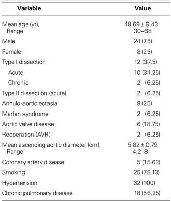
TABLE I. Baseline Characteristics of the 32 Patients
Variable Value
Mean age (yr), 48.69 ± 9.43
Range 30–68 Male 24 (75) Female 8 (25) Type I dissection 12 (37.5) Acute 10 (31.25) Chronic 2 (6.25)
Type II dissection (acute) 2 (6.25)
Annulo-aortic ectasia 8 (25)
Marfan syndrome 2 (6.25)
Aortic valve disease 6 (18.75)
Reoperation (AVR) 2 (6.25)
Mean ascending aortic diameter (cm), 5.82 ± 0.79
Range 4.2–8
Coronary artery disease 5 (15.63)
Smoking 25 (78.13)
Hypertension 32 (100)
Chronic pulmonary disease 18 (56.25)
AVR = aortic valve replacement
Data are presented as number (percentage) or as mean ± SD (range).
performed in 10 of these patients, a valve-sparing graft interposition in 4, and a supracoronary graft interposi-tion in the other 4. Coronary artery bypass grafting was necessary in 5 of these patients. Circulatory arrest was established at the end of the proximal repair, as de-scribed above. The distal anastomosis was at the level of the ostium of the BC artery, excluding the clamped aortic tissue in 14 patients. Hemiarch replacement was performed in 3 patients and total arch replacement in one.
In all patients, cardiac arrest was established via retro-grade and coronary ostial anteretro-grade administration of blood cardioplegic solution. When all repair was done and CPB was terminated, the graft on the BC artery was cut close to the anastomosis, and the stump was closed with a continuous suture. Operative techniques are summarized in Table II.
Results
Postoperatively, none of the 32 patients had a major neurologic deficit. Five displayed early postoperative
agitation, which resolved within the first 24 hours. No sequela related to BC artery cannulation—such as bleeding or brachial ischemia—was observed. There were 2 in-hospital deaths (6.25%). One was that of a 68-year-old man who had severe aortic stenosis and aneurysm of the ascending aorta, left ventricular hyper-trophy, coronary artery disease, and chronic obstructive pulmonary disease. He died on the 11th postoperative day because of low cardiac output, prolonged ventila-tory support, and sepsis. The other patient, who had an acute type I aortic dissection, died of postoperative renal failure and hemodialysis sequelae on the 3rd post-operative day.
The mean follow-up period was 9.43 ± 12.46 months (range, 1–44 mo). All clinical results are summarized in Table III.
Discussion
When surgical repair of the proximal aorta under cir-culatory arrest is considered, there are several choices of access for arterial cannulation. Femoral arteries have for years been the most widely used sites, but the retrograde flow within the aorta carries risks of malperfusion and atheroembolization. Therefore, other access sites are sought. The brachial artery,4-6 the axillary artery,5 and
the BC artery3,7-11 have been used for this purpose. All
3 of these sites have the advantage of providing a more central and physiologic flow, and they enable antegrade cerebral perfusion during circulatory arrest.
Cerebral protection is achieved through the combina-tion of such techniques as hypothermia, cerebral per-fusion, external cranial cooling, and medical ischemic cerebral preconditioning. Hypothermia is the principal and most effective of these for use during CA.12 Several
techniques have been described for cerebral perfusion and in affirmation of its effectiveness. Most inves-tigators have agreed upon the superiority of antegrade
Fig. 1 Anastomosis of the vascular graft to the brachiocephalic
(BC) artery for cannulation. The BC artery is isolated and taped, and an 8-mm Dacron graft is anastomosed in an end-to-side fashion. The aortic cannula is then inserted into the graft and fixed at several points. Through this graft, the surgeon can easily initiate cardiopulmonary bypass and provide antegrade cerebral perfusion during circulatory arrest merely by clamping the proxi-mal BC and left carotid arteries.
TABLE II. Operative Techniques in the 32 Patients
Variable No.
Distal anastomosis
Ascending aorta 26
Hemiarch replacement 5
Total arch replacement 1
Proximal anastomosis
Supracoronary anastomosis 15
Bentall procedure 12
Valve-sparing graft implantation 5
over retrograde cerebral perfusion, in combination with hypothermia. Various instruments can measure the effectiveness of such perfusion, but the simplest method is to confirm a good connection between the 2 sides of the cerebral circulation. After the onset of CA, brief release of the clamp on the left carotid artery enables retrograde flow to be checked and its rate evalu-ated visually. In all our patients, the left carotid blood flow was judged to be sufficient.
When femoral artery cannulation is used, additional cannulas and routes become necessary for ACP. In treat-ing patients with aortic dissection, another disadvantage of femoral cannulation is the need to change arterial flow from the femoral artery to the aortic graft after the distal aortic anastomosis has been completed.
In consideration of these facts, we advocate proximal arterial cannulation for proximal aortic surgery when circulatory arrest is necessary. Brachiocephalic artery cannulation, as one of the choices, does not necessitate an additional incision, does provide more central flow, and can be applied to most patients. This technique, in our judgment, is easier and more practical than the alternative of axillary or brachial artery access.
In regard to the degree of hypothermia during op-eration, we prefer a body temperature of 24 °C for the period of circulatory arrest. The wider use of cerebral perfusion techniques during aortic arch surgery has en-couraged a trend toward the routine use of moderate-to-mild hypothermia (28–35 °C). Even so, some major
centers favor the performance of aortic surgery with deep hypothermic CA, even in the absence of additional per-fusion.13 In 2007, Kamiya and colleagues14 reported on
possible neurologic sequelae to prolonged lower-body CA (>60 min) in the presence of moderate hypother-mia (28 °C). In a subgroup analysis, they observed a 6-fold increase in death and ischemic spinal cord injury. Supported by these and other15 comparisons of recent
approaches to aortic arch surgery, we prefer to stay on the safer side, with hypothermia at less than 25 °C. Banbury and Cosgrove7 were the first to report BC
artery cannulation via a graft. In their paper, published in 2000, they claimed 4 major advantages for this tech-nique: 1) it eliminates the need for a 2nd incision, 2) it provides a higher flow rate without the need for higher pressure, because the BC artery is larger than the axil-lary artery, 3) it enables blood pressure monitoring via the right radial artery during ACP, and 4) it avoids the brachial plexus injuries associated with axillary artery cannulation.
When a graft anastomosis has to be performed for axillary cannulation, the anastomosis can bleed and become a major site of blood loss. On the other hand, any blood loss from the graft or the anastomosis at the BC artery is within the same operative field and can be returned to the reservoir via a simple vent catheter. Di Eusanio and colleagues8 have reported, in their series,
similar advantages of the technique. Direct cannulation of the BC artery with the routinely used arterial cannu-las has also been suggested,9 but that technique carries
a higher risk of sequelae.3 Rerouting of the cannula tip
toward the aortic arch during warming and cooling, and toward the brain during CA, requires extra manipu-lation. Closure of the arteriotomy after decannulation also carries the risk of creating a stenosis.3
In a report from the Texas Heart Institute,11 68
pa-tients were cannulated via the BC artery, all through a side graft. Coselli and colleagues had outstanding re-sults: only one 30-day death (1.5%), 3 strokes (4.4%), and 7 patients with temporary postoperative confu-sion (10.3%). At variance from our practice, they per-fused both cerebral hemispheres in 63 of the 68 patients (having added another perfusion catheter through the left common carotid artery).11
Brachiocephalic artery cannulation can of course have disadvantages in specific situations. When the BC artery is involved in the lesion, as in the case of a dissection or atherosclerotic plaques, it is not suitable for cannulation. In fact, in one of our patients, the BC artery was involved in the dissection, rendering can-nulation impossible at that site. Graft anastomosis for cannulation adds approximately 15 to 20 minutes to the total operative time. Still, in comparison with axillary cannulation from a 2nd incision and with additional grafting, this time loss might be no worse than the alter-native loss. Last, when the diameter of the BC artery is
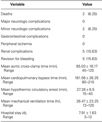
TABLE III. Results of Surgery in the 32 Patients
Variable Value
Deaths 2 (6.25)
Major neurologic complications 0
Minor neurologic complications 2 (6.25)
Gastrointestinal complications 0
Peripheral ischemia 0
Renal complications 5 (15.63)
Revision for bleeding 5 (15.63)
Mean aortic cross-clamp time (min), 85.03 ± 16.17
Range 45–125
Mean cardiopulmonary bypass time (min), 161.56 ± 26.25
Range 80–210
Mean hypothermic circulatory arrest (min), 27.28 ± 6.5
Range 15–40
Mean mechanical ventilation time (hr), 26.47 ± 23.25
Range 12–120
Hospital stay (d), 7.91 ± 1.63
Range 3–12
Data are presented as number (percentage) or as mean ± SD (range).
less than 9 mm, the placement of a side clamp for graft anastomosis could further attenuate distal flow.3 Limitations
These operations were performed in 4 different medical centers by a single surgeon (MU) within the specified time period. Because of the lack of a suitable system for collecting follow-up data and the failure of patients to adhere to follow-up protocols, the long-term results cannot be evaluated.
This study also has the classical limitations of a ret-rospective design. There was no opportunity to com-pare these patients with a similar group of patients who underwent surgery with other arterial cannulation techniques, because the authors used only this method within the specified time period. The follow-up period was short because of very low patient compliance with follow-up protocols. Still, we believe that the early post-operative period provided good insight into the effec-tiveness of this operative technique.
Conclusion
In proximal aortic surgery that necessitates CA, arterial cannulation via a graft anastomosed to the BC artery is safe and effective. It can be applied to a wide variety of patients with ease. It eliminates the need for a 2nd inci-sion, permits ACP, and enables monitoring of right ra-dial arterial pressure during ACP. The larger size of the BC artery enables higher flows at lower blood pressures. The risk of brachial plexus injury that is associated with axillary arterial cannulation is also eliminated.
References
1. Strauch JT, Spielvogel D, Lauten A, Galla JD, Lansman SL, McMurtry K, Griepp RB. Technical advances in total aortic arch replacement. Ann Thorac Surg 2004;77(2):581-90. 2. Kazui T, Washiyama N, Muhammad BA, Terada H,
Yama-shita K, Takinami M, Tamiya Y. Total arch replacement using aortic arch branched grafts with the aid of antegrade selective cerebral perfusion. Ann Thorac Surg 2000;70(1):3-9. 3. Huang FJ, Wu Q, Ren CW, Lai YQ, Yang S, Rui QJ, Xu SD.
Cannulation of the innominate artery with a side graft in arch surgery. Ann Thorac Surg 2010;89(3):800-3.
4. Ozatik MA, Mungan U. Aortic surgery with brachial artery cannulation [in Turkish]. Turk J Thorac Cardiovasc Surg 2011;19 Suppl 2:8-14.
5. Sanioglu S, Sokullu O, Yapici F, Yilmaz M, Arslan IY, Has-taoglu IO, et al. Axillary artery cannulation in surgery of the ascending aorta and the aortic arch [in Turkish]. Turk J Thorac Cardiovasc Surg 2007;15(3):197-201.
6. Tiwari KK, Murzi M, Bevilacqua S, Glauber M. Which can-nulation (ascending aortic cancan-nulation or peripheral arterial cannulation) is better for acute type A aortic dissection sur-gery? Interact Cardiovasc Thorac Surg 2010;10(5):797-802. 7. Banbury MK, Cosgrove DM 3rd. Arterial cannulation of the
innominate artery. Ann Thorac Surg 2000;69(3):957. 8. Di Eusanio M, Quarti A, Pierri MD, Di Eusanio G.
Can-nulation of the brachiocephalic trunk during surgery of the thoracic aorta: a simplified technique for antegrade cerebral
perfusion. Eur J Cardiothorac Surg 2004;26(4):831-3. 9. Ji S, Yang J, Ye X, Wang X. Brain protection by using
innomi-nate artery cannulation during aortic arch surgery. Ann Tho-rac Surg 2008;86(3):1030-2.
10. Stassano P, Musumeci A, Iannelli G, D’Alise G, Mottola M. A new cannula for innominate artery cannulation. J Thorac Cardiovasc Surg 2005;130(3):944-5.
11. Preventza O, Bakaeen FG, Stephens EH, Trocciola SM, de la Cruz KI, Coselli JS. Innominate artery cannulation: an alter-native to femoral or axillary cannulation for arterial inflow in proximal aortic surgery. J Thorac Cardiovasc Surg 2013;145(3 Suppl):S191-6.
12. Ergin MA. Technical innovations to facilitate cerebral pro-tection during operations on the aortic arch: a historical per-spective and current application [in Turkish]. Turk J Thorac Cardiovasc Surg 2011;19 Suppl 2:1-7.
13. Dumfarth J, Ziganshin BA, Tranquilli M, Elefteriades JA. Cerebral protection in aortic arch surgery: hypothermia alone suffices. Tex Heart Inst J 2013;40(5):564-5.
14. Kamiya H, Hagl C, Kropivnitskaya I, Bothig D, Kallenbach K, Khaladj N, et al. The safety of moderate hypothermic lower body circulatory arrest with selective cerebral perfusion: a propensity score analysis. J Thorac Cardiovasc Surg 2007; 133(2):501-9.
15. Misfeld M, Mohr FW, Etz CD. Best strategy for cerebral pro-tection in arch surgery - antegrade selective cerebral perfusion and adequate hypothermia. Ann Cardiothorac Surg 2013;2 (3):331-8.