Full Terms & Conditions of access and use can be found at
https://www.tandfonline.com/action/journalInformation?journalCode=wjtd20
Journal of Trauma & Dissociation
ISSN: 1529-9732 (Print) 1529-9740 (Online) Journal homepage: https://www.tandfonline.com/loi/wjtd20
Lateralization of Neurobiological Response in
Adolescents with Post-Traumatic Stress Disorder
Related to Severe Childhood Sexual Abuse: the
Tri-Modal Reaction (T-MR) Model of Protection
Tuba Mutluer, Vedat Şar, Çiğdem Kose-Demiray, Harun Arslan, Sibel Tamer, Serap Inal & Anıl Ş. Kaçar
To cite this article: Tuba Mutluer, Vedat Şar, Çiğdem Kose-Demiray, Harun Arslan, Sibel Tamer, Serap Inal & Anıl Ş. Kaçar (2018) Lateralization of Neurobiological Response in Adolescents with Post-Traumatic Stress Disorder Related to Severe Childhood Sexual Abuse: the Tri-Modal Reaction (T-MR) Model of Protection, Journal of Trauma & Dissociation, 19:1, 108-125, DOI: 10.1080/15299732.2017.1304489
To link to this article: https://doi.org/10.1080/15299732.2017.1304489
Accepted author version posted online: 10 Mar 2017.
Published online: 12 Apr 2017. Submit your article to this journal
Article views: 428
View Crossmark data
ARTICLE
Lateralization of Neurobiological Response in Adolescents
with Post-Traumatic Stress Disorder Related to Severe
Childhood Sexual Abuse: the Tri-Modal Reaction (T-MR)
Model of Protection
Tuba Mutluer, MDa, VedatŞar, MDb, Çiğdem Kose-Demiray, PhDc, Harun Arslan, MDd, Sibel Tamer, MDe, Serap Inalf, and Anıl Ş. Kaçarg
aDepartment of Child and Adolescent Psychiatry, Koc University Hospital, Istanbul, Turkey;
bDepartment of Psychiatry, Koc University School of Medicine (KUSOM)Istanbul, Turkey;cDepartment
of Psychology, Istanbul Arel University, Istanbul, Turkey;dDepartment of Radiology, Van Yuzuncu Yıl
University School of Medicine, Van, Turkey;eDepartment of Neurology, Sincan Koru Hospital, Ankara,
Turkey;fPsychology Unit, Van Regional Research and Education Hospital, Van, Turkey;gDepartment of
Psychiatry, Koc University School of Medicine (KUSOM), Istanbul, Turkey
ABSTRACT
This study inquires into neurobiological response to stress and its clinical correlates among adolescents with post-traumatic stress disorder (PTSD). Structural magnetic resonance imaging (MRI) measures of cerebral anatomy were carried out on 23 female adolescents with PTSD related to severe childhood sexual abuse and 21 matched healthy controls. Clinician Administered PTSD Scale for Children and Adolescents, Adolescent Dissociative Experiences Scale, Childhood Trauma Questionnaire, Schedule for Affective Disorders and Schizophrenia for School Age Children, Beck Depression Scale, and a set of neuro-cognitive tests were administered to all participants. Compared to controls, PTSD group bilaterally had smaller amygdala, hippocampus, ante-rior cingulate, and thinner prefrontal cortex but normal thalamus. Further analyses within the PTSD group suggested an association between symptoms of PTSD and sizes of right brain structures including smaller amygdala but larger hippocampus and anterior cingulate. Thinner right prefrontal cortex and larger right thala-mus seemed to be related to denial and response prevention, respectively. Being related to both hemispheres, dissociative amnesia was negatively associated with proportion of the right amygdala to right thalamus and to both left and right prefrontal cortex. Suggesting a neuro-protective effect against traumatic stress at least through adolescence, depersonalization –derealiza-tion and identity altera–derealiza-tion were correlated with thicker left pre-frontal cortex. Unlike the lateralization within PTSD group, correlations between regions of interest were rather symmetrical in controls. The graded response to stress seemed to be aimed at mental protection by lateralization of brain functions and possibly diminished connection between two hemispheres. A Tri-Modal Reaction (T-MR) Model of protection is proposed.
ARTICLE HISTORY Received 3 August 2016 Accepted 12 February 2017 KEYWORDS Complex PTSD; dissociation; MRI; neurobiology; PTSD; sexual abuse
CONTACTTuba Mutluer, MD [email protected] Department of Child and Adolescent Psychiatry, Koc University Hospital, Davutpaşa St. 4, Topkapi 34010, Istanbul, Turkey.
The study was conducted in Regional Research and Education Hospital, Van, Turkey 2018, VOL. 19, NO. 1, 108–125
https://doi.org/10.1080/15299732.2017.1304489
Chronic traumatization in childhood, at critical times of brain development, can cause neurodevelopmental deficits that may lead to harm in emotional, cognitive, and behavioral spheres (Solomon & Heide,2005). In chronic stress, activation of the hypothalamic–pituitary–adrenal axis results in increased cortisol level leading to volume reductions in hippocampus, amygdala, and anterior cingulate (Bremner, 2006). Bilateral reduction in frontal cortex gray matter volume was also associated with childhood sexual abuse (SA) (Sheffield, Williams, Woodward, & Heckers,2013). Despite consensus about particular neurobiological sequelae of chronic traumatization, inconsistencies in findings continue to feed ambiguities. For example, two studies on women with a history of childhood SA reported no significant differences in amygdala volumes (Andersen et al.,2008; Bremner et al.,
1997). Better linking of neurobiological data to clinical observations would assist researchers in navigating through scientific endeavors.
Not only the childhood adversity itself, but the subsequent psychopathology, preexisting features, and the phase of the trauma-generated response may influ-ence the findings. Andersen et al. (2008) proposed that hippocampus was affected only if the trauma was lived between 3 and 5 or 11 and 13 years of age, corpus callosum 9–10, and frontal cortex 14–16 years. In a meta-analysis, hippocampus was bilaterally smaller in adults with childhood maltreatment-related PTSD compared to healthy controls, but not in children with maltreatment-related PTSD, suggesting that these deficits may not be apparent until adulthood (Woon & Hedges, 2008). Indeed, in a study on children with SA-related PTSD (De Bellis, Hall, Boring, Frustaci, & Moritz, 2001), there was no evidence of a smaller hippocampus.
Compared to adults, childhood trauma reports of adolescents address a relatively near period of life and the neurobiological response to stress is in its earlier yet expanding phase. The present study inquired a group of adolescents who had PTSD related to severe SA. We investigated the volumes of selected brain regions in comparison with those of the healthy controls. Additionally, clinical data were gathered from, and neuropsychological tests were adminis-tered to all participants. Beside a comparison between traumatized and control groups, analyses were conducted within the PTSD group to inquire into possible components of trauma-generated response. Finally, we tried to integrate the neurobiological data with clinical features and neuropsychological test results in context of a proposed model.
Method
Participants
The participants were recruited from [redacted for peer review], located in a non-industrialized region of eastern Turkey. All participants were Turkish citizen of Kurdish ethnicity and belonged to low economic status. This state
institution was hosting adolescents who were under governmental custody to stop ongoing SA which was proven by a legal verdict. The study was con-ducted between January 2013 and June 2014. The participants and their legal guardians have given written informed consent. The study was approved by the Ethics Committee of the [redacted for peer review] Hospital.
Psychiatric comorbidity except depressive and dissociative disorders was considered as a reason of exclusion. Two adolescents with a clinical diagnosis of PTSD declined to participate. One adolescent was excluded due to insuffi-cient education. As the final study group, twenty-three female adolescents with PTSD related to SA and 21 healthy female controls matched on age (range = 13–18), education, and family income were assessed. None of the participants was receiving any psycho-pharmacological treatment. Control group was composed of typically developing children without a psychiatric history recruited randomly from a state vocational school in the region. All participants were right handed.
Assessment instruments
All administered instruments were validated in Turkish (Gökler et al., 2004; Hisli, 1989; Karakaya et al., 2007; Şar, Öztürk, & İkikardeş, 2012; Zoroglu, Sar, Tuzun, Tutkun, & Savas, 2002).
Clinician Administered PTSD Scale for Children and Adolescents (DSM-IV): CAPS-CA is a 33-item clinician-administered diagnostic interview (Blake et al.,
1995) for 8–18 years of age.
Adolescent Dissociative Experiences Scale: ADES is a 30-item self-report instrument to assess the severity of dissociative experiences in adolescents (Armstrong, Putnam, Carlson, Libero, & Smith, 1997).
Schedule for Affective Disorders and Schizophrenia for School Age Children-Present and Lifetime Version (DSM-IV): KSADS-PL is a semi-struc-tured interview assessing 32 psychiatric disorders (Kaufman et al., 1997).
Beck Depression Inventory: BDI is a 21-item self-report instrument (Beck, Ward, & Mendelson, 1961). A denial score was also derived from BDI by counting the number of “0” responses.
Childhood Trauma Questionnaire: CTQ is 28-item self-report inventory measuring severity of five types of childhood trauma alongside possible minimization of them (Bernstein et al., 2003).
Childhood Abuse and Neglect Questionnaire: The 11-item CANQ screens childhood traumata including their severity, age of occurrence, and the perpetrator(s).
Neuropsychological Tests: Tower of London (executive planning), Clock Drawing (visuospatial and constructional abilities), Judgement of Line Orientation (visuospatial skills), Stroop TBAG (focused attention, ability of altering the perceptual status when exposed to changing demands, sensitive to
left frontal lobe—in particular the orbitofrontal region-), and Serial Digit Learning (attention /concentration, general memory, information processing).
Procedure
Clinical instruments were administered by a female child and adolescent psy-chiatrist (T.M.), and neuro-cognitive tests were conducted by a female psychol-ogist. All participants underwent an MRI scan at the Van-City, State General Hospital. Images were acquired with a GE Sigma 1.5-T system (GE Medical Systems, Milwaukee). The whole brain was scanned with a 3D inversion recov-ery prepared fast spoiled GRASS (SPGR) T1-weighted sequence. Images were acquired in the coronal, axial, and sagittal plane with 1.5 mm contiguous sections; TR 13.8 ms; TI 450 ms; TE 2.8 ms; flip angle 20°; one data average; and 256 × 256 × 128 pixel matrix. MRI images were transferred to a workstation and displayed using the DISPIM image display software. The images and volumes of the subcortical structures were outlined using a mouse-driven cursor. Thickness of the prefrontal (i.e., covering superior and middle frontal areas) cortical gray matter was indicated using a fixed color scale and recon-structed into a color-coded, three-dimensional surface model (for further details see Schreyer et al.,2000; Wiegand et al.,2004). All images were coded and rated twice by an experienced radiologist blind to the group affiliations. Intra-rater correlation coefficients (ICC) were between 0.91 and 0.97 for all regions of interest. Spearman method was utilized in calculation of all correlations.
Results
Characteristics of the participants
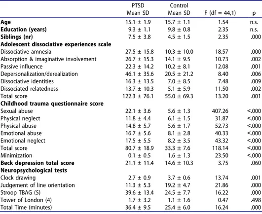
All adolescents with PTSD reported SA. In 14 of them (68.0%), it included coitus. Mean age during the first SA was 12.4 (SD = 0.6, range 6–16). For one participant (4.5%) abuse occurred once, for seven (31.8%) a few times, for five (22.7%) several times, and for nine participants (49.9%) regularly. In 38.0% (n = 9) of them, the perpetrator was either biological father or brother; in 62.0%, this person was the stepfather, stepbrother, or a relative, acquain-tance, or “husband” in an underage “marriage” enforced by parents. Five victims got pregnant due to the SA. Two of them gave birth to their children (one conceived from biological father), and the pregnancy was ended by curettage for the remaining three girls. Of the victims, 90.0% reported at least one suicide attempt, and 19.0% twice or more. Self-mutilation was reported by 62.0% (n = 13) of the group. In addition to SA, 66.7% (n = 14) of the participants reported physical abuse, 76.2% (n = 16) emotional abuse, and 61.0% (n = 13) emotional and/or physical neglect. Dissociation but not depression scores differed PTSD from control group (Table 1).
Structural brain imaging Bilateral findings
Compared to controls, PTSD group had bilaterally smaller hippocampus, amygdala, anterior cingulate cortex, and thinner prefrontal cortex with no difference on thalamus (Table 2). Younger age during the earliest reported SA was correlated with larger right (rho = 0.43 n = 22 p = 0.048) and left (rho = 0.45 n = 22 p = 0.035) anterior cingulate. There was no correlation between any brain region and specific trauma type (CTQ sub-scores not reported) or dissociative amnesia (DA) in PTSD group (Table 3). However, DA was negatively correlated with proportion of right amygdala to right thalamus (rho = −0.44, n = 22, p = 0.041) and to both right and left prefrontal cortex (right rho = −0.47, n = 18, p = 0.048; left rho = −0.54, n = 19, p = 0.017).
Unilateral findings
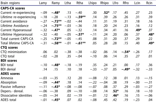
Vast majority of correlations (Table 3 and Figure 1) within the PTSD group pointed to lateralization of the neurobiological response. Smaller right amygdala was associated with current and lifetime avoidance, current hyperarousal and re-experiencing, depression, passive influence experiences, and absorption. Unlike
Table 1.Demographic and clinical characteristics of the participants (ANOVA).
PTSD Mean SD Control Mean SD F (df = 44,1) p Age 15.1 ± 1.9 15.7 ± 1.1 1.54 n.s. Education (years) 9.3 ± 1.1 9.8 ± 0.8 2.35 n.s. Siblings (nr) 7.5 ± 3.8 4.5 ± 1.5 2.35 .000 Adolescent dissociative experiences scale
Dissociative amnesia 27.5 ± 15.8 10.3 ± 10.0 18.57 .000 Absorption & imaginative involvement 26.7 ± 15.3 14.1 ± 9.5 10.73 .002 Passive influence 22.3 ± 14.2 10.2 ± 8.1 12.08 .001 Depersonalization/derealization 46.1 ± 35.6 20.5 ± 21.2 8.40 .006 Dissociative identities 16.3 ± 13.5 7.0 ± 8.5 7.48 .009 Dissociated relatedness 13.7 ± 10.3 5.1 ± 5.9 11.50 .002 Total score 122.3 ± 76.1 55.0 ± 69.3 13.20 .001 Childhood trauma questionnaire score
Sexual abuse 22.1 ± 3.6 5.6 ± 1.3 407.26 ˂.000 Physical neglect 11.8 ± 4.4 6.1 ± 1.5 31.87 ˂.000 Physical abuse 14.8 ± 5.7 5.6 ± 1.7 52.73 ˂.000 Emotional abuse 16.7 ± 5.6 8.1 ± 2.8 40.33 ˂.000 Emotional neglect 17.5 ± 5.5 8.2 ± 3.5 43.32 ˂.000 Total score 80.7 ± 18.9 33.3 ± 7.6 118.14 ˂.000 Minimization 0.1 ± 0.5 1.6 ± 1.3 23.50 ˂.000 Beck depression total score 21.1 ± 11.4 14.6 ± 10.3 3.75 .060 Neuropsychological tests
Clock drawing 2.7 ± 0.9 3.7 ± 0.6 13.74 .001 Judgement of line orientation 11.3 ± 5.3 19.2 ± 4.7 21.86 .000 Stroop TBAG (5) 39.6 ± 13.4 24.5 ± 7.7 16.22 .000 Tower of London (4) 1.7 ± 3.2 1.1 ± 1.6 0.47 .498 Total Time (minutes) 36.4 ± 9.5 25.4 ± 6.0 16.24 .000
in PTSD (Table 4), size of the right amygdala was not related to any measure among controls (Table 5). In PTSD, current re-experiencing was associated with larger right hippocampus and right anterior cingulate. Also correlated with life-time hyperarousal, right anterior cingulate was larger (M = 2.83 SD = 0.42) in coitus group (i.e., more severe abusive act) compared to those with other types (M = 2.34 SD = 0.47) of SA (Mann–Whitney U-test, z = 2.14 p = 0.032).
Thinner right prefontal cortex was associated with larger right amygdala and CTQ minimization of trauma and BDI denial scores. Additionally, right
Table 2.Comparison between PTSD and control groups: Brain regions (ANOVA).
Brain regions PTSD (n = 23) Mean SD Control (n = 21) Mean SD F df = (42,1) p Right amygdala 1.14 0,07 1.22 0,07 15.48 .000 Left amygdala 1.14 0,05 1.22 0.06 20.95 .000 Right thalamus 6.31 0.44 6.36 0.42 0.19 .669 Left thalamus 6.46 0.50 6.49 0.55 0.04 .834 Right hippocampus 2.64 0.23 2.99 0.23 26.05 .000 Left hippocampus 2.60 0.25 3.01 0.25 28.11 .000 Right anterior Cingulate 2.69 0.48 3.04 0.22 9.84 .003 Left Anterior cingulate 2.60 0.50 3.00 0.16 12.38 .001 Left prefrontal cortex 2.80 0.41 3.06 0.24 5.37 .026 Right prefrontal cortex 2.83 0.38 3.10 0.31 5.64 .023
Table 3.Correlational analysis (Spearman) in the PTSD group: Volumetric measures of brain regions and clinical scale scores.
Brain regions Lamy Ramy Ltha Rtha Lhipo Rhipo Lfro Rfro Lcin Rcin CAPS-CA scores Current re-experiencing −.09 −.48* .13 −.40 .30 .52* .17 .45 .27 .23 Lifetime re-experiencing −.18 −.28 −.13 −.59** .14 .39 .26 .26 .31 .39 Current avoidance −.27 −.72** −.02 −.44 .11 .31 .19 .31 .18 .16 Lifetime Avoidance −.24 −.75** −.03 −.36 .18 .20 .24 .23 .16 .12 Current Hyperarousal −.32 −.47* .05 −.32 .14 .34 .41 .16 .49* .37 Lifetime Hyperarousal −.32 −.40 −.05 −.57* −.11 .24 .20 .06 .37 .48* Total current CAPS-CA −.25 −.72** −.05 −.45 .08 .31 .20 .37 .29 .22 Total lifetime CAPS-CA −.31 −.58** −.01 −.61** .05 .28 .28 .15 .40 .49* CTQ scores CTQ minimization .06 .02 −.38 −.38 −.02 .06 .14 −.54* −.26 .17 CTQ total −.02 −.28 .35 −.04 −.10 .06 .14 .35 .27 .01 BDI scores BDI total −.10 −.48* −.18 −.19 .35 .24 .21 .00 .17 .36 BDI denial .14 .15 .10 .18 −.33 −.37 .05 −.49* −.32 −.33 ADES scores Amnesia —.03 —.35 .12 .20 —.08 .12 .38 .01 .13 —.15 Absorption —.09 −.44* .18 .14 —.22 —.04 .38 .19 −.00 —.31 Passive influence —.11 −.43* −.08 —.08 —.07 .08 .37 .29 −.03 —.27 Depers.- dereal. —.06 —.38 .09 —.10 −.08 .14 .52* .16 .18 —.10 Dissociative identities —.05 —.34 .02 —.19 —.01 .12 .46* .16 .15 —.06 ADES total −.01 −.45* .07 .02 −.08 .45 .42 .19 −.23 .04 R: right, L: left, Amy: amygdala, Tha: thalamus, Hip: hippocampus, Fro: prefrontal, Cin: cingulate, ADES:
adolescent dissociative experiences scale, CAPS-CA: clinician administered PTSD scale for children and adolescents, CTQ: childhood trauma questionnaire, BDI: beck depression inventory, Depers: depersonaliza-tion, Dereal: derealizadepersonaliza-tion, *p < 0.05, **p < 0.001.
thalamus was negatively correlated with lifetime hyperarousal and re-experi-encing. As the only region of the left hemisphere involved, left anterior cingulate (in correlation with right anterior cingulate) was associated with current hyperarousal. Participants sexually abused by their biological father or brother (i.e., perpetrator in “closer” relationship with the victim) had smaller left anterior cingulate (M = 2.24 SD = 0.34) compared to that of the victims of (M = 2.81 SD = 0.48) other perpetrators (Mann–Whitney U-test, z = 2.42 p = 0.016) and reported more DA (M = 36.2 SD = 14.61
RIGHT LEFT
Dissociative Amnesia
Primary Reaction
“Inflammation” “The Trauma Ilness” “
Secondary Reaction Tertiary Reaction Alienation” Lifetime and current avoidance Anterior Cingulate Hippocampus Amygdala Hippocampus Lifetime hyper-arousal Lifetime re-experiencing Depression Passive influence and Absorption Dissociative identities (switching) Prefrontal Cortex Current re-experiencing Prefrontal Cortex Thalamus Thalamus Depersonalization Derealization Current hyperarousal Denial Anterior Cingulate Amygdala
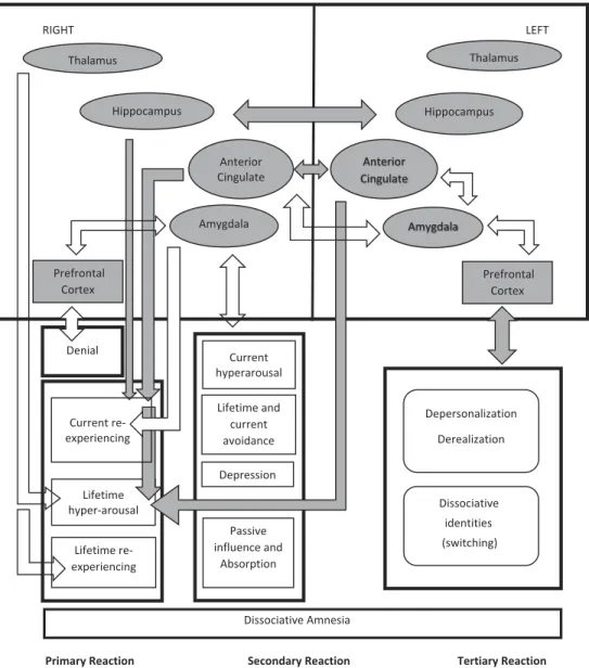
Figure 1.Three modes of trauma generated response (gray arrows demonstrate positive and empty arrows demonstrate negative significant correlations; each arrow ending outside of a quadrangle refers to correlations with all items inside of the quadrangle while those ending inside refers to those with the pointed item only).
versus M = 21.4 SD = 13.99) (z = 2.21 p = 0.027) and absorption (M = 35.89 SD = 11.57 versus M = 20.31 SD = 14.61) (z = 2.11 p = 0.035).
In contrast to the general downsizing of the brain structures in PTSD group, thickness of the left prefrontal cortex was positively correlated with depersonalization, derealization, and identity alteration (ADES) scores. Right anterior cingulate was correlated with smaller left amygdala which was negatively correlated with left prefrontal cortex.
Altered connectivity
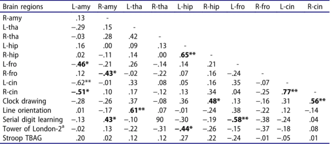
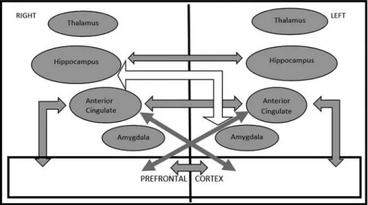
Correlations in controls are shown in Table 5. Like in PTSD, hippocampus and anterior cingulate but not amygdala and thalamus of the two hemi-spheres were significantly correlated with each other. However, in contrast to PTSD, there were significant correlations between left and right prefrontal cortex which were correlated with anterior cingulate. These correlations demonstrated better symmetry and suggested better connectedness between
Table 4.Correlational analyses (Spearman) in PTSD group: brain regions and neuropsychological test scores.
Brain regions L-amy R-amy L-tha R-tha L-hip R-hip L-fro R-fro L-cin R-cin
R-amy .13 -L-tha −.29 .15 -R-tha −.03 .28 .42 -L-hip .16 .00 .09 .13 -R-hip .02 −.11 .14 .00 .65** -L-fro −.46* −.21 .26 −.14 .14 .21 -R-fro .12 −.43* −.02 −.22 .07 .16 −.24 -L-cin −.62** −.01 .33 .08 .05 .16 .35 −.07 -R-cin −.51* .10 .17 −.12 .13 .34 .04 −.25 .77** -Clock drawing −.28 −.26 .37 −.08 .36 .48* .13 −.16 .31 .56** Line orientation .01 −.17 .61** .07 −.01 −.24 .38 −.22 .12 −.14 Serial digit learning −.13 .43* −.10 90 −.30 −.19 −.58** −.38 −.24 .04 Tower of London-2a −.02 .13 −.22 −.31 −.44* −.26 −.15 −.37 −.18 .08 Stroop TBAG .20 .02 .12 .12 .27 .22 −.24 −.01 −.05 .01 R: right, L: left, Amy: amygdala, Tha: thalamus, Hip: hippocampus, Fro: prefrontal, Cin: cingulate,aTower of
London subtest 2, *p < 0.05, **p < 0.001.
Table 5.Correlational analyses (Spearman) in control group: brain regions.
Brain regions Lamy Ramy Ltha Rtha Lhipo Rhipo Lfront Rfront Lcin
Lamy -Ramy .30 -Ltha .37 −.02 -Rtha .12 .13 .41 -Lhipo −.04 .20 −.11 .06 -Rhipo –.49* −.02 −.18 −.17 .70** -Lfront .40 .19 .07 −.02 .35 .21 -Rfront .32 .28 .32 .29 .03 −.06 .69** -Lcin .10 .04 −.05 −.08 .32 .33 .73** .53* -Rcin .33 .17 .07 .04 .33 .22 .71** .69** .73** R: right, L: left, Amy: amygdala, Tha: thalamus, Hip: hippocampus, Fro: prefrontal, Cin: cingulate.
two hemispheres in controls compared to the PTSD group (Figure 2). Unlike in PTSD, right hippocampus was negatively correlated with left amygdala.
Neuropsychological tests
All neuro-cognitive tests were impaired in PTSD except the Tower of London (Table 1). Representing a general impact, impairment of the Stroop TBAG was not in a relationship with any brain region (Table 4). Better performance in Clock Drawing and Serial Digit Learning was corre-lated with two right brain structures: hippocampus and anterior cingulate; that is, regions involved with current re-experiencing and lifetime hyperar-ousal, respectively. Smaller right amygdala and thicker left prefrontal cortex were also associated with impairment in Serial Digit Learning. Judgment of Line Orientation was not associated with right hemisphere structures. It was positively influenced by left thalamus (a structure not affected by PTSD). Smaller left hippocampus was associated with impairment of Tower of London-2.
The proposed model: Tri-modal reaction of protection
Unlike the symmetrical associations between brain structures in controls, analyses conducted within the PTSD group yielded distinct patterns in two hemispheres. In tandem with clinical phenomena and neuro-cognitive test
Figure 2.Statistically significant correlations between regions of interest volumes in the control group (gray and black arrows demonstrate positive and empty arrows demonstrate negative correlation).
results, neurobiological response to developmental trauma seemed to cover three patterns each characterized by an interaction between trauma-related intrusions and operations of controlling the subsequent psychological pain to maintain a “window of tolerance” (Siegel,1999).
The primary mode (“Inflammation”) covered current and lifetime re-experiencing and lifetime hyperarousal. Neurobiologically, this pattern was predominantly driven by right hemisphere structures; that is, smaller amygdala and larger hippocampus and anterior cingulate. Possibly join-ing larger right hippocampus and anterior cjoin-ingulate in “remembering” traumatic memories, smaller right amygdala seemed to be the main driver of this mode. In contrast of this (representing operations of control), denial of trauma was associated with a thinner prefrontal cortex and a larger right thalamus which may dampen the perception of the psychological pain.
The most consistently implicated structure of the secondary mode (“Trauma Illness”) was smaller right amygdala which was correlated with symptoms of lifetime or current, and in fact, Complex PTSD alongside “dissociative depression” (Sar, Akyüz, Öztürk, & Alioğlu, 2013) and passive influence experiences. Smaller right amygdala seemed to represent the chronic and complex quality of this polisymptomatic phase. Participants sexually abused by their biological father or brother reported more DA and absorption compared to that of the victims of other perpetrators alongside having a smaller left anterior cingulate (as the only region of the left hemi-sphere involved). Hence, the latter phenomenon may possibly serve in alleviating the stress by neuro-biologically facilitating the “attachment to the perpetrator” who was also a “caretaker” (Ross, 1997). Namely, a larger left anterior cingulate (in correlation with the right cingulate) was associated with current hyperarousal.
The tertiary mode (“Alienation”) was characterized by core dissociative phenomena representing the dissociative subtype of PTSD or dissociative identity disorder (DID) (American Psychiatric Association, 2013); that is, depersonalization, derealization, and identity alteration (Sar, Alioğlu, & Akyuz, in press; Sar et al., in press). In contrast to the previous ones, this pattern was related to the left hemisphere with possibly diminished connec-tions between two hemispheres. Namely, in contrast to the general volume decrease in structures, thickness of left prefrontal cortex was correlated with core dissociative phenomena, suggesting a possible neuro-protective phe-nomenon at the cost of cognitive impairment in Serial Digit Learning. As valid for control group as well and possibly representing the extention of response to the left hemisphere, right anterior cingulate was negatively correlated with left amygdala. The latter was also negatively correlated with left prefrontal cortex; that is, the impact of smaller left amygdala was responded by deeper dissociation.
Being associated with several brain regions, DA seemed to be involved with all three modes. Although there was no significant correlation between right amygdala and DA directly, such correlation existed in relation (propor-tion) of the right amygdala to both right and left prefrontal regions and to right thalamus. While smaller right prefrontal region (denial) was associated with diminished correlation with DA, larger left prefrontal region (dissocia-tion) and larger right thalamus (less pain) were associated with more DA. Discussion
The findings may be interpreted in two ways: First, effects of PTSD and child-hood adversity on the whole brain and regions of interest (inter-group differ-ences). Second, analyses conducted in PTSD group only (within-group differences). PTSD related to childhood trauma downsized hippocampus, amyg-dala, anterior cingulate, and prefrontal cortex of both hemispheres with no impact on thalamus. Within the PTSD group, however, a systematic lateraliza-tion tendency was identified. A “Tri-Modal Reaction (T-MR) Model of Protection” was proposed after contextualization of the neurobiological findings with clinical and neuro-psychological assessment. Being in accordance with basic principles of trauma treatment composed of stabilization, trauma proces-sing, and integration (Van Der Hart, Nijenhuis, & Steele,2006), this model is also inspiring for further research on Eye Movement Desensitization and Re-processing (EMDR) treatment covering bilateral stimulation of the brain (Laugharne et al.,2016) and neurobiologically informed mindfulness therapies adressing inter-hemispheric equanimity (Siegel,1999).
In the present study, smaller hippocampus and amygdala but larger ante-rior cingulate of the right hemisphere (“Emotional Brain”) were associated with symptoms of both“Simplex” and Complex PTSD (Sar,2011). Amygdala was the most consistently implicated structure. Many researchers have hypothesized that the amygdala is hyper-responsive in PTSD (Shin, Rauch, & Pitman,2006) and alterations in amygdala functional connectivity pointed to a disruption of the innate alarm network (Rabellino et al., 2016). The effects of glucocorticoids and other neurotransmitters or neuromodulators may explain downsizing of the structures over time (Driscoll et al., 2003). Amygdala volumes were inversely associated with time spent in institutions (Mehta et al.,2009) and positively associated with age at adoption in severely deprived children /adolescents (Tottenham et al., 2010). Another study also described negative correlation between severity of SA scores and right amyg-dala volume (Veer et al., 2015). In consideration of these reports, the relationship between smaller right amygdala and more PTSD symptoms can be attributed to the chronicity and early onset of traumatization as previously shown for patients with DID (Vermetten, Schmahl, Lindner, Loewenstein, & Bremner,2006).
Schore (2009) underlined that the infant’s psychobiological reaction to
trauma comprised two response patterns: the active state of sympathetic hyperarousal characterized by increased secretion of cortico-tropin releasing factor (i.e., brain’s major stress hormone which creates a hypermetabolic state) and the parasympathetic dissociative reaction characterized by inhibi-tion and metabolic shutdown. According to Schore (2009), Schutz (2005) noted that “the right hemisphere operates a distributed network for rapid responding to danger and other urgent problems. It preferentially processes environmental challenge, stress, and pain and manages self-protective responses such as avoidance and escape.” Schore (2009) added that “the right brain is fundamentally involved in an avoidant-defensive mechanism for coping with emotional stress, including the passive survival strategy of dissociation.” The present study pointed to a bilateral impact of PTSD on the brain with a predominant role of the right hemisphere in primary and secondary modes of reaction. However, unlike proposed by Shore, core symptoms of dissociation seemed to be related to left brain, to the left pre-frontal cortex in particular.
While providing neurobiological underpinnings of a dissociative subtype of PTSD, Lanius et al. (2010) described two types of reaction to traumatic stress: overmodulation (inhibition) and undermodulation (arousal) of emotions. Representing “undermodulation” in the present study, those participants with PTSD who had a larger right hippocampus reported current re-experiencing and those with a larger right anterior cingulate reported lifetime hyperarousal more frequently. Operations of control consisted of more than one component as well: Denial (thinner right prefrontal cortex), avoidance (smaller right amygdala with impaired Serial Digit Testing), and alienation (thicker left prefrontal cortex with impaired Clock Drawing). Representing the distinctness of the components, these regions were not correlated with each other in size. Associated with a thinner right prefrontal cortex, denial seemed to represent the worst scenario. As a further component of the altered awareness of trauma, neurobiological findings about DA seemed to be related to all three modes (Şar, Alioğlu, Akyüz, Karabulut,2014).
The impact was reversed for hippocampus and prefrontal cortex in the left hemisphere (“Rational Brain”). The left prefrontal cortex was involved with core symptoms of dissociation representing dissociative subtype of PTSD or DID (American Psychiatric Association, 2013). Considering its relationship with a thicker left prefrontal cortex, we propose that, unlike denial and avoidance, dissociation (i.e.,“alienation”) may have a neuro-protective effect (Ross, Goode, & Schroeder, 2015) at least through adolescence. Although the thicker left pre-frontal cortex is not an absolute neurobiological marker of mental health, the obvious relationship between psychopathology and the downsizing of all eval-uated brain regions in PTSD supported this proposal. Dissociative symptoms cannot be considered as an expression of good mental health either. However,
the possibility of successful treatment (“restitutio ad integrum”) of dissociative disorders by means of psychotherapy (Brand, Classen, McNary, & Zaveri,2009) even at a later time in life and the probable positive natural course of dissociative disorders in a subgroup of adolescents (Sar, Önder, Kilincaslan, Zoroglu, & Alyanak,2014) support the possible role of dissociation in mental survival (Şar &
Öztürk,2007).
Cohen et al. (2006) could not determine the relative importance of specific types of events. In the present study, there was no correlation between specific or total childhood trauma scores and volumes of structures of brain either. However, earlier age and more severe type of SA (i.e., involving coitus) led to larger left anterior cingulate, while the opposite was valid for SA by a perpetrator in a closer relationship with the victim. The latter observation may be related to a relatively blank response (e.g., DA and absorption) due to the “betrayal” (Freyd, 1994) in ongoing attachment (Freyd, Deprince, & Zurbriggen, 2001). On the other hand, in severe and repetitive SA, and with the involvement of fathers and brothers in particular, the abuse may be mixed with severe forms of neglect by the perpetrator(s) or other involved caretakers. Hence, it is difficult to attribute any neurobiolo-gical alteration to SA only.
In their “preliminary” publication on traumatized dissociative patients, Breuer and Freud (1893) stated:“the hysteric suffers mainly from
reminis-cences.” Indeed, traumatic memories seemed to be the main driver of “ill-ness,” in primary and secondary modes in particular. Namely, right anterior cingulate and hippocampus were associated with current re-experiencing. Moreover, with its role as a “hub” embedded in numerous structures of the limbic system alongside its contribution to the integration of emotion, perception, and cognition (including memories of past autobiographical events), amygdala forges the establishment and maintenance of an integrated self (Markowitsch & Staniloiu, 2011) as implied by the association between smaller right amygdala and passive influence phenomena in the secondary mode. Interestingly, not the size of the right amygdala directly, but its proportions to both right and left prefrontal cortex and to right thalamus, was correlated with DA. This still suggested the presence of a relationship between right amygdala and DA which was moderated by the levels of denial, core components of dissociation, and perception of psychological pain.
Both right and left prefrontal cortex were involved with altering awareness of traumatic experiences but not with symptoms of PTSD (Figure 1, see also Depue, Curran, & Banich, 2007). Two previous studies using SPECT on patients with DID also reported bilateral perfusion changes in frontal regions (Sar, Unal, Kiziltan, Kundakci, & Ozturk, 2001; Sar, Unal, & Ozturk, 2007). Suggesting a partial concordance between structural and functional imaging, the second study (Sar et al., 2007) yielded bilateral increased perfusion in prefrontal areas beside bilateral perfusion deficit in inferior (orbito-) frontal
regions seen in both studies. Lack of lateralization in two previous functional imaging studies may be related to the presence of DID (i.e., the most severe type of dissociation) rather than PTSD in all participants.
There were more dense correlations between regional volumes in controls compared to PTSD group including right and left prefrontal regions. Farina et al. (2014) demonstrated that, compared to the controls, dissociative indi-viduals did not show an increase in EEG connectivity after administration of an interview triggering memories of early attachment; that is, the brain’s overall response lacked the integrative reaction shown in healthy controls. Decreased right/left cortical integration has been proposed as associated with childhood SA and/or physical abuse (Teicher, Ito, Glod, Schiffer, & Gelbard,
1994). Corpus callosum is the major neural pathway that connects homo-logous cortical areas of the two cerebral hemispheres both in an excitatory and inhibitory role (Bloom & Hynd,2005). The total corpus callosum area of the abused/neglected patients was smaller than in controls and psychiatric patients who had not been abused or neglect (Teicher et al., 2004). SA was the strongest factor associated with reduced corpus callosum size in girls. In a diffusion tensor imaging (DTI) study, adolescents with childhood SA-related PTSD showed decreased fractional anisotropy (i.e., white matter integrity) in corpus callosum (Rinne-Albers et al.,in press). Abnormalities in the integrity of the corpus callosum were related to anger. Another DTI study documen-ted significantly decreased fractional anisotropy in right anterior corona radiata of dissociative patients (Basmacı-Kandemir et al., in press). An association between bad paternal relationships and lower fractional aniso-tropy in the genu of the corpus callosum was shown in female patients who were maltreated by their fathers. Considering both findings on lateralization and connectivity in the present study, we speculate that diminished connectiv-ity may be part of the “protective” response among traumatized adolescents to “quarantine” left hemisphere while right hemisphere was operating in “frontline.”
All neuro-cognitive tests were impaired in PTSD except the Tower of London (Table 1). Possibly representing bilateral impact, impairment in Stroop TBAG was not associated with any particular brain region. However, Clock Drawing and Serial Digit Learning scores were correlated with right hippocampus and right anterior cingulate; that is, regions involved with current re-experiencing and lifetime hyperarousal, respectively. Smaller right amygdala was also associated with less performance in Serial Digit Learning. Left frontal region had a negative influence on Serial Digit Learning; that is, cognitive impairment by “overmodulation” as cost of dissociation. These findings fit the proposed model’s assumptions about lateralization.
The present study has limitations. Although the findings are consistent and fit clinical phenomenology well, the interpretations should be taken
with caution as they are based on correlations with small numbers. Given that all participants were assessed at one point in time, the evidence for different modes of response may not be considered as strong. While the absolutely severe and objectively documented SA histories maximize the validity and reliability of the data, findings and assumptions cannot be generalized to adult survivors either. Hence, the proposed “Tri-Modal Reaction (T-MR) Model of Protection” but cannot be considered as a fully proven way of thinking yet. Nevertheless, attempts to integrate neurobiological evidence with clinical phenomenology by a simplified modeling are highly desirable as they can guide subsequent elaborations in clinical work and research.
References
American Psychiatric Association. (2013). Diagnostic and statistical manual of mental dis-orders (DSM-5) (5th ed.). Washington, DC: American Psychiatric Association.
Andersen, S. L., Tomada, A., Vincow, E. S., Valente, E., Polcari, A., & Teicher, M. H. (2008). Preliminary evidence for sensitive periods in the effect of childhood sexual abuse on regional brain development. The Journal of Neuropsychiatry and Clinical Neurosciences, 20 (3), 292–301. doi:10.1176/jnp.2008.20.3.292
Armstrong, J. G., Putnam, F. W., Carlson, E. B., Libero, D. Z., & Smith, S. R. (1997). Development and validation of a measure of adolescent dissociation: The adolescent dissociative experiences scale. The Journal of Nervous and Mental Disease, 185 (8), 491– 497. doi:10.1097/00005053-199708000-00003
Basmacı-Kandemir, S., Bayazıt, H., Selek, S., Kılıçaslan, N., Kandemir, H., Karababa, I. F., . . . Çeçe, H. (2016). Tracking down the footprints of bad paternal relationship in dissociative disorders: A DTI study. Journal of Trauma & Dissociation, 17 (3), 371–381.
Beck, A. T., Ward, C., & Mendelson, M. (1961). Beck depression inventory (BDI). Archives of General Psychiatry, 4 (6), 561–571. doi:10.1001/archpsyc.1961.01710120031004
Bernstein, D. P., Stein, J. A., Newcomb, M. D., Walker, E., Pogge, D., Ahluvalia, T., . . . Desmond, D. (2003). Development and validation of a brief screening version of the childhood trauma questionnaire. Child Abuse & Neglect, 27 (2), 169–190. doi:10.1016/ S0145-2134(02)00541-0
Blake, D. D., Weathers, F. W., Nagy, L. M., Kaloupek, D. G., Gusman, F. D., Charney, D. S., & Keane, T. M. (1995). The development of a clinician-administered PTSD scale. Journal of Traumatic Stress, 8 (1), 75–90. doi:10.1002/(ISSN)1573-6598
Bloom, J. S., & Hynd, G. W. (2005). The role of the corpus callosum in interhemispheric transfer of information: Excitation or inhibition? Neuropsychology Review, 15 (2), 59–71. doi:10.1007/s11065-005-6252-y
Brand, B. L., Classen, C. C., McNary, S. W., & Zaveri, P. (2009). A review of dissociative disorders treatment studies. The Journal of Nervous and Mental Disease, 197, 646–654.
doi:10.1097/NMD.0b013e3181b3afaa
Bremner, J. D. (2006). Traumatic stress: Effects on the brain. Dialogues in Clinical Neuroscience, 8 (4), 445.
Bremner, J. D., Randall, P., Vermetten, E., Staib, L., Bronen, R. A., Mazure, C., . . . Charney, D. S. (1997). Magnetic resonance imaging-based measurement of hippocampal volume in
posttraumatic stress disorder related to childhood physical and sexual abuse: A preliminary report. Biological Psychiatry, 41 (1), 23–32. doi:10.1016/S0006-3223(96)00162-X
Breuer, J., & Freud, S. (1893). On the psychical mechanism of hysterical phenomena: Preliminary communication. In J. Strachey (Ed.), The standard edition of the complete psychological works of Sigmund Freud (Vol. II, pp. 1–17). (1893–1895): Studies on Hysteria. New York, NY: Basic Books.
Cohen, R. A., Grieve, S., Hoth, K. F., Paul, R. H., Sweet, L., Tate, D., . . . Hitsman, B. (2006). Early life stress and morphometry of the adult anterior cingulate cortex and caudate nuclei. Biological Psychiatry, 59 (10), 975–982. doi:10.1016/j.biopsych.2005.12.016
De Bellis, M. D., Hall, J., Boring, A. M., Frustaci, K., & Moritz, G. (2001). A pilot longitudinal study of hippocampal volumes in pediatric maltreatment-related posttraumatic stress disorder. Biological Psychiatry, 50 (4), 305–309. doi:10.1016/S0006-3223(01)01105-2
Depue, B. E., Curran, T., & Banich, M. T. (2007). Prefrontal regions orchestrate suppression of emotional memories via a two-phase process. Science, 317 (5835), 215–219. doi:10.1126/ science.1139560
Driscoll, I., Hamilton, D. A., Petropoulos, H., Yeo, R. A., Brooks, W. M., Baumgartner, R. N., & Sutherland, R. J. (2003). The aging hippocampus: Cognitive, biochemical and structural findings. Cerebral Cortex, 13, 1344–1351. doi:10.1093/cercor/bhg081
Farina, B., Speranza, A. M., Dittoni, S., Gnoni, V., Trentini, C., Vergano, C. M., . . . Della Marca, G. (2014). Memories of attachment hamper EEG cortical connectivity in dissocia-tive patients. European Archives of Psychiatry and Clinical Neuroscience, 264 (5), 449–458. doi:10.1007/s00406-013-0461-9
Freyd, J. J. (1994). Betrayal trauma: Traumatic amnesia as an adaptive response to childhood abuse. Ethics & Behavior, 4 (4), 307–329. doi:10.1207/s15327019eb0404_1
Freyd, J. J., Deprince, A. P., & Zurbriggen, E. L. (2001). Self-reported memory for abuse depends upon victim-perpetrator relationship. Journal of Trauma & Dissociation, 2 (3), 5– 15. doi:10.1300/J229v02n03_02
Gökler, B., Ünal, F., Pehlivantürk, B., Kültür, E. Ç., Akdemir, D., & Taner, Y. (2004). Reliability and validity of schedule for affective disorders and schizophrenia for school age children-present and lifetime version-turkish version (K-SADS-PL-T). Turkish Journal of Child and Adolescent Mental Health, 11 (3), 109–116.
Hisli, N. (1989). Beck Depresyon Envanterinin üniversite öğrencileri için geçerliği, güvenirliği. Türk Psikoloji Dergisi, 7 (23), 3–13.
Karakaya, I., Memik, N. Ç., Ağaoğlu, B., Aker, A. T., Şişmanlar, Ş. G., Öç, Ö. Y., & Coşkun, A. (2007). Çocuk ve gençler için klinisyen tarafından uygulanan travma sonrası stres bozukluğu ölçeği (tssb-öçe) geçerlik güvenirlik çalışması. Turkish Journal of Child Adolescent Mental Health, 14 (3), 125–132.
Kaufman, J., Birmaher, B., Brent, D., Rao, U., Flynn, C., Moreci, P., . . . Ryan, N. (1997). Schedule for affective disorders and schizophrenia for school-age children-present and lifetime version (K-SADS-PL): Initial reliability and validity data. Journal of the American Academy of Child & Adolescent Psychiatry, 36 (7), 980–988. doi: 10.1097/00004583-199707000-00021
Lanius, R. A., Vermetten, E., Loewenstein, R. J., Brand, B., Schmahl, C., Bremner, J. D., & Spiegel, D. (2010). Emotion modulation in PTSD: Clinical and neurobiological evidence for a dissociative subtype. American Journal of Psychiatry, 167 (6), 640–647. doi:10.1176/ appi.ajp.2009.09081168
Laugharne, J., Kullack, C., Lee, C. W., McGuire, T., Brockman, S., Drummond, P. D., & Starkstein, S. (2016). Amygdala volumetric change following psychotherapy for posttrau-matic stress disorder. The Journal of Neuropsychiatry and Clinical Neurosciences, 28 (4), 312–318. doi:10.1176/appi.neuropsych.16010006
Markowitsch, H. J., & Staniloiu, A. (2011). Amygdala in action: Relayingbiological and social significance to autobiographical memory. Neuropsychologia, 49, 718–733. doi:10.1016/j. neuropsychologia.2010.10.007
Mehta, M. A., Golembo, N. I., Nosarti, C., Colvert, E., Mota, A., Williams, S. C., . . . Sonuga-Barke, E. J. (2009). Amygdala, hippocampal, and corpus callosum size following severe early institutional deprivation: The English and Romanian adoptees study pilot. Journal of Child Psychology and Psychiatry, 50, 943–951. doi:10.1111/j.1469-7610.2009.02084.x
Rabellino, D., Densmore, M., Frewen, P. A., Théberge, J., McKinnon, M. C., & Lanius, R. A. (2016). Aberrant functional connectivity of the amygdala complexes in PTSD during conscious and subconscious processing of trauma-related stimuli. Plosone, 11, e0163097. doi:10.1371/journal.pone.0163097
Rinne-Albers, M. A. W., Van Der Werff, S. J. A., Van Hoof, M.-J., Van Lang, N. D., Lamers-Winkelman, F., Rombouts, S. A., & Van Der Wee, N. J. A. (2016). Abnormalities of white matter integrity in the corpus callosum of adolescents with PTSD after childhood sexual abuse: A DTI study. European Child and Adolescent Psychiatry, 25 (8), 869–878.
Ross, C. A. (1997). Dissociative identity disorder. Diagnosis, clinical features, and treatment of multiple personality. New York, NY: Wiley Press.
Ross, C. A., Goode, C., & Schroeder, E. (2015). Hippocampal volumes in a sample of trauma patients: A possible neuro-protective effect of dissociation. The Open Psychiatry Journal, 9 (1), 7– 10.
Sar, V. (2011). Developmental trauma, complex PTSD and the current proposal of DSM-5. European Journal of Psychotraumatology, 2, 5662. doi:10.3402/ejpt.v2i0.5622
Sar, V., Akyüz, G., Öztürk, E., & Alioğlu, F. (2013). Dissociative depression among women in the community. Journal of Trauma & Dissociation, 14 (4), 423–438. doi:10.1080/ 15299732.2012.753654
Sar, V., Alioğlu, F., & Akyuz, G. (in press). Depersonalization and derealization in self-report and clinical interview: The spectrum of borderline personality disorder, dissociative dis-orders, and healthy controls. Journal of Trauma & Dissociation.
Sar, V., Alioğlu, F., Akyuz, G., & Karabulut, S. (2014). Dissociative amnesia in dissociative disorders and borderline personality disorder: Self-rating assessment in a college popula-tion. Journal of Trauma & Dissociation, 15 (4), 477–493. doi:10.1080/ 15299732.2014.902415
Sar, V., Alioğlu, F., Akyüz, G., Tayakısı, E., Ogulmus, F. E., & Sonmez, D. (in press). Awareness of identity alteration and diagnostic preference between borderline personality disorder and dissociative disorders. Journal of Trauma & Dissociation.
Sar, V., Önder, C., Kilincaslan, A., Zoroglu, S. S., & Alyanak, B. (2014). Dissociative identity disorder among adolescents: Prevalence in a university psychiatric outpatient unit. Journal of Trauma & Dissociation, 15 (4), 402–419. doi:10.1080/15299732.2013.864748
Şar, V., & Öztürk, E. (2007). Functional dissociation of the self: A sociocognitive approach to trauma and dissociation. Journal of Trauma & Dissociation, 8 (4), 69–89. doi:10.1300/ J229v08n04_05
Şar, V., Öztürk, E., & İkikardeş, E. (2012). Validity and reliability of the Turkish version of Childhood Trauma Questionnaire. Turkiye Klinikleri Journal of Medical Sciences, 32 (4), 1054–1063. doi:10.5336/medsci.2011-26947
Sar, V., Unal, S. N., Kiziltan, E., Kundakci, T., & Ozturk, E. (2001). HMPAO SPECT study of regional cerebral blood flow in dissociative identity disorder. Journal of Trauma & Dissociation, 2 (2), 5–25. doi:10.1300/J229v02n02_02
Sar, V., Unal, S. N., & Ozturk, E. (2007). Frontal and occipital perfusion changes in dissociative identity disorder. Psychiatry Research: Neuroimaging, 156 (3), 217–223. doi:10.1016/j.pscychresns.2006.12.017
Schore, A. (2009). Relational trauma and the developing right brain an interface of psycho-analytic self psychology and neuroscience. Self and systems. Annals of the New York Academy of Sciences, 1159, 189–203. doi:10.1111/j.1749-6632.2009.04474.x
Schreyer, A. G., Fielding, J. R., Warfield, S. K., Lee, J. H., Loughlin, K. R., Dumanli, H., & Kikinis, R. (2000). Virtual CT cystoscopy: Color mapping of bladder wall thickness. Investigative Radiology, 35, 331–334. doi:10.1097/00004424-200005000-00008
Schutz, L. E. (2005). Broad-perspective perceptual disorder of the right hemisphere. Neuropsychology Review, 15 (1), 11–27. doi:10.1007/s11065-005-3585-5
Sheffield, J. M., Williams, L. E., Woodward, N. D., & Heckers, S. (2013). Reduced gray matter volume in psychotic disorder patients with a history of childhood sexual abuse. Schizophrenia Research, 143 (1), 185–191. doi:10.1016/j.schres.2012.10.032
Shin, L. M., Rauch, S. L., & Pitman, R. K. (2006). Amygdala, medial prefrontal cortex, and hippocampal function in PTSD. Annales of New York Academy of Sciences, 1071, 67–79. doi:10.1196/annals.1364.007
Siegel, D. (1999). The developing mind. New York, NY: Guilford.
Solomon, E. P., & Heide, K. M. (2005). The biology of trauma implications for treatment. Journal of Interpersonal Violence, 20 (1), 51–60. doi:10.1177/0886260504268119
Teicher, M., Ito, Y., Glod, C., Schiffer, F., & Gelbard, H. (1994). Early abuse, limbic system dysfunction, and borderline personality disorder. In K. Silk (Ed.), Biological and neurobe-havioral studies of borderline personality disorder (pp. 177–207). Washington, DC: American Psychiatric Association Press.
Teicher, M.H., & Dumont, N.L., Ito, Y., Vaituzis, C., Giedd, J.N., Andersen, S.L. (2004). Childhood neglect is associated with reduced corpus callosum area. Biological Psychiatry, 56, 80–85.
Tottenham, N., Hare, T. A., Quinn, B. T., McCarry, T. W., Nurse, M., Gilhooly, T., . . . Casey, B. J. (2010). Prolonged institutional rearing is associated with atypically large amygdala volume and difficulties in emotion regulation. Developmental Science, 13, 46–61. doi:10.1111/j.1467-7687.2009.00852.x
Van Der Hart, O., Nijenhuis, E. R., & Steele, K. (2006). The haunted self: Structural dissocia-tion and the treatment of chronic Ttraumatizadissocia-tion. New York, NY: Norton.
Veer, I. M., Oei, N. Y., Van Buchem, M. A., Spinhoven, P., Elzinga, B. M., & Rombouts, S. A. (2015). Evidence for smaller right amygdala volumes in posttraumatic stress disorder following childhood trauma. Psychiatry Research: Neuroimaging, 233 (3), 436–442. doi:10.1016/j.pscychresns.2015.07.016
Vermetten, E., Schmahl, C., Lindner, S., Loewenstein, R. J., & J Douglas Bremner, M. (2006). Hippocampal and amygdalar volumes in dissociative identity disorder. American Journal of Psychiatry, 163 (4), 630–636. doi:10.1176/ajp.2006.163.4.630
Wiegand, L. C., Warfield, S. K., Levitt, J. J., Hirayasu, Y., Salisbury, D. F., Heckers, S., . . . Shenton, M. E. (2004). Prefrontal cortical thickness in first-episode psychosis: A magnetic resonance imaging study. Biological Psychiatry, 55, 131–140. doi:10.1016/j. biopsych.2003.07.009
Woon, F. L., & Hedges, D. W. (2008). Hippocampal and amygdala volumes in children and adults with childhood maltreatment-related posttraumatic stress disorder: A meta-analysis. Hippocampus, 18 (8), 729–736. doi:10.1002/hipo.v18:8
Zoroglu, S. S., Sar, V., Tuzun, U., Tutkun, H., & Savas, H. A. (2002). Reliability and validity of the Turkish version of the adolescent dissociative experiences scale. Psychiatry and Clinical Neurosciences, 56 (5), 551–556. doi:10.1046/j.1440-1819.2002.01053.x