83 Asian Journal of Neurosurgery Vol. 10, Issue 2, April‑June 2015 sinus, tethered cord syndrome, diastomatomyelia, lumbosacral lipoma, neurenteric cyst, lipomeningomyelocele, and anterior meningocele.
Before prenatal ultrasonography, SB was a pathology that could only be diagnosed at the time of birth. On the other hand, there are differences in the incidence of SD with respect to race, familial and genetic factors, and diet, as well as prominent regional differences.[1] While there is a decrease in this incidence
depending on the antenatal early diagnosis and performing therapeutic abortion methods when needed, there is again an increase due to globalization and accompanying migration.[4,5]
The embryo pathogenesis of SD is still unclear. The combined defects involving each three germ layers are present during the etiopathogenesis. As a result of defects that occur during neural tube development between day 20 and day 60 after ovulation, only a minor defect may be present in the posterior vertebra elements depending on its time, or they may appear as complex lesions accompanied by additional pathologies, which progress with widespread neural disorders.[6] In
addition, organ anomalies may be observed in the central nervous system and/or other systems.
Patients with intrauterine diagnosis who are monitored may be the patients who were diagnosed with congenital neural
Introduction
Spina bifida (SB) is a congenital deformity that is frequently seen in infancy. While its incidence varies between different societies, it is seen in 0.7–5/1000 live births.[1‑3] According
to the studies carried out in Turkey, this rate is between 1.5 and 2.3/1000 live births.[2,3] Neural tube defect, spinal
dysraphism (SD), and SB are synonymous. Dysraphisms can be examined in two distinct forms, referred to as either open or closed. The open form can be classified as meningomyelocele, meningocele, hydrocerin meningomyelocele, and myeloschisis, whereas the closed form can be classified as spinal dermal
Address for correspondence:
Dr. Huseyin Canaz, Spina Bifida Center, Florence Nightingale Bilim University, Sisli, Istanbul, Turkey.
E‑mail: mailto: [email protected]
The role of urodynamic studies in the diagnosis and
treatment of patients with spina bifida
I. Alatas, H. Demirci1, Huseyin Canaz, O. Akdemir2, S. Baydin3, K. Ozel
Spina Bifida Center, Florence Nightingale Bilim University, 2Department of Neurosurgery, GOP Taksim Research and Training
Hospital, 3Department of Neurosurgery, Kanuni Sultan Suleyman Research and Training Hospital, Istanbul, 1Department of
Neurosurgery, Cizre State Hospital, Cizre, Turkey
Aim: Spina bifida (SB) is a congenital deformity that is frequently seen in infancy. Surgical treatment and clinical follow‑up
of patients with the diagnosis of SB are important to provide education to the patients and their relatives, to increase patient survival, to ensure that they have a more comfortable life. Neuro‑urological problems are highly important for the patients in terms of both social and medical.
Materials and Methods: The medical records of patients who underwent surgery for SB and tethered cord syndrome at our
clinic in the past year were retrospectively evaluated. The results of urodynamic studies of the patients were evaluated. The results of patients who underwent control urodynamic studies during the follow‑up period were compared with the previous results, and their clinical courses were determined.
Results: The most frequent urodynamic changes in patients were hyperactive detrusor activity and detrusor sphincter
dyssynergy preoperatively.
Conclusion: A significant improvement was observed when the results of postoperative urodynamic studies were evaluated
in patients who underwent surgery for tethered cord.
Key words: Diagnosis, spina bifida, treatment, urodynamic study
ORIGINAL ARTICLE
ABSTRACT
Access this article online
Quick Response Code:
Website:
www.asianjns.org
DOI:
10.4103/1793-5482.154872
Alatas, et al.: Urodynamic studies in spina bifida
84
Asian Journal of Neurosurgery Vol. 10, Issue 2, April‑June 2015
tube defects at birth or those who revealed clinical symptoms at older ages. Surgical treatment and clinical follow‑up of patients with a diagnosis of SB are important to provide education to patients and their relatives, to increase patient survival, to ensure that they have a more comfortable life, and to ensure their adaptation to the social environment.[7‑10]
Early closure of the primary defect is not only important for survival, but also for neuro‑urological prognosis of children with open SB. Closing the spinal lesion in the 1st day of the life
is demonstrated to provide an opportunity for proper urinary system functions.[4]
There are two major aims behind the neuro‑urological management of children with myelodysplasia who do not have neurological deficits at lower extremities: (1) To protect, in particular, the kidneys, urinary tracts, and bladder from irreversible functional and morphological injury, and (2) to decrease the urinary symptoms to a minimum or to treat them to increase the quality‑of‑life of the children so that they can participate in social life.[4]
Retethering is observed in approximately one‑third of the children operated for SD. This is an important cause underlying the dynamic nature of the disease.[11] When left untreated,
retethering may be significant, and neuropathic bladder may deteriorate the neurourologic malfunction prognosis. On the other hand, retethering may also appear as NM as the first symptom in a considerable group of patients.[12,13]
Unfortunately, the diagnostic criteria for spinal cord tethering in children with myelodysplasia are not well‑defined. Urological, neurological, or orthopedic deterioration during the follow‑up continue to be the sole diagnostic indicator.
Materials and Methods
The medical records of patients who underwent surgery for SB and tethered cord syndrome at our clinic in the past year were retrospectively evaluated. The results of urodynamic studies which were performed during the monitoring and treatment of patients were examined. The results of patients who underwent control urodynamic studies during the follow‑ up period were compared with the previous results, and their clinical courses were determined.
Results
Neuro‑urological follow‑up of 41 patients who underwent surgery for open SB in the early period, and their results of early postoperative urodynamic studies were evaluated. Twenty‑one patients were male, and 20 patients were female. The abnormal urodynamic findings of the patients are extremely active detrusor activity (EAD) was observed in three patients, EAD + detrusor sphincter dyssynergy (DSD) was observed in 31 patients, detrusor underactivity + sphincter underactivity
was observed in two patients, hypoactive bladder + DSD was observed in one patient, and normal detrusor + DSD was observed in four patients [Table 1]. According to the second postoperative control, urodynamics results of 4 out of 42 patients, improvement from normal bladder + DSD to EAD was observed in one patient, improvement from EAD + DSD to DSD was observed in one patient, and improvement from EAD + DSD to EAD was observed in one patient. One patient did not have urodynamic change [Table 2].
Urodynamic study was done to all patients with a standard method. An urodynamic device (Dyno®, Aymed, Istanbul,
Turkey) was used to measure the urodynamic parameters. A urethral cystometry catheter was introduced to the bladder to measure vesical pressures, and a rectal balloon catheter was introduced to the rectum to measure intraabdominal pressures. Perianal patch electrodes were used to measure sphincter activities. Detrusor pressures were calculated automatically by the device with substraction of vesical pressure from intraabdominal pressure giving the detrusor pressure according to the basic principle, Pvesical = Pabdominal + Pdetrusor. Sterile serum physiologic solution at room temperature was infused at a rate of 10% of expected bladder capacity ml/min and calculations were made. Any increased contractility during the filling phase of the urodynamic study was interpreted as overactive bladder activity, and any increased contractility of sphincter during the voiding phase of the study was interpreted as overactive sphincter activity and detrusor sphincter dyssynergia.
Urodynamic studies were done postoperatively in all SB patients as their lesions were closed in the newborn period.
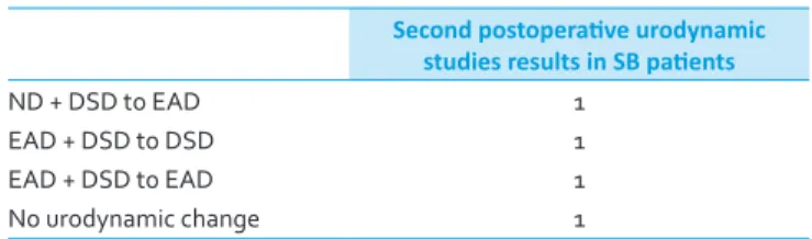
Table 2: Second postoperative urodynamic study results Second postoperative urodynamic
studies results in SB patients
ND + DSD to EAD 1
EAD + DSD to DSD 1
EAD + DSD to EAD 1
No urodynamic change 1
EAD – Extremely active detrusor activity; DSD – Detrusor sphincter dyssynergy; ND – Normal detrusor; SB – Spina bifida
Table 1: Early postoperative urodynamic study results Early postoperative urodynamic
studies results in SB patients
EAD 3
EAD + DSD 31
DU + SU 2
HB + DSD 1
ND + DSD 4
EAD – Extremely active detrusor activity; DSD – Detrusor sphincter dyssynergy; DU – Detrusor underactivity; SU – Sphincter underactivity; HB – Hypoactive bladder; ND – Normal detrusor; SB – Spina bifida
Alatas, et al.: Urodynamic studies in spina bifida
85 Asian Journal of Neurosurgery
Vol. 10, Issue 2, April‑June 2015 Preoperative urodynamic studies could be done in all tethered
cord patients. Routinely, their control urodynamic studies were done 3 months and 9 months after tethered cord release operation as a part of our follow‑up protocol.
Neuro‑urological follow‑up of 25 patients who underwent surgery for tethered cord (6 of them diastematomyelia, 4 of them fatty filum, 15 of them tight filum) between the same dates was performed, and their pre‑and postoperative urodynamics results were evaluated. 11 patients were male, and 14 patients were female. In the urodynamics results of patients who underwent surgery for tethered cord, EAD was observed in seven patients, EAD + DSD was observed in seven patients, DSD was observed in four patients, and normal urodynamics were observed in seven patients. According to the postoperative urodynamics results of four patients, an improvement from EAD to DSD was observed in one patient, improvement from EAD to normal urodynamics was observed in one patient, and improvement from EAD + DSD to decreased intra‑bladder pressure compared to preoperative urodynamics was observed in one patient. There was no change in the normal urodynamics result in one patient.
Discussion
Various urination disorders develop as a result of abnormal development of the spinal tube and anomalies in the innervation of the detrusor smooth muscle and urethral sphincter. The mechanical tethering of the spinal cord results in the impairment of the blood flow in the nervous tissue, and subsequent ischemia causes progressive neurological deficit.[11]
Leg deformities caused by loss of sense and loss of strength and muscle imbalance are present. There are two major aims behind the neuro‑urological management of children with myelodysplasia who do not have neurological deficits: (1) To protect, in particular, the kidneys, urinary tracts, and bladder from irreversible functional and morphological injury, and (2) to decrease the urinary symptoms to a minimum or to treat them to increase the life quality of children so that they can participate in society.[11] However, only NM may appear
as the first symptom during retethering in a majority of
the patients.[12] The result of the retethering surgery is not
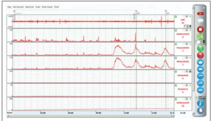
always predictable. The success of any secondary retethering surgery depends on good timing. By performing surgery prior to ischemia and irreversible injury, the innervation of the lower urinary system is protected. Therefore, close urological monitoring continues to be the most valuable tool as better outcomes are obtained with early diagnosis [Figures 1 and 2]. In a study that supported this hypothesis, secondary tethering was observed in 56 out of 401 children (14%) with SD Urological deterioration was observed in 58% of the patients with secondary tethering and neuro orthopedic deterioration was observed in 42% of the patients with secondary tethering. When preoperative and postoperative month six urological findings were compared, diagnosis and treatment before the age of 7 resulted in an improvement in the results of the urodynamics, as well as clinical symptoms and findings of the patients.[13]
Conclusion
An improvement in the urodynamics of three of four patients who were operated on for tethered spinal cord syndrome and who underwent second control urodynamics was observed. While the number of patients and urodynamics studies is still insufficient, early‑ and late‑stage urodynamics studies during the clinical follow‑up of patients, who underwent SB and tethered cord surgery, are important to determine the development of neuropathic bladder, tethered cord, and/or retethering, and these studies should be performed using larger patient groups.
References
1. Prevalence of neural tube defects in 20 regions of Europe and the impact of prenatal diagnosis, 1980‑1986. EUROCAT Working Group. J Epidemiol Community Health 1991;45:52‑8.
2. Mandiracioglu A, Ulman I, Lüleci E, Ulman C. The incidence and risk factors of neural tube defects in Izmir, Turkey: A nested case‑control study. Turk J Pediatr 2004;46:214‑20.
3. Sarac M, Ozel SK, Kaplan M, Kazez A. Spina Bifida: The Current Condition in Eastern Anatolia. XXVth National Congress of Turkish
Association of Pediatric Surgeons; 2007 October, 22‑27.
4. Ozek MM, Cinalli G, Maixner W, editors. The Spina Bifida
Figure 1: Normal urodynamic test Figure 2: This urodynamic test resulted extremely active detrusor
activity and detrusor sphincter dyssynergy
Alatas, et al.: Urodynamic studies in spina bifida
86
Asian Journal of Neurosurgery Vol. 10, Issue 2, April‑June 2015
Management and Outcome. Milan, Italy: Springer; 2008. p. 381‑97. 5. Thiagarajah S, Henke J, Hogge WA, Abbitt PL, Breeden N, Ferguson JE.
Early diagnosis of spina bifida: the value of cranial ultrasound markers. Obstet Gynecol 1990;76:54‑7.
6. Nakatsu T, Uwabe C, Shiota K. Neural tube closure in humans initiates at multiple sites: Evidence from human embryos and implications for the pathogenesis of neural tube defects. Anat Embryol (Berl) 2000;201:455‑66.
7. Barnes MA, Wilkinson M, Khemani E, Boudesquie A, Dennis M, Fletcher JM. Arithmetic processing in children with spina bifida: Calculation accuracy, strategy use, and fact retrieval fluency. J Learn Disabil 2006;39:174‑87.
8. Juranek J, Salman MS. Anomalous development of brain structure and function in spina bifida myelomeningocele. Dev Disabil Res Rev 2010;16:23‑30.
9. Mayes SD, Calhoun SL. Frequency of reading, math, and writing disabilities in children with clinical disorders. Learn Individ Differ 2006;16:145‑57.
10. Mitchell LE, Adzick NS, Melchionne J, Pasquariello PS, Sutton LN,
Whitehead AS. Spina bifida. Lancet 2004;364:1885‑95.
11. Yamada S, Iacono RP, Andrade T, Mandybur G, Yamada BS. Pathophysiology of tethered cord syndrome. Neurosurg Clin N Am 1995;6:311‑23.
12. Tarcan T, Bauer S, Olmedo E, Khoshbin S, Kelly M, Darbey M. Long‑term followup of newborns with myelodysplasia and normal urodynamic findings: Is followup necessary? J Urol 2001;165:564‑7. 13. Tarcan T, Onol FF, Ilker Y, Simsek F, Ozek M. Does surgical release of
secondary spinal cord tethering improve the prognosis of neurogenic bladder in children with myelomeningocele? J Urol 2006;176:1601‑6.
How to cite this article: Alatas I, Demirci H, Canaz H, Akdemir O,
Baydin S, Ozel K. The role of urodynamic studies in the diagnosis and treatment of patients with spina bifida. Asian J Neurosurg 2015;10:83‑6.
Source of Support: Nil, Conflict of Interest: None declared.
Author Help: Online submission of the manuscripts
Articles can be submitted online from http://www.journalonweb.com. For online submission, the articles should be prepared in two files (first page file and article file). Images should be submitted separately.
1) First Page File:
Prepare the title page, covering letter, acknowledgement etc. using a word processor program. All information related to your identity should be included here. Use text/rtf/doc/pdf files. Do not zip the files.
2) Article File:
The main text of the article, beginning with the Abstract to References (including tables) should be in this file. Do not include any informa-tion (such as acknowledgement, your names in page headers etc.) in this file. Use text/rtf/doc/pdf files. Do not zip the files. Limit the file size to 1 MB. Do not incorporate images in the file. If file size is large, graphs can be submitted separately as images, without their being incorporated in the article file. This will reduce the size of the file.
3) Images:
Submit good quality color images. Each image should be less than 4096 kb (4 MB) in size. The size of the image can be reduced by decreas-ing the actual height and width of the images (keep up to about 6 inches and up to about 1800 x 1200 pixels). JPEG is the most suitable file format. The image quality should be good enough to judge the scientific value of the image. For the purpose of printing, always retain a good quality, high resolution image. This high resolution image should be sent to the editorial office at the time of sending a revised article. 4) Legends:
Legends for the figures/images should be included at the end of the article file.