Address for Correspondence / Yazışma Adresi: Serkan Akbulut, MD Ankara University Faculty of Medicine, Department of General Surgery Surgical Oncology, Ankara, Turkey E-mail: [email protected]
©Telif Hakkı 2019 Gazi Üniversitesi Tıp Fakültesi - Makale metnine http://medicaljournal.gazi.edu.tr/ web adresinden ulaşılabilir. ©Copyright 2019 by Gazi University Medical Faculty - Available on-line at web site http://medicaljournal.gazi.edu.tr/
doi:http://dx.doi.org/10.12996/gmj.2019.16
Dermatofibrosarcoma Protuberans: A Rare Cause of Giant Breast Mass
Dermatofibrosarkoma Protuberans: Dev Meme Kitlesinin Nadir Bir Sebebi
Serkan Akbulut
1, Özgür Külahçı
2, Kağan Gökçe
31 Ankara University Faculty of Medicine, Department of General Surgery Surgical Oncology, Ankara, Turkey 2 Adana City Hospital Department of Pathology, Adana, Turkey
3 Muğla Sıtkı Koçman University Education and Research Hospital Surgical Oncology, Mugla, Turkey
ABSTRACT
Dermatofibrosarcoma protuberans (DFSP) is a rare soft tissue tumor of dermis and subcutaneous tissue, mostly seen in trunk and extremities. Here we present a case of DFSP in the breast of a young women. A 26 years old female admitted to hospital with a 7 cm painless mass at right breast. Trucut biopsy reported spindle cell tumor. PET/CT and thorax CT revealed no metastases. Breast conserving surgery and sentinel lymph node biopsy was done. Pathological examination report was DFSP and reactive lymphadenopathy. Due to high recurrence rates in DFSP cases, wide local excision for acceptable oncologic margins and follow up is necessary. Key Words: Dermatofibrosarcoma protuberans, spindle cell, breast tumor
Received: 12.04.2017 Accepted:09.20.2018
ÖZET
Dermatofibrosarkoma protuburens (DFSP) genellikle gövde ve extremitede görülen, dermis ve cilt altı dokulardan kaynaklanan nadir bir yumuşak doku tümörüdür. Burada genç bir kadının memesinde DFSP vakası sunulmaktadır. Sağ memede ağrısız 7 cm lik kitle ile 26 yaşında kadın hastanemize başvurdu. Trucut biyopsi sonucu iğsi hücreli tümör olarak raporlandı. PET/BT ve toraks BT’ de metastaz saptanmadı. Meme koruyucu cerrahi ve sentinel lenf nodu biyopsisi yapıldı. Patoloji sonucu DFSP ve reaktif lenf nodları olarak bildirildi. DFSP vakalarında yüksek rekürens riski nedeni ile onkolojik olarak kabul edilir geniş sınırlarla rezeksiyon ve yakın takip gereklidir.
Anahtar Sözcükler: Dermatofibrosarkoma protuberans, iğsi hücre, meme tümörü
Geliş Tarihi: 04.12.2017 Kabul Tarihi:20.09.2018
INTRODUCTION
Dermatofibrosarcoma protuberans (DFSP), first reported by Darier and Ferrand in 1924 (1), is a rare soft tissue tumor of dermis and subcutaneous tissue. It is seen in all ages but particularly between the second and fifth decades. Although the lesion can be seen at any part of the body, mostly trunk and extremities are involved (2). Breast involvement is rare and here we present DFSP in the breast of a young woman.
CASE REPORT
A 26 years old woman admitted to our hospital with a 7 cm painless mass at her right breast just under the incision scar of a previous segmental mastectomy (patient mentioned ) 6 years ago. The pathological evaluation of the biopsy material was benign according to the patient has mentioned. Breast USG, mammography and MRI (Figure 1) depicted a 7x6x5 cm well circumscribed mass at right breast and reactive lymphadenopathies at the right axilla.
Figure 1. MRI view of 7x6x5 cm well circumscribed mass of right breast and reactive lymphadenopathies.
Trucut biopsy reported a spindle cell tumor. In FDG-PET/CT and thorax CT, there was no metastases. Therefore level 1 oncoplastic breast conserving surgery and sentinel lymph node biopsy were done. Intraoperative frozen section analysis reported acceptable surgical margins (2 cm) were achieved with no lymph node metastases. Final pathology report was DFSP with achieved acceptable surgical margins and reactive lymphadenopathies (Figure 2-3-4). No tumor recurrence was seen during sonographic follow-up over 20 months after surgery.
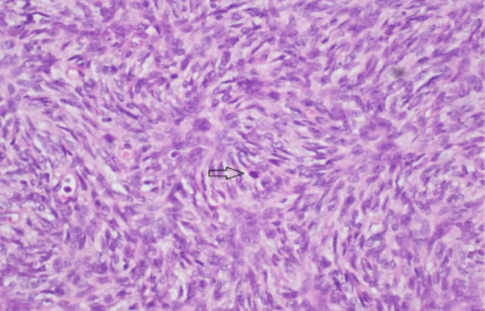
Figure 2. DFSP, adipose tissue in the storiform pattern of spindle cells.
Figure 3. DFSP, storiform pattern of spindle cells with a few mitotic figures.
Figure 4. DFSP, diffuse and strong positivity with CD 34 staining.
DISCUSSION
Up to date few breast DFSP cases have been reported and differential diagnoses of these lesions are difficult (2). In the evaluation of breast lesions, trucut biopsy has one of the top priority. As in presented case, the spindle cell lesions of breast, fibroadenoma, cystosarcoma phylloides, hamartoma, adenomyoepithelioma, pseudoangiomatous stromal hyperplasia, metaplastic carcinoma, sarcomas should be kept in mind (3) and intraoperative frozen section assessment should be done for the achievement of accurate surgical margins and examination of lymph node involvement.
In histological examination of DFSP, spindle cells arranged in storiform pattern, honeycomb infiltration of lipomatous tissue can be seen. Mitose rates are low and in 84-100 % of lesions diffuse CD 34 staining are seen (4).
DFSP is a locally aggressive tumor which invades subcutaneous tissue and adjacent structures. Recurrence rate is high whereas metastasis is rare but if it occurs lungs and lymph nodes are usually affected (5). In our case, the patient had a history of segmental mastectomy 6 years ago because of a lesion at the same localization. Probably she had DFSP, and this is the local recurrence of the lesion.
Surgical treatment for DSFP is wide local excision and Mohs micrographic surgery. Different recurrence rates are reported for both procedures (2,6). Surgical excision with a margin of at least 2 to 3 cm is recommended for treatment because the local recurrence rate is 20% to 50% in cases of incomplete resection (7). Wide local excision with clear margins was performed for this case. For selected patients with DSFP in breast, who likely to default follow-up modified radical mastectomy can be performed. Ismail A. S. Burud et al. recommend mastectomy with axillary clearance for the patients who have the involvement of the resected margins, high mitotic figures, risk of recurrence, previous breast surgeries and strong family history of breast cancer (8).
In 90 % of cases, there is translocation between 17. and 22. chromosomes causing overproduction of platelet derived growth factor β (PDGF β) and stimulating DFSP cell growth (9). This started the use of imitanib, C-kit and PDGF-R inhibitor adjuvant and neoadjuvant therapies for DFSP (6). We vigorously recommended the patient medical oncology consultation, but she did not admit.
CONCLUSION
Breast involvement of DFSP is very rare however it should be kept in mind in the differential diagnosis. In this report DFSP in the breast of a young woman is presented. Due to high recurrence rates, wide local excision for acceptable oncologic margins and follow up is necessary. Keeping in mind modified radical mastectomy can be an option for selected patients.
Conflict of interest
No conflict of interest was declared by the authors. REFERENCES
1.Darier J. Dermatofibromes progressifs et recidivants ou fibrosarcomes de la peau. Ann Dermatol Syph. 1924;5:545-62.
2.Pohlodek K, Mečiarová I, Grossmann P, Kinkor Z. Dermatofibrosarcoma protuberans of the breast: A case report. Oncol Lett. 2017;14(1):993-8. 3.Tse GM, Tan PH, Lui PC, Putti TC. Spindle cell lesions of the breast--the pathologic differential diagnosis. Breast Cancer Res Treat. 2008;109(2):199-207.
4.Al-Rahbi S, Al-Lawati T, Al-Kharusi S, Thomas S, Al-Harrasi K. Dermatofibrosarcoma Protuberans: A Rare Malignancy of the Breast. Oman Med J. 2015;30(5):378-81.
5.Bogucki B, Neuhaus I, Hurst EA. Dermatofibrosarcoma protuberans: a review of the literature. Dermatol Surg. 2012;38(4):537-51.
6.Fontecilla NM, Kittler NW, Geskin L, Samie FH, Niedt G, Imahiyerobo T, Schwartz G, Ingham M, Lewin JM. Recurrent dermatofibrosarcoma protuberans treated with neoadjuvant imatinib mesylate followed by Mohs micrographic surgery. JAAD Case Rep. 2017;3(6):467-469.
7.Sin FN, Wong KW. Dermatofibrosarcomaprotuberans of the breast: a case report. Clin Imaging. 2011;35:398–400.
8.Burud IAS, How NS, CheeWei G, Roslina S. Dermatofibrosarcoma Protuberance of the Breast:a Diagnostic Challenge Indian J Surg 2017;79(2):169–172.
9.Dragoumis DM, Katsohi LA, Amplianitis IK, Tsiftsoglou AP. Late local recurrence of dermatofibrosarcoma protuberans in the skin of female breast. World Journal of surgical oncology. 2010;3;8:48.