Case Report
An Unusual Presentation of Kawasaki Disease: Gallbladder
Hydrops and Acute Cholestatic Hepatitis
B. O. Kılıç , S¸. Baysun, T. C. G¨oks¸en, ˙I. Akınbing¨ol, and Z. Arslan
Departments of Pediatrics, Faculty of Medicine, TOBB University of Economics and Technology, Ankara, Turkey Correspondence should be addressed to B. O. Kılıç; [email protected]
Received 24 March 2018; Accepted 12 April 2018; Published 3 July 2018 Academic Editor: William B. Silverman
Copyright © 2018 B. O. Kılıç et al. This is an open access article distributed under the Creative Commons Attribution License, which permits unrestricted use, distribution, and reproduction in any medium, provided the original work is properly cited. Kawasaki disease is the most common vasculitis of childhood. In its classical form, at least four of five diagnostic criteria including cervical lymphadenopathy (1.5 cm or more), nonsuppurative conjunctivitis, intraoral mucosal changes, edema in hands and feet, and maculopapular rash are required with prolonged fever over 5 days. Atypical cases which are different from the classical type or incomplete cases which does not include all the diagnostic criteria can be seen. The typical Kawasaki disease is a self-limiting disease with fever lasting for an average of two weeks. In such patients who have not been diagnosed and whose treatment has been delayed, coronary artery aneurysm, myocardial depression, arrhythmia, and vascular complications may increase morbidity and mortality. We would like to present a rare case of an atypical patient with gallbladder hydrops and acute cholestatic hepatitis.
1. Case
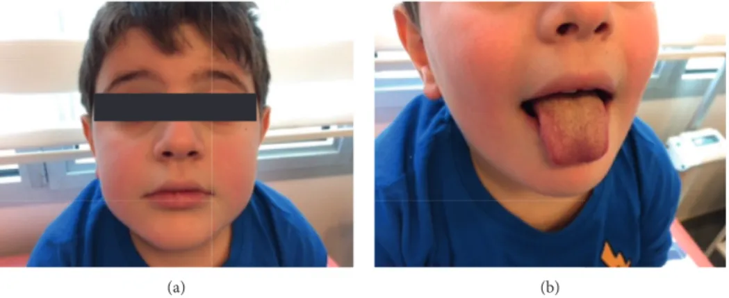
A 7-year-old boy presented due to ongoing fever and ab-dominal pain for 5 days. He had vomiting once and watery stool twice on the same day. On physical examination, he had fever of 39°, restlessness, bilateral nonpurulent
con-juctival hyperemia, redness of the lip and tongue, poly-morphic rash in the face and trunk, and obvious abdominal tenderness in the right upper quadrant (Figure 1).
In laboratory examination, hemoglobin was 13 g/dl, leu-kocyte was 15700/mm3, thrombocyte was 93000/mm3, CRP was 171 mg/dl (0–5 mg/dl), erytrocyte sedimentation rate was 75 mm/h, aspartate aminotransferase was 109 U/L (15–50 U/L), alanine aminotransferase was 202 U/L (10–50 U/L), total bili-rubin was 3.49 mg/dl (0.3–1.35 mg/dl), direct biluribine was 3.42 mg/dl (0.05–0.5 mg/dl) and sodium was 126 meq/l (130–150 meq/L). Adenovirus was negative in nasal swab and stool. Stool microscopy was normal. Serology tests were negative for hepatitis A, B, and C. Abdominal ultrasonography showed acute cholangitis/cholecystitis, thickening of the gall-bladder wall, hydrops, and intrahepatic bile duct stasis.
Echocardiography (ECHO) showed minimal pericardial effusion, and mild mitral and tricuspid regurgitation in the
left ventricle. Troponin I value (HST) was found to be 22.3 ng/L (normal value < 0.2).
The patient was considered to have incomplete Kawasaki disease, and he was given a single dose of immunuglobulin (IVIG) with a 12-hour intravenous infusion of 2 g/kg and ace-thylsalicylic acid (ASA) of 50 mg/kg/day divided into 4 doses. Cultures were taken, and treatment with ceftriaxone 80 mg/kg for enteric fever and cholecystitis was initiated. The next day, antibiotic was stopped because of negative results of microbial cultures. Forty-eight hours after the patient’s fever returned to normal, aspirin was reduced to only one dose of 3–5 mg/kg.
The patient’s platelet count increased to 676,000/mm3 in the second week. Coronary artery involvement was not observed in the first echocardiography. Troponin I level also fell below 1.5 ng/L. In the third week, sedimentation and CRP values returned to normal and ASA treatment was terminated.
2. Discussion
In addition to fever as a classical symptom of Kawasaki disease, our patient had nonpurulent conjunctivitis, redness
Hindawi
Case Reports in Medicine
Volume 2018, Article ID 4930234, 2 pages https://doi.org/10.1155/2018/4930234
of lip and tongue, and polymorph rash, with no coronary artery involvement at the ECHO. Febrile cholestatic hepa-titis with gallblader hydrops was thought to be the symptoms for atypical Kawasaki disease. Diarrhea, vomiting, abdom-inal pain, hepatic dysfunction, and bile duct hydrops had been reported at different rates among nonspecific symp-toms even though they are not the part of the diagnostic criteria [1].
In a case-control study, approximately 30% of the 280 patients had moderate transaminase elevation and occa-sionally obstructive jaundice since gallblader hydrops was observed [2]. In a cohort study from Italy with 219 kawasaki disease cases, incomplete Kawasaki presentation was seen in 9 of 10 children with severe abdominal discomfort, and it was shown that the gastrointestinal system symptoms were not recognized early in Kawasaki disease [3]. However, acute cholestasis and gallblader hydrops were also reported as atypical baseline findings [4].
In another study, Kawasaki disease was detected in 6% of the patients with fever and abdominal pain [5].
In a Kawasaki disease series with 35 cases, reported from Turkey, acute cholestasis was not reported as a baseline finding [6]. However, in another study of 23 patients, hydrops of gallblader was reported in 1 case and trans-aminase elevation in 7 cases [7]. Kaman et al. also reported 2 cases of atypical kawasaki disease with acute febrile jaundice [8].
Vasculitis-associated inflammation and obstruction in the liver and gallbladder are thought to be the cause of increased transaminase levels and cholestasis. It is also known that the sodium level below 135 meq/l increases the risk for coronary artery disease, and the risk is higher in atypical cases than in classical cases. In addition to the atypical clinical presentation of our patient, hypona-tremia and high transaminases increased the risk for complication developed that is why IVIG treatment was started on the sixth day of fever, and no complication was observed.
With this case report, we would like to remind that gallblader hydrops and cholestasis can be nonclassical early findings of Kawasaki disease, and if considered, early IVIG treatment can rapidly improve the clinical findings and prevent complications.
Conflicts of Interest
The authors declare that they have no conflicts of interest.
References
[1] A. L. Baker, M. Lu, L. L. Minich et al., “Associated symptoms in the ten days before diagnosis of Kawasaki disease,” Journal of Pediatrics, vol. 154, no. 4, pp. 592.e2–595.e2, 2009.
[2] J. C. Burns, W. H. Mason, M. P. Glode et al., “Clinical and epidemiologic characteristics of patients referred for evaluation of possible Kawasaki disease,” Journal of Pediatrics, vol. 118, no. 5, pp. 680–686, 1991.
[3] F. Zulian, F. Falcini, L. Zancan et al., “Acute surgical abdomen as presenting manifestation of Kawasaki disease,” Journal of Pediatrics, vol. 142, no. 6, pp. 731–735, 2003.
[4] P. Valentini, E. Ausili, A. Schiavino et al., “Acute cholestasis: atypical onset of Kawasaki disease,” Digestive and Liver Disease, vol. 40, no. 7, pp. 582–584, 2008.
[5] M. Eladawy, S. R. Dominguez, M. S. Anderson, and M. P. Glod´e, “Kawasaki disease and the pediatric gastroen-terologist: a diagnostic challenge,” Journal of Pediatric Gas-troenterology and Nutrition, vol. 56, no. 3, pp. 297–299, 2013. [6] B. Aldemir-Kocabas¸, A. Karbuz, C. Karadeniz et al., “Another face of Kawasaki disease,” Turkish Journal of Pediatrics, vol. 56, no. 4, pp. 392–398, 2014.
[7] H. ¨Ozdemir, E. Ciftçi, A. Tapısız et al., “Clinical and
epide-miological characteristics of children with Kawasaki disease in Turkey,” Journal of Tropical Pediatrics, vol. 56, no. 4, pp. 260–262, 2010.
[8] A. Kaman, T. Aydın-Teke, Z. G. Gayretli-Aydın et al., “Two cases of Kawasaki disease presented with acute febrile jaun-dice,” Turkish Journal of Pediatrics, vol. 59, no. 1, pp. 84–86, 2017.
(a) (b)
Figure 1: Nonpurulent conjunctivitis, malar rash, and mucosal changes on the lip and on the tongue.
Stem Cells
International
Hindawi www.hindawi.com Volume 2018 Hindawi www.hindawi.com Volume 2018MEDIATORS
INFLAMMATIONofEndocrinology
International Journal ofHindawi www.hindawi.com Volume 2018 Hindawi www.hindawi.com Volume 2018
Disease Markers
Hindawi www.hindawi.com Volume 2018 BioMed Research InternationalOncology
Journal of Hindawi www.hindawi.com Volume 2013 Hindawi www.hindawi.com Volume 2018Oxidative Medicine and Cellular Longevity
Hindawi
www.hindawi.com Volume 2018
PPAR Research
Hindawi Publishing Corporation
http://www.hindawi.com Volume 2013 Hindawi www.hindawi.com
The Scientific
World Journal
Volume 2018 Immunology Research Hindawi www.hindawi.com Volume 2018 Journal ofObesity
Journal of Hindawi www.hindawi.com Volume 2018 Hindawi www.hindawi.com Volume 2018 Computational and Mathematical Methods in Medicine Hindawi www.hindawi.com Volume 2018Behavioural
Neurology
Ophthalmology
Journal of Hindawi www.hindawi.com Volume 2018Diabetes Research
Journal ofHindawi
www.hindawi.com Volume 2018
Hindawi
www.hindawi.com Volume 2018 Research and Treatment
AIDS
Hindawi
www.hindawi.com Volume 2018
Gastroenterology Research and Practice
Hindawi www.hindawi.com Volume 2018