Right Atrium’s Pump And Reservoir Functions are Preserved But
Conduit Function Impaired in Patients with Secondary
Pulmonary Hypertension with Different Etiologies and
Severities
Değișik Etiyolojilere Bağlı ve Değișen Ciddiyetteki İkincil Pulmoner Hipertansiyonda Sağ Atriyumun Pompalama ve Depolama Fonksiyonları Korunmakta Ancak İletim Fonksiyonu Bozulmaktadır
Orçun Çiftci
1, Necla Özer
2, Enver Atalar
2, Kenan Övünç
2, Serdar Aksöyek
21 Başkent University, Faculty of Medicine, Department of Cardiology,
Ankara/Turkey
2 Hacettepe University, Faculty of Medicine, Department of Cardiology,
Ankara/Turkey
Objective: Pulmonary hypertension (PHT) may result in right heart failure and death. Changes in right atrial function and size have been poorly studied in PHT. We aimed to determine how right atrial functions and size are affected in patients with secondary PHT with different severities and etiologies. Materials and Method: This study enrolled a total of 83 subjects with PHT secondary to left heart diseases excluding left ventricular systolic dysfunction, connective tissue disorders, and chronic respiratory diseases, and 49 healthy age- and sex-matched healthy controls. All subjects underwent 2-dimensional echocardiography and strain rate measurement to quantify right atrial size and function. Subjects with PHT were compared with the controls and with each other.
Results: Subjects with PHT had significantly larger right atrial diameters and a significantly lower right atrial ejection fraction compared to controls. Although right atrial ejection fraction worsened in moderate and severe PHT, right atrial lateral wall late diastolic strain rate was preserved in different PHT severities. With increasing PHT severity, there occurred an increase in the right atrium’s reservoir/conduit ratio where right atrial emptying worsened but right atrial filling was preserved. The three PHT etiologies were comparable with respect to right atrial size and function.
Conclusion: Our findings suggest that right atrium preserves its systolic function and behaves like more of a reservoir than a conduit in PHT. These changes together may help to increase right ventricular preload to sustain pulmonary and systemic output. PHT etiologies do not appear to exert any differential effect on right atrial function and size in PHT.
Key Words: Pulmonary hypertension, right atrium, echocardiography, strain rate
Amaç: Pulmoner hipertansiyon (PHT) sağ ventrikül yetersizliği ve ölüme yol açabilen bir hastalıktır. PHT’de sağ atriyal fonksiyon ve boyutlardaki değișim hakkında az çalıșma vardır. Biz bu çalıșmada, değișik ciddiyet ve etiyolojilere bağlı sekonder PHT’de sağ atriyal fonksiyon ve boyutların nasıl değiștiğini belirlemeyi amaçladık.
Gereç ve Yöntem: Bu çalıșmaya, sol ventrikül sistolik disfonksiyonu olmayan sol kalp hastalıkları, bağ dokusu hastalıkları ve kronik respiratuar hastalıklara sekonder 83 PHT hastası ve 49 yașa ve cinsiyete göre eșleștirilmiș sağlıklı kontrol alındı. Tüm deneklerde 2 boyutlu ekokardiyografi ve strain hızı ölçümü ile sağ atriyal depolama, iletim ve pompa fonksiyonları ve 2 boyutlu ekokardiyografik boyutlar değerlendirildi. PHT olan hastalar kontrol grubu ile ve kendi içlerinde karșılaștırıldı.
Bulgular: PHT olan hastalarda kontrol grubuna oranla anlamlı șekilde sağ atriyal çaplar daha büyük ve sağ atriyal ejeksiyon fraksiyonu daha düșüktü. Orta ve ciddi PHT’de sağ atriyal ejeksiyon fraksiyonu kötüleșse de, sağ atriyal lateral duvar geç diastolik (atriyal sistolik) strain hızı farklı PHT ciddiyetlerinde korunmuș olarak bulundu. Artan PHT ciddiyeti ile birlikte, sağ atriyumun boșalma kabiliyetinin azaldığı ancak dolumunun sağlam kaldığı depolama/iletme oranında artıș tespit edildi. Üç PHT etiyolojisinin sağ atriyal boyut ve fonksiyon yönünden farklılık göstermedikleri görüldü.
Sonuç: Sonuçlarımız göstermektedir ki, sekonder PHT’de sağ atriyum sistolik fonksiyonu korunmakta ve sağ atriyum kanı ileten bir boșluktan daha çok depolayan bir boșluk haline gelmektedir. Bu değișiklikler hep birlikte sağ ventrikül önyükünün korunması ve böylece pulmoner ve sistemik debinin devam ettirilmesine yardım ediyor olabilirler. PHT etiyolojileri sağ atriyal fonksiyon ve boyutlarına etki etmiyor görünmektedir.
Anahtar Sözcükler: Pulmoner hipertansiyon, sağ atriyum, ekokardiyografi, strain hızı
DOI: 10.1501/Tıpfak_000000994 Araștırma Makalesi / Research Article
Received: Dec. 18, 2017 Accepted: March 06, 2018 Corresponding Author:
Orçun Çiftci
E-mail: [email protected] Phone: +90 (532) 594 68 22 Faks: +90 (312)
Bașkent University Faculty of Medicine, Department of Cardiology. Yukarı Bahçelievler Mahallesi, Mareșal Fevzi Çakmak Cd. No:45, 06490 Çankaya/Ankara
Pulmonary hypertension (PHT) is a cluster of heterogeneous disorders characterized by increments in pulmonary artery pressures leading to progressive right heart dilatation and dysfunction. It is unknown how right atrium reacts to chronic elevations in right ventricular pressures. The morphological and functional changes to the right atrium in PHT have been sparsely studied because the assessment of right atrial functions is problematic owing to the amorphous structure of this chamber and the complexity of the anatomical relationship between right atrium, right ventricle, and systemic veins (1). Understanding right atrial changes in pulmonary hypertension may aid in future therapeutic options on this disease. Tissue Doppler echocardiography and strain measurement offer novel opportunities to evaluate functional changes to heart chambers including the atria in various disorders. The aim of the present study was to compare 2-dimensional and strain rate measurement characteristics between patients with secondary pulmonary hypertension and healthy controls as well as between patients with different PHT etiologies and severities.
Materials and Method
This study was approved by Hacettepe University Faculty of Medicine, Local Ethics Committee for Medical, Surgical, and Pharmacological Studies (No: LUT 07-55), and all patients gave informed consent. This study included a total of 83 patients with secondary pulmonary hypertension older than 18 years of age who presented to Hacettepe University Faculty of Medicine, Department of Cardiology. Twenty-nine (34.9%) patients were male and 54 (65.1%) were female. Pulmonary hypertension was defined as a pulmonary artery systolic pressure of ≥40 mmHg measured on transthoracic echocardiography. Three major groups of pulmonary hypertension were enrolled. The first group was pulmonary hypertension associated with left heart diseases excluding left
ventricular systolic dysfunction. The second group was associated with connective tissue disorders. The third group was pulmonary hypertension associated with respiratory diseases and/or chronic hypoxemia. The control group consisted of 49 healthy age- and sex-matched subjects (14 (29%) males and 35 (71%) females) without any systemic disorder with a
normal transthoracic echocardiography and a normal
systolic pulmonary artery pressure (< 40 mmHg). Pulmonary hypertension severity was graded as follows: mild pulmonary hypertension (systolic pulmonary artery pressure 40-49 mmHg), moderate pulmonary hypertension (systolic pulmonary artery pressure 50-69 mmHg), and severe pulmonary hypertension (systolic pulmonary artery pressure ≥70 mmHg) (2,3). Patients with the following conditions were excluded: left ventricular systolic dysfunction and/or left ventricular dilatation (left ventricular ejection fraction < 40% and/or left ventricular end-diastolic diameter > 58 mm and/or left ventricular end-systolic diameter > 45 mm); rheumatic mitral stenosis with rheumatic tricuspid involvement;
intraventricular and/or atrioventricular conduction defects
including a QRS duration of greater than 120 ms, complete right bundle branch block (RBBB), complete left bundle branch block (LBBB), bifascicular and trifascicular blocks, advanced 1° AV block (PR> 240 ms), Mobitz type 1 or type 2 II° AV block, and complete AV block; atrial fibrillation, atrial flutter, atrial tachycardia, frequent supraventricular or ventricular extrasystolic beats or extrasystolic beats in the form of bigemini, trigemini, couplets, triplets, and nonsustained or sustained ventricular tachycardia; and a history of open or closed heart surgery, especially at the atrial level.
All subjects underwent echocardiographic examination according to the American Society of Echocardiography guidelines (4,5). Subjects were examined in lateral decubitis position using “Vingmed
System Five GE ultrasound, Horten, Norway” echocardiography device using a 2.5-3.5 Mhz transducer. Parasternal long axis, parasternal short axis, apical 4-chamber, and subcostal views were employed to acquire images of cardiac chambers. All subjects underwent B-mode (two-dimensional) and color tissue Doppler
examinations. Chamber
quantifications were performed
according to the latest chamber quantification guideline (4).
Systolic pulmonary artery pressure was quantified according to the Modified Bernoulli Equation which states that ΔP = 4 V² where ΔP denotes the pressure gradient between the right ventricle and right atrium at mid systole, and V denotes the velocity of the tricuspid regurgitant jet measured by continuous wave (CW) Doppler. While measuring ΔP, utmost care was taken to ensure keeping the angle between the ultrasound beam and the tricuspid regurgitant jet as close to 0 degrees as possible by selecting the most suitable acoustic window possible, which was the apical four chamber view in the majority of patients, as well as having an as dense tricuspid regurgitant jet as possible on continuous wave Doppler spectral scale to discern the apex of the regurgitant jet clearly.
Once the pressure gradient was calculated between the two chambers, right ventricular systolic pressure was calculated by adding the right atrial pressure to ΔP according to the formula RVSP: 4 (VTR)² + RAP, where RVSP denotes right ventricular systolic pressure and RAP denotes right atrial pressure. Estimated right atrial pressure obtained from inferior vena cava diameter and the response of the latter to inspiration, as described elsewhere (4,5). Provided that a patient does not have pulmonary or right ventricular outflow tract obstruction, calculated RVSP is considered to be equal to the systolic pulmonary artery pressure (sPAP). Two- dimensional right atrial measurements were done from the apical 4-chamber window. Right atrial 2-dimensional diamaters included two
right atrial orthogonal diameters, namely right atrial long axis and mid-cavitary diameters measured at end-diastole (6). Right atrial volumes were also measured on apical 4-chamber window using one-plane disc algorithm. According to this algorithm, the largest (RAV1) and the smallest (RAV2) right atrial volumes were measured and right atrial ejection fraction (RAEF) was calculated based
on the formula RAEF: [(RAV1-
RAV2) / RAV1] *100. Largest right atrial volume provides information about the reservoir function of the right atrium and the smallest volume about the conduit function.
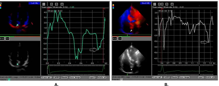
Right atrial strain rate measurements were performed using apical 4-chamber window. Two dimensional color tissue Doppler samples used for strain rate measurement were recorded at end-expiration to prevent motion artefacts and in a way to encompass at least 3 cardiac cycles. All recordings were recorded with the two-dimensional gray-scale image at a frame rate of 150/sec and a depth of 10.8 mm. Sample volumes were placed to mid segments of right atrial lateral wall and interatrial septum. Sample volumes were placed to the inner side of atrial myocardium to minimalize the angle between Doppler flow and longitudinal contraction direction. Right atrial strain rate analysis included three measurements, namely “S”, “E”, and “A” waves. The first one corresponds to ventricular systolic strain rate, second one corresponds to ventricular early diastolic strain rate, and the third one corresponds to late diastolic (atrial systolic) strain rate (7) (Figure 1). The off-line analysis of the recordings and calculations were performed using
Echopac 6.3.6. GE software package. Statistical analysis
Statistical Package for Social Sciences (SPSS) 11.5 for Windows software package (Chicago, Il, USA) was used for all statistical analyses. The distribution of quantitative variables was tested using the Kolmogorov-Smirnov test. The descriptive statistics
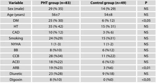
Table 1. Demographic and clinical characteristics of pulmonary hypertension and control groups
Variable PHT group (n=83) Control group (n=49) P
Sex (male) 29 (% 35) 14 (% 29) NS Age (years) 56±7 54±8 NS DM 25 (% 30) 6 (% 12) <0,05 HT 35 (% 42) 15 (% 31) NS CAD 10 (% 12) 3 (% 6) NS Smoking 24 (%29) 15 (%31) NS NYHA 1 (1-3) 1 (1-2) NS BB 8 (%10) 6 (%12) NS CCB 28 (%34) 11 (%22) NS ACEI 18 (%22) 6 (%12) NS ARB 19 (%23) 3 (%6) <0,01 Diuretic 23 (%28) 9 (%18) NS Digoxin 8 (%10) 0 (%0) <0,05
DM= Diabetes Mellitus, HT= Hypertension, CAD= Coronary Artery Disease, NYHA= New York Heart Association Class, BB= Beta blocker, CCB= Calcium Channel Blocker, ACEI= Angiotensin Converting Enzyme Inhibitor, ARB= Angiotensin Receptor Blocker, NS= non-significant
Table 2. Distribution of the PHT subjects by PHT etiology and severity PHT associated with left heart
diseases excluding left ventricular systolic dysfunction (n=39) Connective tissue disease-associated PHT (n=23) Chronic respiratory disease-associated PHT(n=21) MS (n=16) MR (n=8) AS (n=2) HFPEF (n=13) Scleroderma (n=18) SLE (n=2) RA (n=1) MCTD (n=1) Sjogren’s syndrome (n=1) COPD (n=21) Mild PHT (n=20; 24%) Moderately severe PHT (n=11;13%) Severe PHT (n=8; 10%) Mild PHT (n=12; % 14) Moderately severe PHT (n=6; 7%) Severe PHT (n=5; 6%) Mild PHT (n=10; 12%) Moderately severe PHT (n=4; 5%) Severe PHT (n=7; 8%) MS: Mitral stenosis, MR: Mitral regurgitation, AS: Aortic stenosis, HFPEF: Heart failure with preserved ejection fraction, SLE: Systemic lupus erytematosus, RA: Rheumatoid arthritis, MCTD: Mixed connective tissue disease, COPD: Chronic obstructive pulmonary disease, PHT: pulmonary hypertension
Table 3. Comparison between PHT and control groups according to two-dimensional right atrial parameters and right atrial strain rates
Parameter PHT group n=83 Control group n=49 P
RA Mid Diameter (cm) 3,7 (2,8-8,2) 3,6 (2,6-4,5) < 0,05 RA Long Diameter (cm) 5,0 (3,4-7,5) 4,2 (3,2-4,9) < 0,001 RAV1 (cm³) 44,0 (28,1-198,4) 39,4 (19,7-67,3) NS RAV2 (cm³) 22,5 (18,0-82,0) 17,1 (13,3-29,4) < 0,05 RAEF (%) 48 (±23) 58 (±08) < 0,01 RAS (1/s) 5,0 (±0,6) 5,5 (±0,5) < 0,01 RAE (1/s) 4,0 (2,4-5,4) 4,6 (3,1-5,8) < 0,001 RAA (1/s) 3,5 (2,3-4,3) 3,5 (2,5-6,7) NS IASS (1/s) 5,3 (±0,7) 5,8 (±0,6) < 0,001 IASE (1/s) 4,2 (3,0-7,4) 4,3 (3,6-6,3) NS IASA (1/s) 3,7 (2,6-5,2) 4,0 (2,0-6,2) < 0,05 RA= Right atrium, RAV1= largest right atrial volume, RAV2= smallest right atrial volume, RAEF: right
atrial ejection fraction, RAS= Right atrial lateral wall ventricular systolic strain rate, RAE= Right atrial lateral wall ventricular early diastolic strain rate, RAA= Right atrial lateral wall atrial systolic strain rate, IASS= Interatrial septum ventricular systolic strain rate, IASE= Interatrial septum ventricular early diastolic strain rate, IASA= Interatrial septum atrial systolic strain rate.
included mean ± standard deviation for normally distributed quantitative data, median (min-max) for non-normally distributed quantitative data, and number and percentage for nominal variables. The overall PHT and control groups were compared with respect to demographic and clinical properties, and right atrial size and functional measurements. PHT groups of different eitologies and severities were compared with one another and with the control group with regard to the same measurements. The comparisons between the PHT and control groups were performed with the independent samples t-test for normally distributed quantitative variables and Mann-Whitney-U test for non-normally distributed quantitative variables. The PHT groups of differing severities and etiologies were compared using the one-way analysis of variance (ANOVA) with Tukey’s Honestly Significant Difference test for inter-group differences for normally distributed variables. Kruskal-Wallis test and Mann-Whitney U test with Bonferroni correction to determine the significantly different pairs were used for the comparison of non-normally distributed variables. Chi-square test was used to test the nominal variables. Correlation analyses using Pearson’s and Spearman’s correlation analyses were performed between 2-dimensional right atrial measurements and right atrial strain rate measurement parameters. A p value of less than 0.05 was considered statistically significant for all statistical comparisons.
RESULTS
The demographic and clinical properties of the PHT and control groups were shown on Table 1. There were no significant differences between the two groups with respect to mean age, gender distribution, and comorbidities except for diabetes mellitus that was significantly more common in the PHT group. Table 2 shows the distribution of the PHT subjects by PHT etiology and severity. Accordingly, there were 39 (47%)
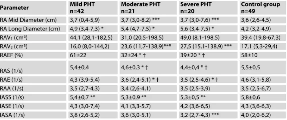
Table 4. Comparison of differing PHT severities regarding right atrial two-dimensional echocardiographic parameters and right atrial strain rate parameters
Parameter Mild PHT n=42 Moderate PHT n=21 Severe PHT n=20 Control group n=49 RA Mid Diameter (cm) 3,7 (0,4-5,9) 3,7 (3,0-8,2) *** 3,7 (3,0-7,6) *** 3,6 (2,6-4,5) RA Long Diameter (cm) 4,9 (3,4-7,3) * 5,4 (4,7-7,5) * 5,6 (3,4-7,5) * 4,2 (3,2-4,9) RAV1 (cm³) 44,1 (28,1-182,5) 31,0 (20,5-198,5) 49,0 (8,1-198,5) 39,4 (19,8-67,3) RAV2 (cm³) 16,0 (8,0-144,2) 23,6 (11,7-138,9)*** 27,5 (15,1-138,9) *** 17,1 (5,3-29,4) RAEF (%) 61±22 32±24 * † 39±20 * † 58±10 RAS (1/s) 5,4±0,4 4,6±0,3 * † 4,4±0,4 * † 5,5±0,5 RAE (1/s) 4,3 (3,9-5,4) 3,6 (2,4-5,1) * † 3,5 (2,5-4,6) * † 4,6 (3,1-5,8) RAA (1/s) 3,5 (2,7-4,3) 3,4 (2,6-4,1) 3,5 (2,5-3,9) 3,5 (2,5-6,7) IASS (1/s) 5,4±0,7 ** 5,3±0,9 ** 5,3±0,5 ** 5,8±0,6 IASE (1/s) 4,3 (3,0-7,4) 4,1 (3,3-5,7) 4,2 (3,6-6,5) 4,3 (3,6-6,3) IASA (1/s) 3,8 (2,6-5,2) 3,6 (3,0-5,1) 3,2 (2,7-4,3) *** 4,0 (2,0-6,2)
RA= Right atrium, RAV1= largest right atrial volume, RAV2= smallest right atrial volume, RAEF: right
atrial ejection fraction, RAS= Right atrial lateral wall ventricular systolic strain rate, RAE= Right atrial lateral wall ventricular early diastolic strain rate, RAA= Right atrial lateral wall atrial systolic strain rate, IASS= Interatrial septum ventricular systolic strain rate, IASE= Interatrial septum ventricular early diastolic strain rate, IASA= Interatrial septum atrial systolic strain rate. * p < 0,001 vs controls, ** p < 0,05 vs controls, *** p < 0,01 vs controls, † p< 0,01 vs mild PHT
Table 5. Comparison of right atrial two-dimensional echocardiographic parameters and right atrial strain rate analyses according to the PHT etiology
Parameter PHT associated with left heart diseases excluding left ventricular systolic dysfunction n=39 Connective tissue disease-associated PHT n=23 Respiratory disease-associated PHT n= 21 Control group n=49 RA Mid diameter (cm) 3,7 (2,3-5,9)* 3,7 (2,2-6,3) 3,3 (2,7-8,2) 3,6 (2,6-4,5) RA long diameter (cm) 5,3 (±0,9)* 5,0 (±0,5)** 5,4 (±1,4)* 4,2 (±0,3) RAV1 (cm³) 46,0 (18,4-182,5) 40,5 (20,5-133,6) 38,8 (8,1-198,5) 39,4 (19,8-67,3) RAV2 (cm³) 24,5 (18,4-144,2)* 15,5 (11,7-98,2) 14,5 (10,9-138,9) 17,1 (9,4-29,4) RAEF (%) 0,42 (0,12-0,95)* 0,56 (0,20-0,95) 0,46 (0,26-0,95) 0,58 (0,44-0,81) RAS (1/s) 5,0 (4,0-5,8)** 5,0 (4,1-6,1) ℓ 4,8 (3,3-6,2) ℓ 5,4 (4,0-7,0) RAE (1/s) 4,1 (2,5-5,4) ℓ 4,0 (3,4-5,1) ℓ 4,0 (2,4-4,8)* 4,6 (3,1-5,8) RAA (1/s) 3,5 (2,5-4,3) 3,5 (2,6-4,1) 3,5 (3,1-4,0) 3,5 (2,5-6,7) IASS (1/s) 5,3 (±0,7)* 5,5 (±0,8) 5,3 (±0,5)** 5,8 (±0,6) IASE (1/s) 4,2 (3,0-6,5) 4,3 (3,3-6,2) 4,3 (3,3-7,4) 4,3 (3,6-6,3) IASA (1/s) 3,6 (±0,6) 3,8 (±0,5) 3,5 (±0,4) 3,9 (±0,7)
RA= Right atrium, RAV1= largest right atrial volume, RAV2= smallest right atrial volume, RAEF: right atrial ejection fraction, RAS= Right atrial lateral wall ventricular systolic strain rate, RAE= Right atrial lateral wall ventricular early diastolic strain rate, RAA= Right atrial lateral wall atrial systolic strain rate, IASS= Interatrial septum ventricular systolic strain rate, IASE= Interatrial septum ventricular early diastolic strain rate, IASA= Interatrial septum atrial systolic strain rate. * p < 0,001 vs controls , ** p<0,01 vs controls, ℓ p< 0.008 vs controls (Bonferroni correction)
Table 6. Correlation analyses between right atrial late diastolic (atrial systolic) strain rates and right atrial diameters, volumes, and ejection fraction
Parameter RAA (1/s) IASA (1/s)
RA Mid diameter (cm) r = -0,439 * r = NS
RA long diameter (cm) r = -0,402 * r = NS
RAV1 (cm³) r = -0,410 * r = NS
RAV2 (cm³) r = -0,513 * r = NS
RAEF (%) r = 0,425 * r = NS
RA= Right atrium, RAV1= largest right atrial volume, RAV2= smallest right atrial volume, RAEF: right
atrial ejection fraction, RAA= Right atrial lateral systolic strain rate, IASA= Interatrial septal systolic strain rate.
Figure 1. Right atrial strain rate measu rements. S denotes ventricular systolic, E denotes early diastolic, and A denotes late diastolic (atrial systolic) strain rate.
A. B.
Figure 2. Right atrial strain rate measurements from the lateral right atrial wall in a control subject (A) and a subject with PHT (B). Whereas the ventricular systolic (RAS) and early diastolic (RAE) strain rates were reduced in the subject with PHT, the late diastolic (atrial systolic) strain rate was preserved in both subjects.
A. B.
Figure 3. Interatrial septal strain rate measurements from the interatrial septum in a control subject (A) and a subject with PHT (B). Whereas the ventricular systolic (IASS) and early diastolic (IASE) strain rates were reduced in the subject with PHT, the late diastolic (atrial systolic) strain rate was preserved in both subjects.
subjects with PHT associated with left heart diseases excluding left ventricular systolic dysfunction, 23 (27.7%) subjects with PHT associated with connective tissue diseases, and 21 (25.3%) subjects with chronic respiratory disease-associated PHT. Overall, 42 (51%) subjects had mild, 21 (25%) had moderately severe, and 20 (24%) had severe pulmonary hypertension. Table 3 shows the comparison of the PHT and control groups in terms of right atrial 2-dimensional and strain rate parameters. The PHT group had significantly greater both right atrial diameters than the control group. The PHT group also had a significantly greater smallest right atrial volume (RAV2) and a significantly lower right atrial ejection fraction. Right atrial lateral wall systolic (S) and early diastolic (E) strain rates were significantly lower in the PHT group whereas late diastolic strain rate (A) was similar in both groups. Interatrial septal strain rate analysis revealed significantly lower systolic (S) and late diastolic (A) strain rates in the PHT group whereas early diastolic strain rate (E) did not differ significantly between the two groups (Table 3,
Figures 2, 3).
The comparison of right atrial size and function between different PHT severities was shown on Table 4. On 2-dimensional echocardiography, right atrial size increased and right atrial ejection fraction was reduced in moderately severe and severe PHT subgroups compared to the controls and the mild PHT subgroup. Lateral right atrial systolic strain rate and early diastolic strain rate were significantly lower in moderate and severe PHT subgroups compared to control and mild PHT groups whereas lateral right atrial late diastolic (atrial systolic) strain rate was not significantly different across the groups. Interatrial septal systolic strain rate was significantly lower in all PHT severities compared to controls whereas there were no difference in early diastolic strain rate among groups. Severe PHT subgroup had a significantly lower interatrial septal
late diastolic strain rate compared to controls.
The comparisons according to the PHT etiology were shown on Table 5. Right atrial long diameter was comparable in all PHT etiologies and greater than controls . All PHT etiologies had decreased RAS and RAE values compared to controls. In conctrast, RAA values were not significantly different in any group compared to one another or controls. Correlation analyses revealed that right atrial lateral wall late diastolic (atrial systolic) strain rate, but not the interatrial septum late diastolic (atrial systolic) strain rate, was significantly correlated with right atrial diameters, right atrial volumes, and right atrial ejection fraction. (Table 6).
DISCUSSION
Right atrium, like its left-sided counterpart, is suggested to play an important role in maintaining cardiac output by supplying right ventricle with adequate preload during entire diastolic period. Ideally, the right atrium must transmit the blood from central veins to the right ventricle under low filling pressures without permitting peripheral blood pooling or hepatic congestion (1). Right atrium has a “reservoir” function, an early “conduit” function, and a “booster”, “kick”, or late conduit function (1). The former permits right atrium to store blood coming from central veins and allows the heart to adapt blood volume changes. The second function helps blood moving from right atrium to right ventricle at early and mid diastole and makes up most part of the ventricular preload. The third function, in which active right atrial contraction takes place, occurs in late diastole and right atrium pumps the remaining blood into the right ventricle, finalizing right ventricular preload. All three functions must operate intact for the right ventricle to pump blood effectively to the pulmonary circulation. In case of failure of one or more functions of right atrium, there
occurs right ventricular under filling, a drop in cardiac output, and peripheral and/or hepatic congestion, a condition also known as congestive right ventricular failure. Right atrial dysfunction may result from volume overload, as in case of tricuspid insufficiency, atrial septal defect, and total or partial venous return anomalies, or pressure overload such as pulmonary hypertension, tricuspid stenosis, and cor triatriatum dexter. Studies evaluating right atrial functions in pulmonary hypertension are limited. Right atrial contribution to right ventricular performance in pulmonary hypertension is under investigated. It has been shown that, right ventricular dilatation and systolic dysfunction almost inevitably results in right atrial dilatation. It has been reported that right atrial dilatation is a poor prognostic finding in pulmonary hypertension (8). PHT causes an increase in right ventricular afterload, which results in right ventricular myocardial remodeling, hypertrophy, and diastolic dysfunction (9-11). Since ventricular stiffness distorts right ventricular diastolic filling, the right atrial contribution to diastolic filling becomes essential in advanced PHT cases (11). It has been shown that an increase in left ventricular afterload and resulting left ventricular diastolic dysfunction causes an increase in left atrial size and functions (12). Such an increase in right atrial functions may be expected in pulmonary hypertension. It has been postulated that, by the time right ventricle becomes dilated and develops systolic dysfunction, right atrial volumes and systolic function increases to help the right ventricle cope with pressure overload (13).
Our study demonstrated that the moderately severe and severe PHT subgroups had a significantly reduced right atrial ejection fraction and a significantly increased smallest right atrial volume compared to both controls and mild PHT, suggesting a disturbance of right atrial conduit function, where blood transmission from the right atrium to the right ventricle is impaired. As the
impairment in conduit function of the right atrium may result from an impairment of both early (ventricular filling) and late (atrial kick) conduit functions, we used right atrial strain rates to check right atrial systolic function. The right atrial late diastolic strain rate (A) was neither different between PHT and the controls, nor between subgroups with differing PHT severity and the controls. In contrast, right atrial lateral wall early diastolic strain rate was significantly lower in PHT cases compared to controls, particularly in moderately severe and severe PHT. These findings suggest that in PHT the right atrial deformation rate, in other words right atrial kick function, remains relatively intact. In contrast, right ventricular filling is impaired in PHT due to a loss of elastic properties of the right ventricle, which is largely responsible from the loss of atrial conduit function. All PHT subgroups and the control group had similar largest right atrial volumes, suggesting that atrial reservoir function is preserved irrespective of PHT severity. Relative conservation of right atrial reservoir function in face of an impaired right atrial early conduit function (i.e a high right atrial reservoir/conduit ratio) suggests that as PHT becomes more severe, right atrium becomes more compliant, possibly to aid right ventricular filling and avoid systemic venous congestion. Some former studies have reported an increased right atrial reservoir/conduit ratio in conditions characterized by chronic right ventricular pressure strain. Cioffi et. Al. (13) showed an increase in right atrial size and a supranormal right atrial systolic function with increasing pulmonary hypertension severity in subjects with normal right ventricular functions, which suggested an increase in right atrial systolic performance and reservoir function in PHT. They also detected a positive correlation between right atrial ejection force and right ventricular systolic function, suggesting that intact, or even supranormal, right atrial kick function and increased right atrial size (albeit at the expense of
increased wall stress) are needed to sustain normal right ventricular filling and pump function. Gaynor et al (11) showed in dogs that the right atrium increased its contractility in response to a chronically increased right ventricular afterload induced by pulmonary artery banding. Gaynor et al (1) reported that right atrial reservoir functions rather than conduit functions became predominant in right ventricular pressure overload in sheep. Similar findings for left atrium have also been shown where an increase in cardiac output was mediated by an increase in left atrial reservoir/conduit ratio (14-17). Hoit et al. (18) showed that left atrial reservoir function increased by 19% and atrial kick function increased by almost two-fold in normal left atria in dogs with heart failure. In contrast, they showed that rapid pacing-induced failing left atria demonstrated a decrease in reservoir function of 30%, an increase in conduit function of 33% and a completely lost atrial kick function. The transition from reservoir function to conduit function, however, was not sufficient to sustain cardiac output which decreased by 20%. Recently, Roca et al (19) reported intact right atrial active kick functions independent of PHT severity among patients with PHT.
We found no differences in right atrial two-dimensional echocardiographic indices and right atrial strain rates between different PHT etiologies. This may suggests that the pulmonary hypertensive process rather than the disease causing pulmonary hypertension causes right atrial alterations in pulmonary hypertension disease course. Interatrial septum is a shared wall between left and right heart and therefore both cardiac sides can affect interatrial septal behavior both in normal and pulmonary hypertensive subjects. Indeed, in our study interatrial late diastolic strain rate was not significantly correlated with right atrial diameters, volumes and ejection fraction in correlation analyses. On the other hand, right atrial lateral wall late diastolic strain
rate was significantly correlated the aforementioned indices (Table 5). These findings suggest that right atrial lateral wall late diastolic strain rate more closely reflects right atrial functions, particularly the systolic one. Our study has some limitations. First, it was a cross-sectional study without follow-up data. Second, we did not employ invasive catheterization and determine invasive right heart pressures but rather used echocardiographically measured systolic pulmonary artery pressures. Nevertheless, as stated in methods section, echocardiographically measured systolic pulmonary artery pressures shows a good correlation with the ones measured invasively (20,21), provided that the patients have good acoustic windows, which was the case in our study. Third, we compared groups based on systolic artery pressure solely rather than using invasively derived mean pulmonary artery pressure. However, systolic pulmonary artery pressure remains the major tool to diagnose, prognosticate, and treat majority of patients with pulmonary hypertension with a broad range of etiologies, and invasive catheterization may be complicated in some patients with pulmonary hypertension and severe comorbidities. Fourth, the right atrium is a curved thin-walled structure, and this may result in an error in estimation of strain rate from an apical 4 chamber view. Fifth, we included healthy subjects as the control group instead of subjects with the studied diseases but without pulmonary hypertension, which prevented us from explaining whether the studied diseases affected atrial size and function per se or via pulmonary hypertension.
In conclusion, right atrial systolic functions remain relatively intact and it becomes more of a reservoir than a conduit in pulmonary hypertension. These alterations may help right ventricle sustain pulmonary and in-turn systemic output by increasing right ventricular preload.
REFERENCES
1. Gaynor SL, Maniar HS, Prasad SM, et al. Reservoir and conduit function of right atrium: impact on right ventricular filling and cardiac output. Am J Physiol Heart Circ Physiol 2005;288(5):H2140-5. 2. Gorcsan J 3rd, Edwards TD, Ziady GM,
et al. Transesophageal Echocardiography to Evaluate Patients With Severe Pulmonary Hypertension for Lung Transplantation. Ann Thorac Surg 1995 59: 717-722
3. Galiè N, Humbert M, Vachiery JLet al. 2015 ESC/ERS Guidelines for the diagnosis and treatment of pulmonary hypertension: The Joint Task Force for the Diagnosis and Treatment of Pulmonary Hypertension of the European Society of Cardiology (ESC) and the European Respiratory Society (ERS): Endorsed by: Association for European Paediatric and Congenital Cardiology (AEPC), International Society for Heart and Lung Transplantation (ISHLT). Eur Respir J. 2015 Oct;46(4):903-75. doi: 10.1183/ 13993003.01032-2015. Epub 2015 Aug 29.
4. Lang RM, Badano LP, Mor-Avi V et al. Recommendations for cardiac chamber quantification by echocardiography in adults: an update from the American Society of Echocardiography and the European Association of Cardiovascular Imaging. J Am Soc Echocardiogr. 2015 Jan;28(1):1-39.e14. doi: 10.1016/j.echo. 2014.10.003.
5. Quiñones MA, Otto CM, Stoddard M, et al. Recommendations for quantification of Doppler echocardiography: a report from the Doppler Quantification Task Force of the Nomenclature and Standards Committee of the American Society of Echocardiography. J Am Soc Echocardiogr. 2002 Feb;15(2):167-84. 6. Rudsky LG, Lai WW, Afilalo J, et al.
Guidelines for the Echocardiographic Assessment of the Right Heart in Adults: A Report from the American Society of Echocardiography Endorsed by the
European Association of Echocardiography, a registered branch of the European Society of Cardiology, and the Canadian Society of Echocardiography. J Am Soc Echocardiogr 2010;23:685-713.
7. Telagh R, Hui W, Abd El Rahman M, et al. Assessment of Regional Atrial Function in Patients with Hypertrophic Cardiomyopathies Using Tissue Doppler Measurement. Pediatr Cardiol. 2007 Sep 21 ; [Epub ahead of print].
8. Raymond RJ, Hinderliter AL, Willis PW, et al. Echocardiographic predictors of adverse outcomes in primary pulmonary hypertension. J Am Coll Cardiol 2002;39(7):1214-9.
9. Leeuwenburgh BP, Steendijk P, Helbing WA, et al. Indexes of diastolic RV function: load dependence and changes after chronic RV pressure overload in lambs. Am J Physiol Heart Circ Physiol 2002;282(4):H1350-8.
10. Chen EP, Craig DM, Bittner HB, et al. Pharmacological strategies for improving diastolic dysfunction in the setting of chronic pulmonary hypertension. Circulation 1998;97(16):1606-12.
11. Gaynor SL, Maniar HS, Bloch JB, et al. Right atrial and ventricular adaptation to chronic right ventricular pressure overload. Circulation 2005;112(9 Suppl):I212-8.
12. Cioffi G, Stefenelli C. Comparison of left ventricular geometry and left atrial size and function in patients with aortic stenosis versus those with pure aortic regurgitation. Am J Cardiol 2002;90(6):601-6.
13. Cioffi G, de Simone G, Mureddu G, et al. Right atrial size and function in patients with pulmonary hypertension associated with disorders of respiratory system or hypoxemia. Eur J Echocardiogr 2007;8(5):322-31.
14. Suga H. Importance of atrial compliance in cardiac performance. Circ Res 1974;35(1):39-43.
15. Hoit BD, Shao Y, Tsai LM, et al. Altered left atrial compliance after atrial appendectomy. Influence on left atrial and ventricular filling. Circ Res 1993;72(1):167-75.
16. Nishikawa Y, Roberts JP, Tan P, et al. Effect of dynamic exercise on left atrial function in conscious dogs. J Physiol 1994;481 ( Pt 2):457-68.
17. Tabata T, Oki T, Yamada H, et al. Role of left atrial appendage in left atrial reservoir function as evaluated by left atrial appendage clamping during cardiac surgery. Am J Cardiol 1998;81(3):327-32. 18. Hoit BD, Shao Y, Gabel M. Left atrial
systolic and diastolic function accompanying chronic rapid pacing-induced atrial failure. Am J Physiol 1998;275(1 Pt 2):H183-9.
19. Roca GQ, Campbell P, Claggett B, et al. Right Atrial Function in Pulmonary Arterial Hypertension. Circulation Cardiovascular measurement. 2015;8(11):e003521.
doi:10.1161/CIRCMEASUREMENT.11 5.003521.
20. Bossone E, Avelar E, Bach DS, Gillespie B, Rubenfire M, Armstrong WF. Diagnostic value of resting tricuspid regurgitation velocity and right ventricular ejection flow parameters for the detection of exercise-induced pulmonary arterial hypertension. Int J Card Measurement. 2000;16:429-436.
21. Denton CP, Cailes J, Philips G, Wells AU, Black CM, Bois RM. Comparison of Doppler echocardiography and right heart catheterization to assess pulmonary hypertension in systemic sclerosis. Br J Rheumatol 1997;36:239-243.