East J Med 23(4): 341-343, 2018 DOI: 10.5505/ejm.2018.46693
*Corresponding Author: Zeynep Gizem Kaya İslamoğlu, Department of Dermatology, Faculty of Med icine, Selcuk University, Konya, Turkey
E-mail: [email protected], Mobile phone: 0(506) 691 00 47 Received: 09.04.2018, Accepted: 06.07.2018
CASE REPORT
Cutenous sarcoidosis, mimicking acne in a patient
and the importance of dermoscopy in different
diagnosis
Zeynep Gizem Kaya İslamoğlu1*, Abdullah Demirbaş2
1Selçuk University Department Of Dermatology, Konya, Turkey 2Selçuk University Department Of Dermatology, Konya, Turkey
Introduction
Cutaneous granulomatosis is a heterogeneous group of skin diseases whose pathophysiological mechanism is still poorly understood. It is a granulomatous inflammatory reaction to a wide variety of stimuli, including infections, systemic inflammations, neoplasia, metabolic disorders, and chemicals. This group includes cutaneus sarcoidosis (1). Sarcoidosis is a systemic disease characterized by non-caseating granulomas with an unknown etiology. It most often affects the lungs and intrathoracic lymph nodes. Cutaneous involvement occurs in 20-35% of cases and usually may be the only organ affected. Specific sarcoidal skin manifestations are very heterogeneous (2). Lesions may occur as macules, papules, plaques, nodules, ulcers, infiltrated scars, subcutaneous lesions infiltrated erythroderma and scarring alopecia (3). Sarcoidosis has also been reported to mimic other diseases. Recently, reports of cutaneous sarcoidosis mimicking herpes zoster, chronic cutaneous lupus erythematous, ichthyosis, psoriasis and leg ulcers have been reported (4). There are a few studies and case reports in the literature on dermatoscopy of cutaneous sarcoidosis. Here, we report a case of
cutaneous sarcoidosis mimicking acne and the importance of dermoscopy in diagnosis.
Case Report
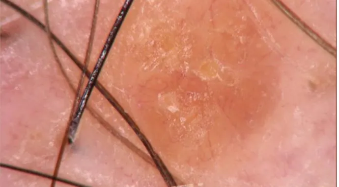
A-50-year old man presented to our clinic for multiple erythematous maculopapules on his front of chest. Initially, the patient was diagnosed as acne two years ago and received oral doxycycline 100mg, sodium sulfacetamide lotion and topical clindamicin treatment but no response was observed. There wasn’t any additional disease or family history. Dermatological examination showed multiple papules with erythema on the chest (Figure-1). Dermoscopy findings showed that, orange and yellowish background and fine linear vessels going from the periphery to the center (Figure-2). In terms of clinical and dermoscopic features, granulomatous diseases and especially sarcoidosis were considered in the patient. A biopsy was performed for the diagnosis of the lesions. Dermal non-caseating granulomas with few lymphoid cells around epithelioid cells were showed in histological examination of a cutaneous biopsy. The chest X-ray and tomography revealed no
ABSTRACT
Sarcoidosis is a multi-systemic granulomatous disease of unknown aetiology. After the lungs, the skin is the second most common organ involved. Cutaneous involvement occurs in up to 30% of patients and skin findings are often the initial presenting symptom. Plaques and papules are the most commonly observed cutaneous lesions. Sarcoidosis can be mimicing other diseases. Dermoscopy is a non-invasive method that allows skin lesions to be visualized. In recent years, it is used for increasing diagnostic accuracy in many skin diseases. Sarcoidosis is one of these diseases. We present a case of 50 year old man, with erythematous maculopapules over the front of chest for about two years. He was treated like acne but had no benefit from therapies. Cutaneus sarcoidosis was diagnosed at the end of t he dermoscopic, clinical and histopatological examination of patient. Here, we emphasized that, sarcoidosis can interfere with many skin diseases and dermoscopy is a non-invasive method that can be used before biopsy in the differential diagnosis.
Kaya İslamoğlu and Demirbaş / Cutenous sarkoidosis, acne and dermoscopy
East J Med Volume:23, Number:4, October-December/2018 342
Fig.1. Multiple papules on the chest
abnormalities. Tuberculin skin test was negative. Laboratory tests were normal. The patient's cardiac, ocular involvement, parotid gland, bone grafts and gastrointestinal examinations showed no sarcoidosis. The patient was diagnosed with cutaneous sarcoidosis based on clinical, dermoscopic and histopatological findings. Systemic steroid therapy was given to the patient. Significant regression was observed in lesions after 1 month (Figure-3).
Discussion
Cutaneous lesions in sarcoidosis are divided into two types: specific lesions in which histopathologic examination of caseating granulomas and non-specific skin lesions in which histopathological granulomas are not observed histologically. Erythema nodosum is the most common cutaneous lesion of sarcoidosis and can be seen in 25% of cases, but not a specific lesion of sarcoidosis. The most common specific lesion of sarcoidosis is papules (5). In our patient we saw papules, too. Papuler sarcoidosis is often seen in the periocular and nasolabial areas (6). Whereas in our patient, the localisation was not compatible with the literature.
Cutaneous sarcoidosis is also known as one of the great imitators in dermatology, together with syphilis and malignant melanoma (1). Lupus vulgaris, foreign body reaction, pseudolymphoma, tuberculoid leprosy,
Fig.2. Orange and yellowish background on dermoscopy
Fig. 3. One month after treatment
granuloma annulare, atypical mycobacterial infection, deep fungal infections, syphilis, lupus miliaris facia, discoid lupus erythematosus and granulomatous rosacea should be considered in differential diagnosis (7). Our patient's lesions were very similar to acne lesions. However, lesions are resistant to treatment and dermoscopic results required biopsy.
The spectrum of inflammatory diseases that can be diagnosed using a dermoscope has markedly increased. Granulomatous skin diseases are a group of inflammatory dermatoses that are characterized pathologically by granuloma formation. Translucent orange areas, white scar-like depigmentation and white scales may be more suggestive of cutaneous sarcoidosis (8). The spectrum of inflammatory diseases that can be diagnosed using a dermoscope
Kaya İslamoğlu and Demirbaş / Cutenous sarkoidosis, acne and dermoscopy
East J Med Volume:23, Number:4, October-December/2018 343
has markedly increased. Granulomatous skin diseases are a group of inflammatory dermatoses that are characterized pathologically by granuloma formation. Few studies evaluated the use of dermoscopy in diagnosing cutaneous sarcoidosis (9). One of them reported that; translucent orange areas, white scar-like depigmentation and white scales may be more suggestive of cutaneous sarcoidosis (8). In other work, Pellicano et al. reported seven cases of cutaneous sarcoidosis, showed presence of yellow translucent globules and vessels on dermoscopy in all patients, and additionally five of them showed central scar like areas (9). Our results showed similar dermoscopic findings.
In conclusion, cutenous sarcoidosis should be considered in unhealed papulopustular lesions. It may be misdiagnosed as acne or the other papulopustular skin diseases since it is a great imitator. Before biopsy, it must be kept in mind that dermoscopy is a non-invasive method that can be used in differential diagnosis.
Acknowledgments
There is no financial and material support.
References
1. Terziroli Beretta-Piccoli B, Mainetti C, Peeters MA, Laffitte E. Cutaneous Granulomatosis: a
Comprehensive Review. Clin Rev Allergy Immunol 2018; 54: 131-146.
2. Tchernev G, Patterson JW, Nenoff P, Horn LC. Sarcoidosis of the skin: a dermatological puzzle: important differential diagnostic aspects and guidelines for clinical and histopathological recognition. J Eur Acad Dermatol Venereol 2010; 24: 125-137.
3. Marchell RM, Judson MA. Cutaneous sarcoidosis. Semin Respir Crit Care Med 2010; 31: 442-451. 4. Noe MH, Rosenbach M. Cutaneous sarcoidosis.
Curr Opin Pulm Med 2017; 23: 482-486.
5. Judson MA. Sarcoidosis: clinical presentation, diagnosis, and approach to treatment. Am J Med Sci 2008; 335: 26-33.
6. Tekin NS. Sarkoidoz. Turk J Dermatol 2012; 6: 80-86.
7. Tchernev. Cutaneous sarcoidosis: the great imitator: etiopathogenesis, morphology, differential diagnosis, and clinical management. Am J Clin Dermatol 2006; 7: 375-382.
8. Ramadan S, Hossam D, Saleh MA. Dermoscopy could be useful in differentiating sarcoidosis from necrobiotic granulomas even after treatment with systemic steroids. Dermatol Pract Concept 2016; 6: 17-22.
9. Pellicano R, Tiodorovic-Zivkovic D, Gourhant JY, et al. Dermoscopy of cutaneous sarcoidosis. Dermatology 2010; 221: 51-54.