Ankara Üniv Yet Fak Derg, 50, 251-252, 2003
Kısa Bilimsel Çalışma / Short Communication
Synovİal sarcoma İn a dog
Tolga GÜVENÇ, Zafer ÖZYILDIZ, Ümit H. MİLLİ
Department of Pathology, Faculty of Yeterinary Medicine, Ankara University, Ankara
Summary: A case of synovial sarcoma in left tarsal joint and metastases of lung at a 10-year-old male mixed breed dog was described. Macroscopically, the tumour mass was 5x4x1.5 cm in size and nodules, 0.5-to-3 cm in size, were seen on the lungs. In microscopical examination, the mass, composed of synovioblastic cells, was diagnosed as synovial sarcoma. Similar tumour cells were seen in the lungs.
Key words: Dog, synovial sarcoma
Bir köpekte sinovial sarkom
Özet: iO yaşlı melez erkek köpeğin sol tarsal ekleminde gözlenen sinovial sarkom olgusu ve akciğer metastazları tanımlandı. Makroskobik olarak, sol tarsal eklem üzerindeki tümör kitlesi 5x4x1.5 cm ölçülerindeydi ve akciğer üzerinde 0.5-3 cm boyutlarında nodüller gözlendi. Mikroskobik incelemede, sinovioblastik hücrelerden oluştuğu gözlenen kitleye sinovial sarkom tanısı kondu. Ben-zer tümör hücreleri akciğerlerde de gözlendi.
Anahtar kelimeler: Köpek, sinovial sarkom
Synovial sareoma is a rarely diagnosed neoplasm of dog (5). It has been reported most eommonly in aged, large breed male dogs, whieh affeets the stifle and elbow (4). The tumour spread by loeal extension along fascial planes and tendon sheats and by via vaseular ehannels is believed to arise from primitiye mesenehymal preeursor eelIs outside the synovial membrane of joints and bursae (4,5). MicroscopiealIy, synovial sareoma is charaeterized by the s)'novioblastie eomponent and fibrosarcomatous eomponent (1-5). Both eelIular elements are seen in biphasic synovial sarcomas. On the other hand, monophasic synovial sarcomas are eomposed of onlyone of these eelIular elements, usualIy the fibroblastic eomponent (6). Cleft formation, giant eelIs and nodular tumour projeetions of synovium might be frequent1y seen as a histologie features of the tumour (1-6).
The aim of this study is to determine the pathological findings of synovial sareoma encountered in a lO-year-old male mixed breed dog.
At necropsy, the tumour mass on the left tarsal joint was grayish in color, 5x4x1.5 cm in size and in the lung s white in color, round to ovoid in shape masses were observed. Tissue speeimens were fixed in 10% buffered
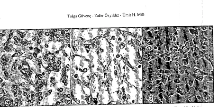
formaldehyde solution and embedded in paraffin wax. Seetions were eut at 5-6 ı.ım and stained with haematoxylin and eosin. Immunohistochemieally, vimentin, desmin and eytokeratin were used to eharaeterization of the tumour celIs. Microscopieally, the neoplasm was composed of synovioblastic elements (Figure la). Cytoplasms of synovioblastie eelIs were eosinophilic and nudei of these eelIs were large, pleomorphie and vesieular. Tumour eelIs were separated by thin fibrous septa. Giant eelI formations were not seen and mitotic figures were less in number. Immunohistochemieally, tumour eelIs were virrientin and cytokeratin positive but desmin negatiye (Figure lb and c). The cytokeratin staining was limited to small dusters of eelIs but diffusely positive for vimentin. Tumour metastases, composed of similar celIs, were seen in the lung s (Figure 2). According to these histopathological and immunohistoehemieal features, tumour was diagnosed as a synovial sareoma.
In the literature, giant celIs, deft formations and nodular tumour projections of synovium has been reported (1,2,4,6). However, in this ease none of these findings were presenL Most of observed synovial sareomas were biphasic (1,2), but this case was monophasie synovial sareoma.
252 Tolga Güyenç - Zafer Özyıldız - Ümit H. Milli
Figure 1. a. SynoYioblastie tumour eeııs on the left tarsal joint. HxE, x260. b. Vimentin positiye tumour eeııs, aYidin-biotin peroxidase teehnique. x260. e. Cytokeratin positiye tumour eeııs, aYidin-biotin peroxidase teehnique. x260.
Figure 2. Metastatic tumour eeııs in the lung. HxE, x60.
Monophasic synovial sarcomas are usually composed to fibrablastic components (5,6). In the present case, only synovioblastic components were seen. Synovial sarcomas are often locally invasive and metastasis to heart, lungs, kidneys, spleen, hver and regional lymph nodes has been reported (1,2,4). In the present case, only lung metastasis was observed. 1. 2. 3. 4. 5. 6.
References
Griffith JW, Frey RA, Sharkey FE (1987): Synovial
sar-~ i
eoma ofthejaw in a dog. J Comp Bath, 97, 361-364.
, i
Lipowitz AJ, Fetter AW, Walkı;r MA (19791): Synovial
sareoma of the dog. JAVMA, 174,76-81.
i
Misdorp W, Van der Heul RO (1976): Tumours ofbones
andjoints. BuJI WHO, 53, 265-282.
i
Mitchell M, Hurov LI (1979): Synovial sareoma in a dog.
JA VMA, 175, 53-55.
i
Pool RR (1990): Tumors and Tumor Like Lesiolı1s of Joints
and Adjacent Sofi Tissues. i34-143. In: JE Moulton (ed), Tumors in Domestie Animals. 3rd ed. Uniyenlity of Ca-Jifornia Press, Berkeley. •
i
Vail DM, Powers BE, Getzy DM!, Morrison WB, McEn-tee MC, O'Keefe DA, Norris AM, WithrowlSJ (1994):
Evaluation of prognostic factorsfor dogs with s)inovial sar-eoma: 36 eases (1986-1991). JAVMA, 205,1300-1307.
.
i
Geliş tarihi: 23.12.2002/ Kabul tarihi: 31.12.2002
Correspondence address:
Dr. Tolga Güvenç
Ankara Üniversitesi, Veteriner Fakültesi Patoloji Anabilim Dalı