Digestive tract helminths of Turkish ibex
(Capra aegagrus aegagrus Erxleben, 1877)
Bahadır GÖNENÇ
1, Hasan EMİR
2, Olimpia IACOB
31Ankara University, Faculty of Veterinary Medicine, Department of Parasitology, Ankara; 2Republic of Turkey Ministry of Forestry
and Water Affairs, General Directorate of Nature Protection and Natural Parks, Wildlife Department, Ankara, Turkey; 3Ion Ionescu
de la Brad University of Agricultural Sciences and Veterinary Medicine, Faculty of Veterinary Medicine, Department of Parasitology and Parasitic Diseases, Iași, Romania.
Summary: Turkish ibex or wild goat (Capra aegagrus aegagrus Erxleben, 1877) is known to be distributed in Southeast Europe
and Western Asia. In Turkey, the species is distributed in Eastern, Northeastern and steep mountainous areas of Southeast Anatolia. Antalya province is one of the main areas where Turkish ibex are currently found and where the species has had a long history. Digestive tracts of two male wild goats (Capra aegagrus aegagrus) were examined at necropsy from Antalya province, Turkey for gastrointestinal helminth infections. In total 3917 helminth parasites were collected and identified by morphological characteristics. Seven species of trichostrongylid nematodes were identified. Marshallagia marshalli and Telodorsagia circumcincta were the most abundant abomasal nematodes whereas Nematodirus abnormalis was dominant in the small intestine. This is the first report of helminths from Capra aegagrus aegagrus (sub-species of wild goat). Comparison with previous reports conducted on domestic goat and sheep in the nearby areas suggests that transmission of parasites between wild goat and domestic small ruminants is occurring.
Keywords: Capra aegagrus aegagrus, digestive tract, helminth, trichostrongylidae, Turkish ibex.
Türk yaban keçilerinde (Capra aegagrus aegagrus Erxleben, 1877) sindirim sistemi helmintleri
Özet: Güneydoğu Avrupa ve Batı Asya’da da yayılış gösteren Türk dağ keçisi veya yaban keçisi (Capra aegagrus aegagrus
Erxleben, 1877) Türkiye’de doğu, kuzeydoğu bölgeleriyle güneydoğunun dik yamaçlarında görülmektedir. Uzun süredir ve günümüzde de yayılış gösterdiği Antalya yöresi, Türk dağ keçisinin temel yayılım alanlarından biridir. Çalışmada, Antalya yöresinden elde edilen iki erkek dağ keçisi nekropsi yapılarak mide bağırsak helmintleri bakımından incelenmiş, toplam 3917 helmint toplanarak morfolojik özelliklerine göre tür teşhisleri yapılmıştır. Trichostrongylid nematodlardan 7 türün tespit edildiği araştırmada, en yaygın türler olarak Marshallagia marshalli, Telodorsagia circumcincta’ya abomasum’da, Nematodirus abnormalis’e ise ince bağırsaklarda rastlanmıştır. Bu çalışma ile ilk kez yaban keçisi alt türü olan Capra aegagrus aegagrus’un sindirim sistemi helmint türleri belirlenmiştir. Çalışmanın yapıldığı bölgelere yakın alanlarda evcil keçi ve koyunlar üzerinde daha önce yürütülen araştırmalarla bu çalışmada elde edilen sonuçlar karşılaştırıldığında, Antalya yöresinde yaşayan yaban keçileri ile evcil küçük ruminantlar arasında paraziter enfeksiyonlar bakımından karşılıklı bulaşmanın söz konusu olabileceği sonucuna varılmıştır.
Anahtar sözcükler: Capra aegagrus aegagrus, helmint, sindirim sistemi, trichostrongylidae, Türk yaban keçisi.
Introduction
Wild goat or Turkish ibex (Capra aegagrus
aegagrus Erxleben, 1877) is known to be distributed in
Southeast Europe and Western Asia. In Turkey, this sub-species are distributed in Eastern, Northeastern and steep mountainous areas of South East Anatolia. Antalya province is one of the main areas where Turkish ibex are currently found and where these species has had a long history (11). Epidemic diseases, overgrazing, uncontrolled hunting and progressive destruction of natural habitats play an important role in significant decline of the wild goat population in Turkey. For this reason, hunting of young and female ibexes were prohibited in 1937 and male ibex was prohibited in 1955. The measures taken
over the years led to an increase in the number of wild goat. The most recent estimates suggest existence of 22,615 ibex in Turkey and existence of 6,373 ibex in Antalya province (20). Currently, it is allowed to hunt the ibex according to the decisions of the Central Hunting Commission under the Land Hunting Law Coded 4915 in 2003.
Knowledge of the parasitic diseases of wildlife focus on protecting endangered species (9, 14, 16). Parasite infection can lead to a reduction in population size and may ultimately contribute to extinction of species (19). Therefore, it is important to investigate and document the prevalence of parasites among threatened species. At the same time, parasitological studies are important to
understand ways of the potential transmission and interaction of parasites between species, both wild and
domestic animals (12, 17). Although various
gastrointestinal helminth species have been reported in various ibex species (1, 3, 4, 15), this is the first report of parasites recovered from Capra aegagrus aegagrus according to a search of the available literature. The aim of the present study was to describe the gastrointestinal helminth fauna recovered from two Turkish ibex in Turkey.
Materials and Methods
Study animals and area: Under the Central Hunting
Commission decision, two male Turkish ibex aged 9 and 7 years old were hunted in Antalya province, Turkey between July and August. One of the two goats was hunted
in Düzlerçamı (Çetin deresi) where is located between 370
04' 05.43" N and 300 31' 30.62" E latitudes, the other was
hunted in Finike (Arif köyü) where is located 360 32'
27.08" N 300 04' 30.57" E latitudes. As soon as possible,
after removal of the alimentary tract from the body cavity, the organs were ligated at various anatomical sites and then were brought to Department of Parasitology, Faculty of Veterinary Medicine, University of Ankara, for parasitological examination of their digestive tracts.
Worm recovery and count: For collected worms from
the dissected animals, the digestive system organs were separated by ligating sites. Gut contents were first filtered through 150 µm sieve and then all contents were examined using a stereo microscope. Every parasite recovered from the gastrointestinal organs was cleaned with physiological saline, fixed in hot 70% alcohol, and identified morphologically (2, 4, 13, 21, 22) with a light microscope (Nikon Eclipse E 600). After identification, parasites were micro-photographed (Nikon DXM 1200 F camera) and
preserved in ethanol-glycerin-formalin protective
solution.
Results
The identified helminths from the digestive tract of
Capra aegagrus aegagrus were classified to the phylum
Nemathelminthes, class Nematoda, order Strongylida, superfamily, Trichostrongyloidea family Trichostrongylidae,
genres Marshallagia and Teladorsagia, species:
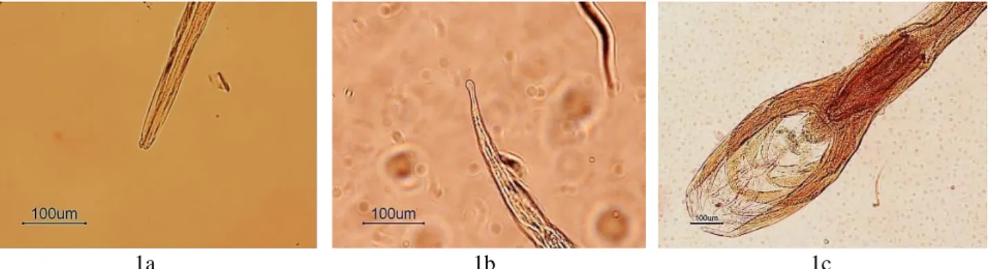
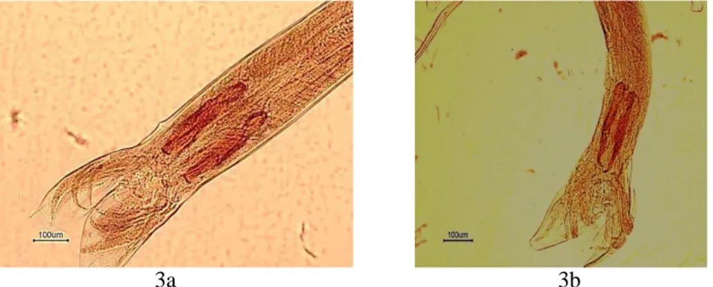
Marshallagia marshalli (Figure 1 a, b, c); Teladorsagia circumcincta (Figure 2 a, b, c, d); T. occidentalis, T. trifurcata (Figure 3 a, b), in the abomasum, also genres Nematodirus and Trichostrongylus, species: N. abnormalis (Figure 4 a, b, c); N. spathiger (Figure 5 a, b), T. colubriformis (Figure 6 a, b, c), in the small intestine.
Figure 1. Marshallagia marshalli a) the stoma b) the tail of female c) caudal end of male. Şekil 1. Marshallagia marshalli a) stoma b) dişi kuyruk bölgesi c) erkek arka uç.
Figure 2. Teladorsagia circumcincta a) the stoma b) the tail of female c) vulvar flap of female d) caudal end of male. Şekil 2. Teladorsagia circumcincta a) stoma b) dişi kuyruk bölgesi c) dişi vulva kapağı d) erkek arka uç.
2a
2b
2c
2d
Figure 3. a) Caudal end of male of Teladorsagia occidentalis b) Caudal end of male of Teladorsagia trifurcate. Şekil 3. a) Teladorsagia occidentalis erkek arka uç b) Teladorsagia trifurcate erkek arka uç.
Figure 4. Nematodirus abnormalis a) the stoma b) the tail of female with a spin c) caudal end of male. Şekil 4. Nematodirus abnormalis a) stoma b) diken taşıyan dişi kuyruk bölgesi c) erkek arka uç.
Figure 5. Nematodirus spathiger a) the stoma b) the caudal end of male. Şekil 5. Nematodirus spathiger a) stoma b) erkek arka uç.
Figure 6. Trichostrongylus colubriformis a) the stoma b) the tail of female c) caudal end of male. Şekil 6. Trichostrongylus colubriformis a) stoma b) diken taşıyan dişi kuyruk bölgesi c) erkek arka uç.
3a
3b
4a
4b
4c
6a
6b
6c
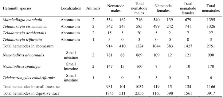
Table 1. The nematode fauna recovered from the digestive tracts. Tablo 1. Sindirim sistemlerinden elde edilen nematodlar.
Helminth species Localization Animals Nematode males Total nematode males Nematode females Total nematode females Total nematodes
Marshallagia marshalli Abomasum 2 554 162 716 540 139 679 1395
Teladorsagia circumcincta Abomasum 2 342 243 585 499 242 741 1326
Teladorsagia occidentalis Abomasum 2 15 5 20 5 2 7 27
Teladorsagia trifurcata Abomasum 1 3 0 3 0 0 0 3
Total nematodes in abomasum 914 410 1324 1044 383 1427 2751
Nematodirus abnormalis Small
intestine 2 781 88 869 109 12 121 990
Nematodirus spathiger Small
intestine 2 147 13 160 7 3 10 170
Trichostrongylus colubriformis Small
intestine 1 3 0 3 3 0 3 6
Total nematodes in small intestine 931 101 1032 119 15 134 1166
Total nematodes in digestive tract 1845 511 2356 1163 398 1561 3917
The number of worms are indicated and their locations in two Turkish ibex are given in Table 1. Mix infections were seen and in total seven nematode species were detected in abomasum and small intestine. No worms were detected in the large intestine of the examined animals.
Discussion and Conclusion
This study showed that Capra aegagrus aegagrus is a host for a number of digestive nematode species. The studied animals were both adult males and it may be that other infections are associated with younger animals. The findings were compared with previous studies on helminth infections in related species in different countries:
Rupicapra R. rupicapra (Alpine chamosis) (7), Capra walie (Walia ibex) (5), Capra ibex ibex (Alpine ibex) (15), Capra pyrenica (Spanish ibex) (1, 18) and West African
Dwarf goat (Nigerian dwarf goat) (6). Marshallagia spp.,
Nematodirus spp., Ostertagia spp. and Trichostrongylus spp., which were also detected in this study, were the most
commonly mentioned helminth genres of these other wild goats’ species.
Helminth parasites of the digestive tract are also widespread among domestic sheep and goats in Turkey (8, 10). Two studies have considered endoparasites of domestic goats and sheep in areas close to where this study was performed (23, 24) and Ostertagia ostertagi,
Teladorsagia circumcincta, T. occidentalis, T. trifurcata, Haemonchus contortus, Marshallagia marshalli, Trichostrongylus axei, T. probolurus, T. vitrinus, Nematodirus abnormalis, N. spathiger, N. filicollis have
all been detected as gastrointestinal helminth infections. The most common species in the abomasum and in small
intestine were M. marshalli and N. abnormalis. The same seven nematode species reported here were found in the abomasum and small intestine of domestic ruminants. It seems that parasites are circulating between wild and domestic ruminants in the study area. Most importantly it may be that the wild goat is acting as a reservoir for some of these infections and may provide a refuge for the parasites away from drug treatment.
This is the first report of the gastrointestinal nematodes fauna of two Capra aegagrus aegagrus in Turkey. The study underlines the need for further studies that may help to design appropriate strategies to the control of helminth parasites in Capra aegagrus aegagrus. It is possible that Turkish ibex can serve as a source of infection of gastrointestinal nematodes for domestic livestock in their living area. In addition, there is a need to further investigate the ecology, seasonality and impact of parasites in wild goat populations in Turkey.
Acknowledgements
The authors thank General Directorate of Nature Conservation and National Parks. We are grateful to Mrs Eileen Harris and Professor David Rollinson of the Natural History Museum, UK for helpful comments and literature support.
References
1. Al Assad S, Morrondo P, Dacal-Rivas V, et al. (2009):
Bronchopulmonary nematode infection of Capra pyrenica in the Sierra Nevada massif, Spain. Vet Parasitol, 164,
340-343.
2. Anderson RC (2000): Nematode Parasites of Vertebrates:
Their Development and Transmission, 2nd ed. CAB
3. Andrews JRH (1973): A host-parasite checklist of
helminths of wild ruminants in New Zealand. New Zeal Vet
J, 21, 43-47.
4. Barji H, Raji AR, Naghibi AG (2011): The comparative
morphology of Marshallagia marshalli and Ostertagia occidentalis (Nematode: Strongylida, Trichostrongylus) by scanning electron microscopy. Parasitol Res, 108,
1391-1395.
5. Bogale B, Chanie M, Melaku A, et al. (2014): Occurrence,
intensity and parasite composition of gastrointestinal helminth parasites in Walia Ibex (Capra walie) at Semien Mountains National Park, Natural World Heritage Site, Northern Ethiopia. Acta Parasitol Glob, 5, 19-25.
6. Chiejina, NS, Jerzy M, Behnke MJ, et al. (2015):
Haemonchotolerance in West African Dwarf goats: contribution to sustainable, anthelmintics-free helminth control in traditionally managed Nigerian dwarf goats.
Parasite, 22, 1-11.
7. Citterio CV, Caslini C, Milani F, et al. (2006): Abomasal
nematode community in an alpine chamois (Rupicapra rupicapra) population before and after a die-off. J Parasitol,
92, 918-27.
8. Doğanay A, Öge S (1997): Türkiye’de koyun ve keçilerde
görülen helmintler. Kafkas Üniv Vet Fak Derg, 3, 97-114.
9. Gebremedhin B, Ficetola GF, Naderi S, et al. (2009):
Combining genetic and ecological data to assess the conservation status of the endangered Ethiopian Walia ibex.
Anim Conserv, 12, 89-100.
10. Güçlü F, Dik B, Kamburgil K, et al. (1996): Konya yöresi
koyunlarında mide-bağırsak nematodlarının yayılışı ve mevsimsel dağılımları. Veterinarium, 7, 50-55.
11. Gündoğdu E, Oğurlu İ (2009): Population ecology of wild
goat Capra Aegagrus Erxleben 1777 in Isparta, Turkey. J
Anim Vet Adv, 8, 2318-2324.
12. Junge RE, Louis EE (2005): Biomedical evaluation of two
sympatric lemur species (Propithecus verreauxi deckeni and Eulemur fulvus rufus) in Tsiombokibo classified forest, Madagascar. J Zoo Wildl Med, 36, 581-589.
13. Lichtenfels JR, Pilitt PA (1983): Cuticular ridge patterns
of Nematodirus (Nematoda: Trichostrongyloidea) parasitic in domestic ruminants of North America, with a key to species. Proc Helminthol Soc Wash, 50, 261-274.
14. Lyles AM, Dobson AP (1993): Infectious-disease and
intensive management-population-dynamics, threatened hosts and their parasites. J Zoo Wildl Med, 24, 315-326.
15. Marreros N, Frey CF, Willisch CS, et al. (2012):
Coprological analyses on apparently healthy Alpine ibex (Capra ibex ibex) from two Swiss colonies. Vet Parasitol,
186, 382-389.
16. Meradi S, Bentounsi B, Zouyed I, et al. (2011): The steppe
species of gastrointestinal nematodes of small ruminants, with a focus on Marshallagia: Climate as a key determinant.
Parasite, 18, 261-269.
17. Morner T (2002): Health monitoring and conservation of
wildlife in Sweden and Northern Europe. Ann N Y Acad
Sci, 969, 34-38.
18. Perez JM, Granados JE, Perez C, et al. (2003): A survey
of the gastrointestinal nematodes of spanish ibex (Capra pyrenica) in a high mountain habitat. J Parasitol, 89,
315-318.
19. Price PW, Westoby M, Rice B, et al. (1986): Parasite
mediation in ecological interaction. Annu Rev Ecol Evol
Syst, 17, 487-505.
20. Sarıbaşak H, Başaran MA, Kaçar S, et al. (2011):
Wildgoat (Capra aegagrus Erxleben, 1777) Population in Antalya-Düzlerçamı Wildlife Progress Area and Evaluation of its Habitat. Batı Akdeniz Orm Araş Enst Tek Bült, 57,
1-62.
21. Soulsby EJL (1965): Textbook of veterinary clinical
parasitology. Vol. I. Helminths. Blackwell, Oxford.
22. Tınar R, Umur Ş, Köroğlu E, et al. (2011): Veteriner
Helmintoloji. Dora Basım Yayın Ltd. Şti, Bursa.
23. Umur Ş, Yukarı BA (2005a): Seasonal activity of
gastro-intestinal nematodes in goats in Burdur region, Turkey.
Turkish J Vet Anim Sci, 29, 441-448.
24. Umur Ş, Yukarı BA (2005b): An abattoir survey of
gastro-intestinal nematodes in sheep in the Burdur region, Turkey.
Turkish J Vet Anim Sci, 29, 1195-1201.
Geliş tarihi: 03.03.2017 / Kabul tarihi: 22.05.2017
Address for correspondence:
Prof.Dr. Bahadır GÖNENÇ
Ankara University, Faculty of Veterinary Medicine, Department of Parasitology
06110, Dışkapı, Ankara, Turkey. e-mail: [email protected]