Could the cytological evaluation of pericardial
effusions illuminate our path?
Perikardiyal efüzyonlarda sitolojik değerlendirme yolumuzu
aydınlatabilir mi?
Cenk EKMEKCİ1, Sümeyye EKMEKCİ2, Yelda DERE3, Yasemen ADALI4, Selim EKİNCİ1, Ali Kemal ÇABUK1, Pınar ÖKSÜZ2, Hatice SOLMAZ1, Öner ÖZDOĞAN1
1Sağlık Bilimleri Üniversitesi Tepecik Eğitim ve Araştırma Hastanesi Kardiyoloji Kliniği, İzmir, Türkiye 2Sağlık Bilimleri Üniversitesi Tepecik Eğitim ve Araştırma Hastanesi Patoloji Bölümü, İzmir, Türkiye 3Muğla Sıtkı Koçman Üniversitesi Tıp Fakültesi, Patoloji Anabilim Dalı, Muğla, Türkiye
4Çanakkale Onsekiz Mart Üniversitesi Tıp Fakültesi, Patoloji Anabilim Dalı, Çanakkale, Türkiye
ABSTRACT
Objective: Pericardial effusion (PE) is a common clinical condition that can develop as a
result of systemic or heart disease. In our study, we synthesized the cytopathological and clinical results of patients who underwent pericardiocentesis due to pericardial effusion.
Method: A total of 213 patients who underwent percutaneous pericardiocentesis between
2007-2017 were included in the study: their cytologic and histopathologic diagnoses were noted and their relations were examined.
Results: Hundred and thirty-two cases were male (61.9%), 81 were female (38.1%) and the
mean of the study population age was 59.9 (min 13-max 97) years. Hundred and sixty-eight patients had benign (78.9%), 10 suspicious (4.6%), 3 non-diagnostic (1.4%) and 32 malig-nant cytologies (15.1%). Benign pericardial effusion was the most common diagnosis. Malignant cytology findings were interpreted as lung carcinoma (n=20: 62.5%), rhab-domyosarcoma (n=1: 3.1%), poorly differentiated adenocarcinoma (n=2: 6.2%), a gastro-intestinal system carcinoma (n=4: 12.5%), undifferentiated epithelial tumor (n=1: 3.1%), breast carcinoma (n=1: 3.1%), and unspecified malignant tumor (n=3: 9%). Four (2.4%) of the 168 patients having diagnosis of benign cytology had previously received diagnosis of a malignant disease, however examination of cytological specimen. Did not reveal any malignancy. Three (30%) of the 10 patients with suspicious cytology, had received diagno-sis of a malignant disease previously.
Conclusion: In developed countries, it is reported that more than 50% of the PE’s are
idiopathic. The percentage of cancer-associated PE’s is 10-25%. In our study, 78.9% of our cases had a benign diagnosis, and 15.1% had malignant PE consistent with the litera-ture findings. Cytological sampling in pericardial fluid is a method that can shed light on the diagnosis of many diseases.
Keywords: Pericardial effusion, pericardiocentesis, cytopathology ÖZ
Amaç: Perikardiyal efüzyon (PE) sistemik veya kalp hastalıklarının bir sonucu olarak
gelişebilen yaygın bir klinik tablodur. Çalışmamızda, perikardiyal efüzyon nedeniyle peri-kardiyosentez uygulanan hastaların sitopatolojik ve klinik sonuçlarını sentezledik.
Yöntem: Çalışmaya 2007-2017 yılları arasında perkütan perikardiyosentez yapılan 213
hasta alınmış, sitolojik ve histopatolojik tanıları not edilmiş ve ilişkileri irdelenmiştir.
Bulgular: Olguların 132’si erkek (%61,9), 81’i kadın (%38,1) olup, yaş ortalaması 59,9
(min. 13 - maks. 97)’dur. Sitolojik bulgulara göre; 168’i benign sitoloji (%78,9), 10’u kuş-kulu sitoloji (%4,6), 3’ü tanısal olmayan (%1,4) ve 32’si malign sitoloji (%15,1) tanısı almıştır. En sık benign perikardiyal efüzyon tanısı mevcuttur.Malign sitoloji tanılı olguların 20’si akciğer karsinomu (%62,5), 1’i rabdomyosarkom (%3,1), 2’si az diferansiye adeno-karsinom (%6,2), 4’ü gastrointestinal sistem ilişkili adeno-karsinom (%12,5), 1’i indifferansiye epitelyal tümör (%3,1), 1’i meme karsinomu (%3,1), 3’ü tiplendirme yapılamayan malign tümör (%9) olarak yorumlanmıştır. Benign sitoloji tanılı 168 hastanın 4 (%2,4)’ünün malig-nite tanısı var olmakla birlikte, sitolojik örneğe yansıyan maligmalig-nite bulgusu yoktur. Kuşkulu sitoloji olarak tanı almış 10 olgunun 3’ünün (%30) malignite tanısı mevcuttur.
Sonuç: Gelişmiş ülkelerde PE’lerin %50’sinden fazlasının idiyopatik olduğu
bildirilmekte-dir. Kanser ilişkili PE’lerin oranı %10-25’tir. Çalışmamızda, literatürle uyumlu olarak %78,9 olgu benign, %15,1 olgu malign PE’dir. Perikardiyal sıvılarda sitolojik örnekleme birçok hastalığın tanısına ışık tutabilen bir inceleme yöntemidir.
Anahtar kelimeler: Perikardial efüzyon, perikardiyosentez, sitopatoloji
Alındığı tarih: 26.02.2018 Kabul tarihi: 08.03.2018
Yazışma adresi: Uzm. Dr. Cenk Ekmekci, Sağlık
Bilimleri Üniversitesi Tepecik Eğitim ve Araştırma Hastanesi, Yenişehir - Konak - İzmir - Türkiye
INTRODUCTION
The pericardium is a double-walled sac contai-ning the heart and roots of the great vessels. Pericardial sac has two layers; serous visceral layer and fibrous parietal layer. Pericardium stabilizes the heart to the mediastinum, protects against infections and provi-des easy movement to the heart (1,2). The composition
of the normal pericardial fluid can be described as an ultrafiltration of the plasma, except for its low prote-in content (3). Some systemic and local disorders such
as coronary artery diseases, malignancies, connective tissue disorders, infections, bleeding disorders and idiopathic causes may lead to pericardial effusion (PE) by disrupting the balance between production and drainage of pericardial fluid (4).
Accurate and early diagnosis is very important in PEs. Pericardiocentesis is the gold standard for the identification of specific etiology. The cause of patho-logic PE is not always clear and the etiology of more than 50% of the cases is unknown. In this study, the clinical and pathological features of PE patients who underwent pericardiocentesis were discussed. There is limited data on cytological evaluation of PE and its etiological role in PE. For this reason, we aimed to report the outcome of pericardial cytology in patients with PE who underwent pericardiocentesis.
MATERIAL and METHOD
In this study, pericardial cytologies of 213 patients who underwent pericardiocentesis due to pericardial effusion between 2007 and 2017 were reviewed. Samples of hemodynamically important pericardial effusions obtained by percutaneous pericardiocente-sis were sent to laboratories for cytopathological and microbiological examination. Patients who had expe-rienced cardiac catheterization, pericardial tampona-de after surgical intervention, and pericardial tampo-nade in the early period of acute myocardial infarcti-on were excluded from the study.
Patients’ clinical, radiological, pathological and laboratory information was obtained from hospital
electronic records. The identified cases were assessed cytologically by 3 pathologists. Patients whose cyto-logical preparations could not be reached due to re-evaluation at different centers were excluded from the study. In terms of evaluation, cases without any cellular competence were accepted as having “non-diagnostic cytology”. Patients who met “non-diagnostic qualification criteria were classified as having benign, suspicious and malignant cytologies according to cytological findings. In case of malignant cytology, cytological typing was made according to whether there is any evidence of cytologic reflection of pri-mary tumor. Patients having malignant cytology were compared clinically and radiologically with regard to the presence or absence of the primary focus, and the interpretation of the cytological material. The results of clinical and radiological methods of all patients were compared with cytological diagnoses.
RESULTS
Hundred and thirty-two cases were male (61.9%), 81 were female (38.1%) and the mean age of the study population was 59.9 (min 13 - max 97) years. The patients had benign (n=168: 78.9%), suspicious (n=10: 4.6%), non-diagnostic (n=3: 1.4%) and malig-nant (n=32: 15.1%) cytologies. Benign pericardial effusion is the most common diagnosis (Table).
Table. Findings Age Gender - Female - Male Pathologic diagnosis Benign Suspicious Malignant Non-diagnostic
Malignant cytology findings - Lung carcinomas
- Gastrointestinal system related carcinomas - Poorly differentiated adenocarcinoma - Undifferentiated malignant epithelial tumor - Rhabdomyosarcom
- Breast carcinoma
- Malignant tumor which were not specified
N (%) 59.9 (min. 13 - max. 97) 81 (38.1 %) 132 (61.9 %) 168 (78.9 %) 10 (4.6 %) 32 (15.1 %) 3 (1.4 %) 20 (62.5%) 4 (12.5 %) 2 (6.2 %) 1 (3.1 %) 1 (3.1 %) 1 (3.1 %) 3 (9%)
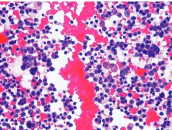
Malignant cytology findings were interpreted as lung carcinoma (n=20: 62.5%) (Figure 1-2), rhabdomyo-sarcoma (n=1: 3.1%), poorly differentiated adenocar-cinoma (n=2: 6.2%), gastrointestinal system related carcinoma (n=4: 12.5%), undifferentiated epithelial tumor (n=1: 3.1%), breast carcinoma (n=1: 3.1%), and unspecified malignant tumor (n=3: 9.4%). Four (2.4%) of the 168 patients diagnosed as benign cyto-logy were diagnosed as malignant previously and there was no malignancy finding in the cytologic specimen. Primary tumor of these cases was lung carcinoma. Three (30%) of the 10 patients having suspicious cytology, had previously received the diagnosis of malignancy. Primary tumors of these 3 cases were Hodgkin’s lymphoma, esophageal and
breast carcinomas.
Most of the patients (n=27: 84.38%) having malignant pericardial fluid were male whilst 5 cases (15.62%) were female. The primary tumors of the female patients were lung carcinoma (n=2), breast carcinoma (n=1) and nonspecific malignant tumors (n=2).
DISCUSSION
Normal pericardial sac contains 10-50 ml of peri-cardial fluid as a plasma ultrafiltrate which functions as a lubricant between the pericardial layers. Deposition over this level is considered to be patho-logical PE. Pericardial diseases can be part of a syste-mic disease or can be isolated. The most common cause of PE is idiopathic and in developed countries its incidence increases up to 50%. Mayosi et al. reported other common causes as cancer (10-25%), infections (15-30%), iatrogenic causes (15-20%) and connective tissue diseases (5-15%) (5,6).
Metastatic tumors, especially lung and breast can-cer, esophageal squamous cell carcinoma, malignant melanoma, thymic carcinoma, kidney, bladder malig-nancies as well as mesenchymal tumors like Ewing’s sarcoma or hematological malignancies (mostly leu-kemia, non-Hodgkin’s lymphoma and Hodgkin’s lymphoma) are the most common causes for patholo-gical PE (7). Primary tumors of the pericardium such
as mesothelioma, fibrosarcoma, lymphangioma, hemangioma, teratoma, neurofibroma and lipomas are rare (8). The third most common cause is infection
and it is often viral. The culprit viral agents include enteroviruses (coxsackieviruses, echoviruses), herpes viruses (EBV, CMV, HHV-6), adenoviruses and par-vovirus B19. Among bacterial agents, Mycobacterium tuberculosis infection is the most frequent cause and it is more common in developing countries. Another common group is autoimmune diseases such as systemic lupus erythematosus, rheumatoid arthritis, scleroderma. Diseases such as amyloidosis, aortic dissection, pulmonary arterial hypertension, hypoth-yroidism, uremic pericarditis, chronic heart failure
Figure 1. Malignant tumor cells (H&E, x400).
may rarely cause PE (9).
In malignancies, pericardial effusion is a common and serious finding (10,11). Gornik and colleagues
reported that 43.8% of the patients who underwent pericardiocentesis had a malignant pericardial effusi-on (11). Mukai et al. (12) reported that malignant
invasi-on of the pericardium can be seen in approximately 10% of all cancer patients and that one third of these patients lost their lives as a result of pericardial effu-sion. The prognosis of the cases which have malig-nant cells in PE fluid were found to be worse than in cases without malignant cells (13). Postmortem studies
have shown that approximately 15-20% of cancer patients have metastatic disease in the heart and peri-cardium (14). There is limited literature on the timing
of malignant pericardial effusion in relation to pri-mary malignancy. Holdener et al. (15) reported that in
a group of patients with malignant pericardial effusi-on, the initial manifestation of the disease was malig-nant pericardial effusion in 50% of patients.
In oncological patients, the pericardial effusion may be caused by several different pathophysiologi-cal mechanisms, such as (a) direct or metastatic spre-ad of the tumor (b) systemic tumor therapy-as a complication of radiation therapy, chemotherapeutic toxicity, or combination therapy (c) opportunistic infections occurring during antineoplastic therapies
(16). In cancer patients, pericardial effusions occur
most commonly (>50%) due to benign lymphatic obstruction caused by the above-mentioned three mechanisms (17). This is why the diagnosis of benign
cytology should not distract the clinician from the likelihood of a cancer diagnosis. In our study, 4 (2.4%) of 168 patients diagnosed with benign cyto-logy had previously received the diagnosis of malig-nancy. In some cases, pericardial effusion may be the first finding of the disease. Among the malignant pericardial effusions, lung cancer was most often diagnosed in accordance with our results. According to previous data in lung cancer patients the presence of malignant cells in pericardial effusions had been associated with worse survival relative to those wit-hout malignant cells (18). Because of all these reasons,
rapid diagnosis and precise discrimination of malig-nant pericardial effusion has therapeutic and prog-nostic importance. Pericardiocentesis is the gold standard for elucidating specific etiology. The main clinical purpose of the cytopathological evaluation is to identify malignant tumor cells and this method has a sensitivity of 66%-100% (19,20). In conclusion,
detec-tion of etiology in pericardial effusions is very chal-lenging and time consuming. Cytopathological exa-mination can illuminate us from a diagnostic point of view and significantly reduce the cost of pertinent examinations thanks to the rapid diagnosis of the disease.
REFERENCES
1. Bonow RO, Mann DL, Zipes DP, Libby P. Braunwald’s Heart Disease, 9th ed., Elsevier Saunders, Philadelphia, 2012. p.1651-2.
2. Jöbsis PD, Ashikaga H, Wen H, Rothstein EC, Horvath KA, McVeigh ER et al.
https://doi.org/10.1152/ajpheart.00967.2007
3. Spodick DH. The Pericardium: A Comprehensive Textbook, Informa Healthcare, London, UK, 1997.
4. Akhter SA. The heart and pericardium. Thoracic Surgery Clinics. 2011;21:205-17.
https://doi.org/10.1016/j.thorsurg.2011.01.007
5. Mayosi BM. Contemporary trends in the epidemiology and management of cardiomyopathy and pericarditis in sub-Saharan Africa. Heart. 2007;93:1176-83.
https://doi.org/10.1136/hrt.2007.127746
6. Mayosi BM, Burgess LJ, Doubell AF. Tuberculous pericardi-tis. Circulation. 2005;112:3608-16.
https://doi.org/10.1161/CIRCULATIONAHA.105.543066 7. Jeong TD, Jang S, Park CJ, Chi HS. Prognostic relevance of
pericardial effusion in patients with malignant diseases. Korean J Hematol. 2012;47:237-8.
https://doi.org/10.5045/kjh.2012.47.3.237
8. Karatolios K, Alter P, Maisch B. Differentiation of malignant from nonmalignant, inflammatory pericardial effusions with biomarkers. Herz. 2009;34:624-33.
https://doi.org/10.1007/s00059-009-3304-8
9. Ristic´ AD, Imazio M, Adler Y, Anastasakis A, Badano LP, Brucato A et al. Triage strategy for urgent management of cardiac tamponade: a position statement of the European Society of Cardiology Working Group on Myocardial and Pericardial Diseases. Eur Heart J. 2014;35:2279-84.
https://doi.org/10.1093/eurheartj/ehu217
10. Halfdanarson TR, Hogan WJ, Moynihan TJ. Oncologic emergencies: diagnosis and treatment. Mayo Clin Proc. 2006;81:835-48.
https://doi.org/10.4065/81.6.835
11. Gornik HL, Gerhard-Herman M, Beckman JA. Abnormal cytology predicts poor prognosis in cancer patients with peri-cardial effusion. J Clin Oncol. 2005;23:5211-6.
12. Mukai K, Shinkai T, Tominaga K, Shimosato Y. The inciden-ce of secondary tumours of the heart and pericardium: a 10 year study. Jpn J Clin Oncol. 1988;18:195-201.
13. He B, Yang Z, Zhao P, Li YJ, Wang JG. Cytopathologic analysis of pericardial effusions in 116 cases: Implications for poor prognosis in lung cancer patients with positive interpretations. Diagn Cytopathol. 2017;45(4):287-93. https://doi.org/10.1002/dc.23671
14. Laham RJ, Cohen DJ, Kuntz RE, Baim DS, Lorell BH, Simons M. Pericardial effusion in patients with cancer: out-come with contemporary management strategies. Heart. 1996;75:67-71.
https://doi.org/10.1136/hrt.75.1.67
15. Holdener EE, Ryser DH, Schaermeli K, Spieler P, Angehrn W, Reutter FW et al. Malignant pericardial effusion-a sign of unfavorable prognosis. Schweiz Med Wochenschr. 1986;116:366-70.
16. Refaat M, Katz W. Neoplastic pericardial effusion. Clin Cardiol. 2011;34:593-8.
https://doi.org/10.1002/clc.20936
17. Wilkes JD, Fidias P, Vaickus L, Perez RP. Malignancy-related pericardial effusion. 127 Cases from the Roswell Park Cancer Institute. Cancer. 1995;76:1377-87.
https://doi.org/10.1002/1097-0142(19951015)76:8<1377:: AID-CNCR2820760813>3.0.CO;2-M
18. El Haddad D, Iliescu C, Yusuf SW, William NW, Khair TH, Song J et al. Outcomes of cancer patients undergoing percu-taneous pericardiocentesis for pericardial effusion. J Am Coll Cardiol. 2015;66:1119-28.
https://doi.org/10.1016/j.jacc.2015.06.1332
19. Imazio M, Adler Y. Management of pericardial effusion. Eur Heart J. 2013;34:1186-97.
https://doi.org/10.1093/eurheartj/ehs372
20. Dragoescu EA, Liu L. Pericardial fluid cytology: An analysis of 128 specimens over a 6-year period. Cancer Cytopathol. 2013;121:242-51.