Is there any association between colonic polyps and gastric
intestinal metaplasia?
Gülhan Kanat Ünler1, Gülsüm Teke Özgür2, Hüseyin Savaş Göktürk1, Hüseyin Korkmaz1, Özgür Hilal Erinanç3 1Department of Gastroenterology, Başkent University School of Medicine, Konya, Turkey
2Department of Family Medicine, Başkent University School of Medicine, Konya, Turkey 3Department of Pathology, Başkent University School of Medicine, Konya, Turkey
INTRODUCTION
Although its incidence varies, gastric cancer is the fourth-most prevalent cancer and the second-most common cause of cancer-related deaths worldwide (1). Intestinal-type gastric cancer, the most common subtype, is thought to develop by the carcinogen-esis cascade defined by Correa (2). In this model, the progression of chronic gastritis is a multistep process of atrophy, intestinal metaplasia (IM), and dysplasia, which may lead to invasive carcinoma (2). Despite ag-gressive and expensive treatments, most patients with gastric adenocarcinoma die during the first year due to delayed diagnosis. Therefore, the early treatment and follow-up of patients with precancerous lesions, includ-ing IM and chronic atrophic gastritis, is important (3). Although guidelines exist for some conditions, such as Barrett’s esophagus, ulcerative colitis, and colonic
pol-yps, there are no clear recommendations for gastric IM, a precancerous gastric lesion. The 2006 American Soci-ety for Gastrointestinal Endoscopy guidelines reported that most patients in the United States had a low risk of cancer progression; therefore, endoscopic surveil-lance for IM was not indicated for average-risk patients (4). According to the 2012 European Society of Gastro-intestinal Endoscopy guidelines, patients with extensive atrophy and metaplasia should be followed up every 3 years (5). However, there are disagreements over the rec-ommended follow-up of IM limited to the antrum and 3-year follow-up interval for extensive metaplasia (6). Helicobacter pylori infection is a major risk factor for the development of gastric IM. A number of studies have reported an association of H. pylori infection with pathogenesis of colonic polyps and colorectal cancer (CRC). This infection is thought to increase the
frequen-Address for Correspondence: Hüseyin Savaş Göktürk E-mail: [email protected] Received: June 18, 2015 Accepted: August 30, 2015
© Copyright 2016 by The Turkish Society of Gastroenterology • Available online at www.turkjgastroenterol.org • DOI: 10.5152/tjg.2016.15212
STOMACH
Or
iginal Ar
ticle
ABSTRACT
Background/Aims: Chronic gastritis progression is a multistep process of atrophy, intestinal metaplasia (IM), and dysplasia, which may lead to invasive carcinoma. In this study, we identified an association of colonic pol-yps with gastric IM in patients undergoing colonoscopy.
Materials and Methods: This retrospective case-control, cross-sectional study was conducted in a tertiary-care institution in Turkey. Pathology and endoscopy reports were reviewed. The study group comprised 400 patients with colonic adenomatous polyps, and the control group comprised 360 patients without colonic adenoma-tous polyps on colonoscopy.
Results: The risk of gastric IM was 1.42-fold higher in the study group (p<0.05). The risk of IM in patients aged ≥50 years with colonic polyps was 3.35-fold higher than in those aged <50 years (p<0.05). The risk of Helico-bacter pylori infection in the study group was 1.07-folder higher than that in the control group (p<0.05). H. pylori infection prevalence was higher only in patients with high-grade colonic polyp dysplasia (p<0.05). There were no statistically significant differences in the proportion of incomplete IM between the groups (p<0.05). Conclusion: This study observed increased rates of gastric IM with colonic polyps. An increased risk of gastric IM was associated with higher grades of polyp dysplasia.
cy of colonic polyps and CRC. The effect of H. pylori infection on colonic polyps and CRC has been investigated in patients undergoing gastroscopy. In this study, we identified an asso-ciation of colonic polyps with gastric IM in patients undergo-ing colonoscopy and determined if the location or number of colonic polyps or the presence of polyp dysplasia can be used to gastric atrophy or gastric IM.
MATERIALS AND METHODS Data collection
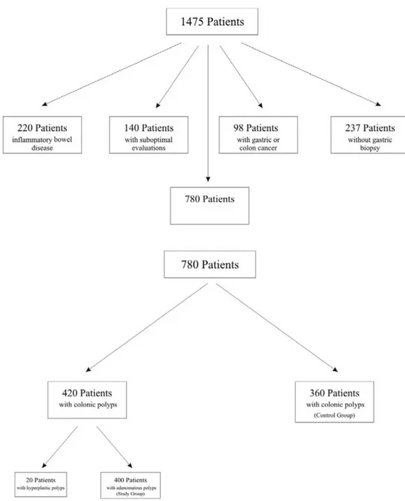
This retrospective case-control, cross-sectional study was con-ducted at Başkent University Medical and Research Center, a tertiary-care institution in Konya, Turkey. Patient pathology and endoscopy reports from January 1, 2010 to December 31, 2013 were reviewed from the electronic database. We analyzed data of 1,475 patients over 18 years of age who underwent gastros-copy and colonosgastros-copy on the same day at the gastroenterol-ogy clinic. Indications for gastrointestinal endoscopy included chronic diarrhea (10%), CRC screening (12%), weight loss (18%), anemia (22%), melena (4%), abdominal pain (21%), abdominal bloating (10%), and a positive fecal occult blood test (3%). Four gastroenterologists who had received the same gastroenterol-ogy fellowship training performed all endoscopies (Evis Exera II system, CLV-180, Olympus, Inc.; Tokyo, Japan).
We excluded 220 patients with inflammatory bowel disease, 237 without antrum or incisura biopsies, 98 with gastric can-cer or CRC, 102 with inadequate bowel preparation, 38 with unsuccessful cecal intubation, and 20 with hyperplastic polyps. Of the remaining 760 patients, demographic data and the pres-ence of H. pylori infection, gastric atrophy, or IM were evaluated in antrum and incisura biopsy specimens. The study group comprised 400 patients with colonic adenomatous polyps, and the control group comprised 360 patients without polyps on colonoscopy. Patient selection is depicted in Figure 1.
Antrum and incisura biopsies were stained using hematoxylin– eosin and Giemsa stains. Metaplasia type was visualized using Periodic acid-Schiff (PAS) and Alcian Blue staining (PAS–Alcian blue stain). The number, location, and size of adenomatous polyps and the presence of dysplasia were also assessed in the study group. The location of colonic polyps was documented and classified as proximal colon, distal colon, or pan-colonic. The proximal colon included the transverse colon, hepatic flex-ure, ascending colon, and cecum. The distal colon included the rectum, sigmoid, descending colon, and splenic flexure. Pol-yps located in both were classified as pan-colonic. Advanced adenomas were defined as large adenomas (≥10 mm in size) or adenomas with histopathologic findings of villous or high-grade dysplasia.
The study was approved by the local Ethics Committee of Başkent University.
Statistical analysis
Data were analyzed using SPSS Statistics for Windows, Version 17.0. (SPSS Inc.; Chicago, IL, USA). The odds ratio (OR) was calcu-lated to determine the association of two binary variables. Fur-thermore, logistic regression analysis was utilized to evaluate the relationships between risk factors of the binary variables; ORs were used to interpret these results. Differences between the means of two continuous variables were analyzed using t-tests, and differences between two proportions were analyzed using z-tests. Statistical analyses were performed using R (3.1.2). P values less than 0.05 were considered statistically significant in all analyses.
RESULTS
A total of 760 adult patients, 400 with colonic adenomatous polyps (study group) and 360 without polyps (control group), were included. Among the 400 patients with colonic polyps, 205 were men and 195 were women. The control group com-prised 360 patients; 145 were men and 215 were women. OR was as 0.64 (p<0.05). Men had a 1.5-fold higher risk of colonic polyps than women (Table 1).
The mean overall age of the 760 patients was 57 years; the mean age for the study and control groups were 58 years (30–87 years) and 56 years (21–93 years), respectively. T-test analysis of these age differences was not statistically significant (p>0.05).
Demographic data and the presence of H. pylori infection, gas-tric atrophy, or IM were first analyzed for all 760 patients. Pa-tients were classified as younger than 50 years or 50 years and older. Associations of risk factors (demographic data and pres-ence of colonic polyps) with gastric IM, gastric atrophy, and H. pylori infection were analyzed by multiple logistic regression analyses (Table 2 and 3).
Or
iginal Ar
ticle
Gender
Male Female Total Patients with colonic polyps (study group) 205 195 400 Patients without colonic polyps (control group) 145 215 360
Total 350 410 760
Table 1. Gender characteristics
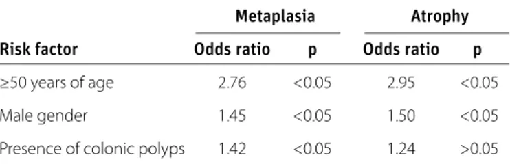
Metaplasia Atrophy
Risk factor Odds ratio p Odds ratio p
≥50 years of age 2.76 <0.05 2.95 <0.05
Male gender 1.45 <0.05 1.50 <0.05
Presence of colonic polyps 1.42 <0.05 1.24 >0.05
IM: intestinal metaplasia
Table 2. Multiple logistic regression analysis for risk factors associated with gastric IM or atrophy
As shown in Table 2, the risk of developing gastric IM in patients aged ≥50 years was 2.76-fold higher than those aged <50 years (OR=2.76, p<0.05). The risk of developing gastric IM in men was 1.45-fold higher than in women (OR=1.45, p<0.05). In addition, the risk of gastric IM in the study group was 1.42-fold higher than that in the control group (OR=1.42, p<0.05).
The risk of gastric atrophy in patients aged ≥50 years was 2.95-fold higher than those aged <50 years (OR=2.95, p<0.05) (Table 2). Similarly, the risk of developing gastric atrophy was 1.5-fold higher in men than in women (OR=1.50, p<0.05). However, the risk of developing gastric atrophy did not significantly differ be-tween the control and study groups (OR=1.24, p>0.05).
Unlike gastric IM and atrophy, the rate of H. pylori infec-tion in patients aged <50 years was 1.15-fold higher than in those aged ≥50 years (Table 3). However, this difference was not statistically significant (OR=1.15, p>0.05). H. pylori infec-tion was 1.30-fold more common among men than among women; this risk difference was also not statistically significant (OR=1.30, p>0.05). The risk of the presence of H. pylori infection in the study group was 1.07-fold higher than that in the control group (OR=1.33, p<0.05).
Risk factors that might be associated with IM and H. pylori in-fection in the study group were also examined. The results showed that the risk of developing gastric IM in patients with
Or
iginal Ar
ticle
colonic polyps aged ≥50 years was 3.35-fold higher than in those aged <50 years (OR=3.35, p<0.05) (Table 4). Although the risk of developing IM was 1.47-fold higher in men with co-lonic polyps than women, the difference was not statistically significant (OR=1.47, p>0.05). The location or number of co-lonic polyps did not play a significant role in the risk of gastric IM in the study group (OR=1.04, p>0.05 and OR=1.13, p>0.05, respectively). The presence of advanced adenoma was associ-ated with a significantly increased risk of gastric IM (OR=1.27, p<0.05).
The presence of H. pylori infection was higher only in patients with high-grade colonic polyp dysplasia (OR=1.32, p<0.05) (Table 5). Patient age or gender and the location and number of colonic polyps were not significantly associated with gastric dysplasia grade (OR=1.00, p>0.05; OR=1.30, p>0.05; OR=1.16, p>0.05; OR=1.05, p>0.05; OR=1.06, p>0.05, respectively). Table 6 shows the number of the patients in both groups with incomplete and complete IM. According to the z-test for pro-portions of two samples, there was no statistically significant difference between the proportions of incomplete IM in these groups (p>0.05).
DISCUSSION
IM is the conversion of the superficial epithelium of oxyntic and antral mucosa into the intestinal epithelium. H. pylori infection is the main factor for the development of gastric IM. H. pylori infection can result in the development of chronic gastritis, glandular atrophy, IM, and dysplasia before carcinoma devel-ops. Colonic polyps may be precursors of CRC through an ad-enoma-carcinoma sequence. The present study from a tertiary hospital investigated the association of IM with colonic polyps that may be precursors of malignancies in the gastrointestinal tract, and also determined if there were signs that might rec-ommend gastroscopy in patients with polyps detected during colonoscopy. Our data showed that the risk of having gastric IM in patients with colonic polyps was than that in patients without colonic polyps (OR=1.42, p<0.05).
The risk of histopathologically determined H. pylori infection in the study group was higher than in the control group in this study, which is consistent with other reports (OR=1.33, p<0.05) (7-10). There was no statistical difference between the mean ages of the study and control groups in this study, but statis-tically more men had colonic polyps. This data showed that male patients had 1.5 times the risk of colonic polyps com-pared to female patients (OR=0.64, p<0.05). The increased rate of colonic polyps in men was also consistent with the literature (11,12). The analysis indicated that the risk of developing gas-tric IM in patients ≥50 years of age was 2.76 times higher than in patients <50 years (OR=2.76, p<0.05). The risk of developing gastric IM was 1.45 times higher in men than in women. This finding was also consistent with other reports (13).
Among common factors, H. pylori infection is most often as-sociated with the pathogenesis of gastric IM and colonic pol-yps. The association of colonic polyps and CRC with H. pylori infection has been reported in many meta-analyses from 2006 to 2013. These previous studies have suggested that H. pylori infection leads to a 1.4–1.6-fold increased risk of colonic ade-noma or CRC. H. pylori infection in patients with CRC or colonic polyps detected by serology and 13C-urea breath tests (7-10).
Recent publications have used immune-histochemical meth-ods to detect H. pylori in colonic polyps and CRC tissues (14). H. pylori-induced gastritis, atrophy, metaplasia, and cancer cas-cade in the gastric mucosa is now well accepted. Atrophy and hyper-gastrinemia developing during this cascade can have a
Or
iginal Ar
ticle
Risk factor Odds ratio p
<50 years of age 1.15 >0.05
Male gender 1.30 >0.05
Presence of colonic polyps 1.33 <0.05
Table 3. Multiple logistic regression analysis for risk factors associated with
H. pylori infection
Risk factor Odds ratio p
≥50 years of age 3.35 <0.05
Male gender 1.47 >0.05
Location of colonic polyps 1.04 >0.05
Number of colonic polyps 1.13 >0.05
Presence of advanced adenoma 1.27 <0.05
Table 4. Multiple logistic regression analysis for risk factors associated with gastric intestinal metaplasia
Risk factor Odds ratio p
≥50 years of age 1.00 >0.05
Male gender 1.30 >0.05
Location of colonic polyps 1.16 >0.05
Number of colonic polyps 1.05 >0.05
Presence of advanced adenoma 1.32 <0.05
Grade of gastric dysplasia 1.06 >0.05
Table 5. Multiple logistic regression analysis for H. pylori infection-associated risk factors in the study group
IM type
Incomplete Complete Total
Patients with colonic polyps 45 60 105
Patients without colonic polyps 25 47 72
Total 70 107 177
IM: intestinal metaplasia
trophic effect on colonic mucosa. They can also contribute to the development of carcinogenicity, with a direct effect on the colonic mucosa. Most hypotheses about the effects of H. pylori infection effect on colonic polyps and CRC etiology are based on hypergastrinemia. Gastrin is the main hormone responsible for stimulation of gastric acid secretion. Additionally, gastrin and its derivatives exert proliferative and anti-apoptotic effects on the early stages of several types of cell tumors. This peptide structure hormone is synthesized as pre-pro-gastrin in the antral G cells. It is first converted to pro-gastrin by peptidase, and then con-verted to its biologically active amidated forms. Related to CRC, it is believed that gastrin has endocrine and paracrine effects on colorectal cells and also has trophic and anti-apoptotic proper-ties. Research in recent years has indicated that pro-gastrin, pre-viously thought to be an inactive form, is responsible for the car-cinogenic effects. Furthermore, gastrin synthesized from tumor tissue also may have an autocrine effect. Increased gastrin levels are correlated with anti-apoptotic bcl-2 activity and increased tumorigenic and mutagenic cyclooxygenase-2 levels (15-18). The second hypothesis regarding the effect of chronic H. pylori gastritis on colonic mucosa is that the infection results in enter-ic infections and bacterial overgrowth due to decreased gas-tric acid secretion. The impairment of gasgas-tric protein digestion due to hypochlorhydria and the fermentation of unabsorbed nutrients reaching the colon with excessive bacterial load may contribute to the development of CRC (19). In addition, the presence of H. pylori infection may be an indication of exposure to poor sanitation standards and frequent intestinal infections during childhood (20).
There is no consensus about the localization of colonic polyps and CRC in the presence of H. pylori infections. Although some literature reports suggest that colorectal adenomas are fre-quently observed in the left colon, other reports indicate that they are more often located in the proximal colon (21-23). These studies report that localization differences could be explained by chromosomal abnormalities during the development of left and right CRC (4). However, a study by Sonnenberg et al. (20) on 156000 patients did not report an association of the presence of H. pylori infection with the localization of colonic polyps or CRC. Similarly, our study did not find an association of H. pylori infection with the location of colonic polyps and gastric IM. The number of polyps did not play a statistically significant role in the risk of developing gastric IM in the study group (OR=1.04, p>0.05 and OR=1.13, p>0.05, respectively). However, increas-ing polyp dysplasia grades were significantly associated with an increased risk of gastric IM (OR=1.27, p<0.05).
After observing that colonic polyps were more frequent in pa-tients with IM, we evaluated the association of gastric IM type with colonic polyps. Although there are reports that gastric cancer more frequently develops on a background of incom-plete IM, several studies have also reported the opposite (24). In our study, the association of the presence of colonic polyps
with IM type was evaluated in biopsies stained with PAS-Alcian blue, but a statistically significant difference was not observed in the prevalence of incomplete or complete IM in the study and control groups.
Several publications have reported that atrophic gastritis is not associated with increased CRC risk (25,26). Our study also found no statistically significant association in the risks of develop-ing gastric atrophy in either the study or the control group (OR=1.24, p>0.05). Previous studies have not reported signifi-cant associations of colonic polyps with gastric atrophy. To our knowledge, there are no other reports on the association of colonic polyps with gastric IM. We believe that this positive as-sociation is related to the long-term course of IM. These data show that the frequent coexistence of colonic polyps and gas-tric IM may not be explained only by H. pylori infection and hy-pergastrinemic atrophic gastritis. A study by Fujimori et al. (27) in Japan reported a steadily decreasing prevalence of H. pylori infection, while the prevalence of CRC increased. Other factors besides H. pylori infection might increase the rate of CRC; how-ever, further investigations are required.
This study had several limitations. Due its retrospective nature, all factors associated with gastrointestinal malignancies could not be further explored. The evaluation of smoking or nutri-tional habits, body mass index, family history of gastrointesti-nal malignancy, socioeconomic status, and use of aspirin and Non-Steroidal Anti Inflammatory Drugs (NSAIDs) might offer a better understanding of the etiopathogenesis of gastrointesti-nal malignancies. Additiogastrointesti-nally, previous antibiotic and proton pump inhibitor use should be evaluated. Insufficient corpus biopsy specimens prevented the classification of atrophy and metaplasia. We used routine biopsies, including antrum and incisura samples. Further studies including multiple gastric bi-opsies are necessary.
In conclusion, this study showed that the rate of gastric IM in-creases in the presence of colonic polyps. The risk of gastric IM increased with higher grades of polyp dysplasia. No association of gastric IM type with colonic polyps. Prospective multicenter studies may better explain the etiopathogenesis and associa-tion of the presence of gastric IM with colonic polyps.
Ethics Committee Approval: N/A. Informed Consent: N/A.
Peer-review: Externally peer-reviewed.
Author Contributions: Concept - G.K.U.; Design - G.K.U.; Supervision
- H.S.G.; Materials - G.K.U.; Data Collection and/or Processing - G.T.O., H.S.G.; Analysis and/or Interpretation - G.K.U., H.S.G., H.K.; Literature Re-view - H.K.; Writer - G.K.U., G.T.O.; Critical ReRe-view - H.S.G.
Conflict of Interest: No conflict of interest was declared by the
au-thors.
Financial Disclosure: The authors declared that this study has
re-ceived no financial support.
Or
iginal Ar
REFERENCES
1. Ferlay J, Shin HR, Bray F, Forman D, Mathers C, Parkin DM. Esti-mates of worldwide burden of cancer in 2008. Int J Cancer 2010;
127: 2893-917. [CrossRef]
2. Correa P. A human model of gastric carcinogenesis. Cancer Res 1988; 48: 3554-60.
3. Dinis-Ribeiro M, Lopes C, da Costa-Pereira A, Moreira-Dias L. We would welcome guidelines for surveillance of patients with gastric atrophic chronic and intestinal metaplasia! Helicobacter
2008; 131: 75-6. [CrossRef]
4. Hirota WK, Zuckerman MJ, Adler DG, et al. ASGE guideline: the role of endoscopy in the surveillance of premalignant condi-tions of the upper GI tract. Gastrointest Endosc 2006; 63: 570-80. [CrossRef]
5. Dinis-Ribeiro M, Areia M, de Vries AC, et al. Management of precan-cerous conditions and lesions in the stomach (MAPS): Guideline from the European Society of Gastrointestinal Endoscopy (ESGE), European Helicobacter Study Group (EHSG), European Society of Pathology (ESP), and the Sociedade Portuguesa de Endoscopia
Di-gestiva (SPED). Endoscopy 2012; 44: 74-94. [CrossRef]
6. Zullo A, Hassan C, Romiti A, et al. Follow-up of intestinal meta-plasia in the stomach: When, how and why. World J Gastrointest
Oncol 2012; 4: 30-6. [CrossRef]
7. Zumkeller N, Brenner H, Zwahlen M, Rothenbacher D. Helico-bacter pylori infection and colorectal cancer risk: a meta-analysis.
Helicobacter 2006; 11: 75-80. [CrossRef]
8. Zhao YS, Wang F, Chang D, Han B, You DY. Meta-analysis of differ-ent test indicators: Helicobacter pylori infection and the risk of
colorectal cancer. Int J Colorectal Dis 2008; 23: 875-82. [CrossRef]
9. Rokkas T, Sechopoulos P, Pistiolas D, Kothonas F, Margantinis G, Koukoulis. The relationship of Helicobacter pylori infection and colon neoplasia, on the basis of meta-analysis. Eur J Gastroenterol
Hepatol 2013; 25: 1286-94. [CrossRef]
10. Hong SN, Lee SM, Kim JH, et al. Helicobacter pylori infection in-creases the risk of colorectal adenomas: cross-sectional study and
meta-analysis. Dig Dis Sci 2012; 57: 2184-94. [CrossRef]
11. Rex DK. Colonoscopy: a review of its yield for cancers and adeno-mas by indication. Am J Gastroenterol 1995; 90: 353-65.
12. Nguyen SP, Bent S, Chen YH, Terdiman JP. Gender as a risk factor for advanced neoplasia and colorectal cancer: a systematic re-view and metaanalysis. Clin Gastroenterol Hepatol 2009; 7: 67681. [CrossRef]
13. Rugge M, Leandro G, Farinati F, et al. Gastric epithelial dysplasia. How clinicopathologic background relates to management.
Can-cer 1995; 76: 376-82. [CrossRef]
14. Kapetanakis N, Kountouras J, Zavos C, et al. Helicobacter pylori infection and colorectal carcinoma: pathologic aspects. J Gastro-intest Oncol 2012; 3: 377-9.
15. Singh P, Sarkar S, Kantara C, MaxwellC. Progastrin Peptides Increase the Risk of Developing Colonic Tumors: Impact on Colonic Stem
Cells. Curr Colorectal Cancer Rep 2012; 8: 277-89. [CrossRef]
16. Chueca E, Lanas A, Piazuelo E. Roleofgastrin-peptidesinBarrett’s and colorectal carcinogenesis. World J Gastroenterol 2012; 18: 6560-70. [CrossRef]
17. D’Onghia V, Leoncini R, Carli R, et al. Circulating gastrin and ghre-lin levels in patients with colorectal cancer: correlation with tu-mour stage, Helicobacter pylori infection and BMI. Biomed
Phar-macother 2007; 61: 137-41. [CrossRef]
18. Konturek SJ, Konturek PC, Hartwich A, Hahn EG. Helicobacter pylori infection and gastrin and cyclooxygenase expression in gastric and
colorectal malignancies. Regul Pept 2000; 93: 13-9. [CrossRef]
19. Inoue I, Kato J, Tamai H, et al. Helicobacter pylori-related chronic gastritis as a risk factor for colonic neoplasms. World J
Gastroen-terol 2014; 20: 1485-92. [CrossRef]
20. Sonnenberg A, Genta RM. Helicobacter pylori is a risk factor for
co-lonic neoplasms. Am J Gastroenterol 2013; 108: 208-15. [CrossRef]
21. Kountouras J, Kapetanakis N, Zavos, C, Romiopoulos I. Impact of Helicobacter pylori infection on normal colorectal mucosa, ad-enomatous polyps and adenocarcinoma sequence. Colorectal
Dis 2014; 16: 390-1. [CrossRef]
22. Zhang Y, Hoffmeister M, Weck MN, Chang-Claude J, Brenner H. Helicobacter pylori Infection and Colorectal Cancer Risk: Evi-dence From a Large Population-based Case-Control Study in
Germany. Am J Epidemiol2012; 175: 441-50. [CrossRef]
23. Inoue I, Mukoubayashi C, Yoshimura N, et al. Elevated risk of colorectal adenoma with Helicobacter pylori-related chronic gas-tritis: A population-based case-control study. Int J Cancer 2011;
129: 2704-11. [CrossRef]
24. González CA, Pardo ML, Liso JM, et al. Gastric cancer occurrence in preneoplastic lesions: a long-term follow-up in a high-risk area
in Spain. Int J Cancer 2010; 127: 2654-60. [CrossRef]
25. Lahner E, Sbrozzi-Vanni A, Vannella L, et al. No higher risk for colorectal cancer in atrophic gastritis-related hypergastrinemia.
Dig Liver Dis 2012; 44: 793-7. [CrossRef]
26. Machida-Montani A, Sasazuki S, Inoue M, et al. Atrophic gastri-tis, Helicobacter pylori, and colorectal cancer risk: a case-control
study. Helicobacter 2007; 12: 328-32. [CrossRef]
27. Fujimori S, Kishida T, Kobayashi T, et al. Helicobacter pylori in-fection increases the risk of colorectal adenoma and adenocar-cinoma, especially in women. J Gastroenterol 2005; 40: 887-93. [CrossRef]
Or
iginal Ar