26
TOXIC EFFECTS OF NITROGEN FERTILIZER ON SERUM PROTEINS AND TISSUE HISTOPATHOLOGY IN TRANSCAUCASIAN BARB,
CAPOETA CAPOETA (GULDENSTTEADT 1773) Muhitdin Yılmaz1
, Seda Vural2, Evren Koç3, Yusuf Ersan1 1
Kafkas University, Faculty of Science and Letters, 36100-Kars 2
Kafkas University, Kağızman Vocational School, 36700-Kars 3
Kafkas University, Faculty of Engineering and Architecture, 36100-Kars e-mail: [email protected]
Abstract
In this study, the effects of nitrogen fertilizer (NH4NO3 + CaMg (CO3)2) on Capoeta capoeta
(Guldensteadt, 1773) were studied with electrophoretic and histopathological methods. The fish caught from Kars Creek (TURKEY) were put into 300 liters tanks and they were enabled to adapt to the medium for 10 days. Then they were divided into 3 groups. The fish in the 1st group were held in normal water, 2nd and 3rd groups were held in the water containing 15 mg/L and 30 mg/L nitrogen fertilizer respectively for 15 days. At the end of this period, blood and tissue samples were taken from the fish for electrophoresis and histopathological examination. Serum samples obtained were carried out in Sodium Dodecyl Sulphate Polyacrylamide Gel Electrophoresis (SDS-PAGE). Tissue samples were fixed in %10 formaldehyde solution, and paraffin blocks were prepared by routine histological methods and section 3-5 µ thickness were performed and all of the sections were stained with hematoxylin and eosin method and investigated under light microscope. In SDS-PAGE of serum proteins, new protein expression as well as thinning and thickness in various protein bands of fish applied nitrogen fertilizer were observed.
In histopathological examination, increasing levels of degeneration, necrosis and mononuclear cell infiltration were observed in the tissues of the liver, intestine, gill and kidney tissues of experimental fish groups in proportion with the increasing of dose.
It is concluded that contamination of nitrogen fertilizer in aquatic environments can have harmful effects on fish. Key words: nitrogen fertilizer; Capoeta capoeta; serum protein; SDS-PAGE; histopathology.
Introduction
Transcaucasian barb, Capoeta capoeta (Guldensteadt, 1773) is a widespread freshwater fish in Kura-Aras River Basin. For this reason, these fish were selected as experimental specimen for the study. Fertilizer is chemical and animal substances used in agricultural fields in order to enhance efficiency and quality of soil (1). Not recycled fertilizers cause pollution to the environment when it is disposed unconsciously (2). Prolonged exposure to extreme usage of fertilizer causes environmental problems such as salinity of soil, heavy metal accumulation, nutrient imbalances, microbial activity disruption, eutrophication in the waters, nitrate accumulation, nitrogen and sulfur emissions to air,
27
thinning in the ozone layer, and the greenhouse effect (3). Nitrogen sources of agricultural areas are involved in the three ways: drainage, filtration and water flow (4). Nitrate filtration is especially linked to soar of cultivation process and fertilization. Fertilizer in soil is converted to nitrate by microorganism nitrification. In this way, nitrate ions reach to ground waters. So nitrogen fertilizer is transmitted to other media.
There are many researches about the effects of excessive nitrogen fertilizer on fish (5-7).
Channa puctatus was applied to fertilizer ammonium sulphate for six months (100 and 500
ppm). Hepatocytes have seem to initial hypertrophy followed by exhaustion as evidenced by degranulation, nuclear pyknosis, and focal necrosis. Thyroid follicles showed various degrees of hypertrophy, hyperplasia, hyperemia, and reduction in colloid content. T he 500 ppm treated fish have been more pronounced changes in liver and thyroid (5). Subchronic toxicity of ammonium sulfate on the skin, liver, kidney, pancreas, and gills of the juvenile rainbow trout (Oncorhynchus mykiss) was analyzed in two-week toxicity tests. Researchers observed histological lesions in skin, gills, liver, pancreas, and trunk kidney of the fish. The most affected organs with exposure to fertilizer were skin, liver, and kidney, respectively (6). About effects on serum protein electrophoresis of fish exposure to fertilizer have not been observed in any study.
Under light of the present information, effects on use of the excess nitrogen fertilizer in agriculture on Capoeta capoeta (Guldensteadt, 1773) were aimed to investigate with the electrophoretic and histopathological methods.
Material and Methods
Thirty six Capoeta capoeta, weighing 112-183 g were caught by electrofishing in Kars Creek, Turkey. Water quality of the river during the collection of fish was as follow; pH 7.7-7.8, dissolved oxygen 5.4-8.6 mg/L, mean conductivity 205 ms/cm2, mean ammonium (NH3)
407 g/L, total phosphate (PO4) 54.7 g/L, nitrate (NO3) 0.223 mg/L and temperature
17.5-18.2ºC.
Fish were divided into three equal groups and placed in 300-L aquariums. Water temperature was adjusted to 18±1ºC with a thermostatic thermometer and dissolved oxygen was supplied at 5±0.3 mg/L concentration in the aquariums. While fishin control group were kept in tap water with no additional fertilizer in it fish in 1st and 2nd groups were placed in aquariums containing 15 mg/L and 30 mg/L nitrogen fertilizer, respectively, for 15 days.
28
Water in tank was refreshed every day and re-applied fertilizer. The fish were fed daily with standard fish-feed.
Sds-Page
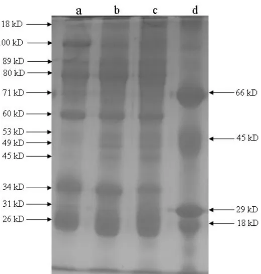
Blood samples were taken from the dorsal aorta of fish and then centrifuged for 10 minutes at +4ºC and 805 g. Total protein amounts of serums were measured by the biuret method(Robert and Michael, 1993). After this process, SDS-PAGE was modified according to Laemmli (1970) and O’Farrell (1975) methods, and serum proteins in SDS-PAGE were run. The gels were then photographed, and molecular weights of proteins were calculated (Weber et al.). Bovine albumin (66 kD), egg albumin (45 kD), carbonic anhydrase (29 kD) and -lactoglobulin (18 kD) were used as standard proteins.
Histopathology
All fish were carefully dissected, and liver, intestine, gill and kidney tissue samples were collected for histopathological examination. All tissue samples were fixed in 10 % phosphate buffered formaldehyde solution for two days. Gills were decalcified with Osteodec (bio-Optica, Italy). Paraffin blocks were prepared and sections of 4-5 µm thickness from samples were obtained. All sections were stained with hematoxylin and eosin, and examined under a light microscope.
Results
During the study, slowdown in the behavior of fish and a reduction in feed intake was observed. Three of the fish applied 30 mg/L nitrogen fertilizer died during the experiment. In SDS-PADE, it was determined that in group applied 15 and 30 mg / L nitrogen fertilizer was thickened in protein bands of 118 kD, 89 kD, 53 kD, 49 kD, 45 kD, 26 kD and it was determined that in group applied 15 and 30 mg / L nitrogen fertilizer was narrowed in protein bands of 100 kD, 60 kD and 34 kD. Also it was determined that in group applied to 15 and 30 mg/L nitrogen fertilizer was inhibited protein band of 31 kD, however, it was determined that in group applied 15 and 30 mg / L nitrogen fertilizer was synthesized a new protein band of 68.5 kD.
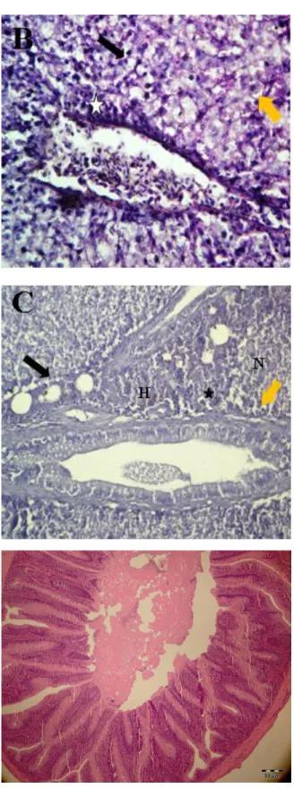
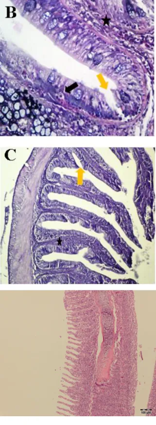
In histopathological examination, the tissue samples taken from control groups were in normal structure. In the liver tissue of the group treated with fertilizers, vacuolar degeneration, necrosis, hyperemia and mononuclear cells filtration in portal regions were observed (Figure 2A, B, C). In intestinal tissue, hydropic degeneration, necrosis and desquamation in epithelium cells at the apex of the villi were determined. mononuclear cell
29
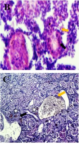
infiltration in the lamina propria was slightly observed (Fig. 3A, B, C). In gill tissues, fusion in secondary lamellae was frequently observed, due to increased dose, necrosis in epithelial cells and clara was observed (Figure 4A, B, C). In kidney tissue, hydropic and / or vacuolar degeneration in the proximal tubule epithelium of cortex, mild mononuclear cell infiltration in hyaline cylinders of some tubules lumina and intertubuler regions was observed (Figure 5A, B, C).
Discussion
In general, use large amounts of nitrogen fertilizer in agriculture causes water pollution due to increased nitrate (8). In addition fertilizers, the another nitrogenous products enters in aquatic environment and various physiological functions of aquatic organisms can be harmful to the deteriorating quality of the water (9). Ammonia in the water penetrates into the fish in 15 min (10) and effects of ammonia firstly occur at cellular and subcellular levels within an hour (11). In histopathological study done on Oncorhynchus mykiss exposure to nitrogen fertilizer, in the epidermis, degenerated/vacuolated epithelial cells, microcystic dilatations, and intracellular edema of mucus cell were observed. Liver had swollen and degenerated hepatocytes without losing adenoid structure. Hematopoietic tissues had occurred necrosis and vacuolar degeneration on proximal tubules of the kidney (6). An another study,
Oncorhynchus mykiss applied to 0.1 mg/L NH3 for 2h, filament and lamella epithelium have
superficially folded, the same concentration after 24 hours, telangiectasia in the filament on the 2 or 3 lamellae has been observed (12). In Ontario (Canada), in the Oncorhynchus mykiss farm in April-May as result of come to toxic levels of ammonia in water was indicated that death of 4000 fish within 48 hours, in pathological examination of fish was reported that telangiectasia in gill lamellae and kidney congestion (13). In a study done on Oreochromis
niloticus exposed to four different ammonia concentration (0.1, 0.2, 0.4 and 0.6 mg / L) for 60
days, depending on the increasing doses, gill tissue proliferation, epithelial hyperplasia and telangiectasia in the secondary lamellae, fusion in intermediate lamella, and vacuolar and pyknotic cells in a large number of primary and secondary lamellae have been observed (14). In the present study, an excessive nitrogen fertilizer application, liver, intestine, gill and kidney tissues received from fish were seen to cell degenerations, mononuclear cell infiltration and necrosis. This study showed that compliance with the above literature.
Effects of cadmium sulphate on the expression of serum proteins of the European chub,
30
CdSO4, a gradual decrease in protein bands was determined. Inhibition of 35.3 kilodalton
(kD) and 100.5 kD proteins in the first group, 1 mg/L and of 44.5 kD and 47.3 kD proteins in the 2nd group, 2 mg/L were detected (15). In a study done on Capoeta capoeta capoeta exposed to 1 and 2 mg/L cobalt-p-hydroxibenzoate for 10 days, compared to control group, over-expression and inhibition of serum proteins with 85.8, 82.6, 73.9,68.5 KD and 23, 15.2 kD molecular weights have been detected in the fish. Researchers claimed that Cobalt-p-hydroxibenzoate was toxic on fish (16).
In the present study, liver, intestine, kidney and gill tissues of Capoeta capoeta exposed to15 and 30 mg/L 33% nitrogen fertilizer were observed to be caused histopathological degenerations. In SDS-PAGE, thickening and thinning in some serum proteins of fish in experimental groups according to control group were observed. In addition, Inhibition and expression of protein bands of 31 kD and 68.5 kD were determined, respectively.
In histopathological and electrophoretic assessments of this study, it is concluded that high levels amounts of nitrogen fertilizers are toxic effect for aquatic organisms.
Acknowledgments
This study (Project number:2011-FEF-10) was supported by Kafkas University’s Scientific Research Project Council.
References
1. Danışman, F. and Bellitürk, K., Harran University Journal of the Faculty of Agriculture, 2007, 11(1/2):7-12.
2. Alloway, B. J., Heavy metals in soils. Chapman and Hall, Second Edition, India, 1995,38-119.
3. Sönmez, I., Kaplan, M., Sönmez, S., West Mediterranean Agricultural Research Institute Derim Journal, 2008,25(2), 24-34.
4. Savcı, S., International Journal of Environment and Sustainable Development, 2012,3(1), 77-80. 5. Ram, R.N. and Sathyanesan, A.G., Ecotoxicology and Environmental Saffety, 1987, l13(2),185-190. 6. Çapkın, E., Birincioğlu, S., Altınok, I., Ecotoxicology and Environmental Saffety, 2009,72, 1999-2004. 7. Sangeetha, S., Sujatha, K., Sentilkumaar, P., Kalyanaraman, V., Eswari, S., Indian Journal of Science
and Technology, 2011,4(7), 770-772.
8. Yüksel, A. N., Journal of the Faculty of Agricultural, Ataturk University, 1979,10(1-2), 179-194. 9. Benli, A. C. K., The Effects of sublethal ammonia concentrations on tissues, growth and blood
parameters of tilapia (Oreochromis niloticus) and mirror carp (Cyprinus carpio). PhD Thesis, 2006, Ankara (Turkey), 1-126.
31
10. Person Le Ruyet, J. P., Boeuf, G., Infante, J. Z., Helgason, S., Roux, A., Comparative Biochemistry and Physiology, 1998, 119A, 511–518.
11. Metcalfe, C. D., Toxicopathic responses to organic compounds. In: Leather- land, J.F., Woo, P.T.K. (Eds.), Fish Diseases and Disorders, Non-Infectious Disorders. CABI, 1998, 133–162.
12. Kirk, R. S., Lewis, J. W., Environmental Technolog ,1993, 14, 577-585. 13. Speare, D. and Backman, S., The Canadian Veterinary Journal, 1988,29, 666.
14. Shebly, E. A. A., Gad, H. A. M., Journal of Microbiology and Biotechnology Research, 2011,1 (4), 183-197.
15. Yılmaz, M., Ersan, Y., Koç, E., Özen, H., Karaman, M., Journal of the Faculty of Veterinary Medicine, 2011, Kafkas University 17 (Suppl A), 131-S135.
16. Yılmaz, M., Ersan, Y., Karaman, M., Özen, H., Koç, E., Necefoğlu, H., Fresenius Environmental Bulletin, 2008, 7( 9a), 1322-1327.
32 Captions Of Figure
Figure 1: Electrophoregram obtained from the SDS-PAGE of the serum proteins of the fish exposed to nitrogen fertilizer. a. Control group, b. 15mg/L dose group. c. 30mg/L dose group,
d. Standard proteins.
Figure 2A. Normal structure of liver tissue. H-E. 10x
33
Figure 2B. Liver tissue exposed to 15 mg/L nitrogen fertilizer. Necrosis (N), Pyknotic degeneration (black arrow), vacuolar degeneration (orange arrow) and Mononuclear cell infiltration (MHI) (star). H-E. 20X
Figure 2C. Liver tissue exposed to 30 mg/L nitrogen fertilizer. Necrosis (N), Hyperemia (H), and Mononuclear cell infiltration (MHI) (star), Pyknotic degeneration (black arrow) and vacuolar degeneration (orange arrow). H-E. 20X
Figure 3A. Intestine tissue in control group. H-E. 20X
34
Figure 3B. Intestine tissue exposed to 15 mg/L nitrogen fertilizer. Desquamation (orange arrow), mononucleear cell infiltration (MHI) in connective tissue. (yıldız) ve hidropik dejenerasyon (siyah ok). H-E. 40X
Şekil 3C. Intestine tissue exposed to 30 mg/L nitrogen fertilizer. Desquamation (orange arrow) and mononuclear cell infiltration (MHI) (arrow). H-E. 20X
Şekil 4A. Gill tissue in control group. H-E. 20X
35
Şekil 4B. Gill tissue exposed to 15 mg/L nitrogen fertilizer. Fusion in secondary lamellia (arrow), necrosis in clara (orange arrow) and necrosis in epitelial cells (N). H-E. 40X
Şekil 4C. Gill tissue exposed to 30 mg/L nitrogen fertilizer. Fusion in secondary lamelia (arrow), necrosis in clara cells (arrow) and necrosis in epitelial cells (N). H-E. 40X
Şekil 5A. Kidney tissue in control group. H-E. 20X
36
Şekil 5B. Kidney tissue exposed to 15 mg/L nitrogen fertilizer. Vacuolar degeneration (orange arrow), mononuclear cell infiltration (MHI) (black arrow) and Hyaline cylinders (star). H-E. 40X
Şekil 5C. Kidney tissue exposed to 30 mg/L nitrojen fertilizer. Hydropic degeneration (black arrow), mononuclear cell infiltration (MHI) (orange arrow) and Hyaline cylinders (star). H-E. 20X