Peripheral Lymphadenopathy in Childhood: Single Center
Study
Çocukluk Çağında Periferik Lenfadenopatiler: Tek Merkezli Çalışma
Yasemin Özkale1; Murat Özkale1
; Tansu Sipahi2
1
Baskent University Faculty of Medicine
,
Department of Pediatrics, ADANA 2Ufuk University Faculty of Medicine, Division Of Child Hematology, ANKARACukurova Medical Journal 2015;40(3):418-429.
ABSTRACTS
Purpose: Lymphadenopathy is defined as an abnormality in the size and/or character of lymph node. In this study we
aimed to describe the clinical and laboratory findings of benign and malignant causes of peripheral lymphadenopathy in childhood.
Material and Methods: Two hundred and twenty four patients who were admitted to the clinic with peripheral LAP were
evaluated. Age, gender, laboratory and radiologic findings, final diagnoses, and duration, localization, size, consistency, spread, and accompanying local and systemic symptoms of LAP were determined. Benign and malignant causes of peripheral lymphadenopathy were compared.
Results: One hundred twenty-six patients (56.0%) were male and 98 patients (44.0%) were female. After the first
evaluation the patients were divided into two groups. The first group included 186 patients with benign causes and the second group included 38 patients with malignant causes. One hundred and sixty four of 224 patients (73.2 %) had localized peripheral lymphadenopathy. The most frequent cause of localized lymphadenopathy in the benign group was acute lymphadenitis (34.8%). The most common cause of localized lymphadenopathy in the malignant group was Hodgkin’s lymphoma (4.3%). Sixty of 224 (26.8%) patients had generalized peripheral lymphadenopathy. The most significant cause of generalized lymphadenopathy in the benign lymphadenopathy group was Epstein–Barr virus (10.0%), whereas Hodgkin’s lymphoma (23.3%) was the most common cause in malign lymphadenopathy group. Localized and generalized lymph node enlargement was most frequently found in cervical region. The most frequent site of involvement among benign and malignant cases was the cervical area. The results revealed that findings such as chronic course, generalized LAP, supraclavicular, cervical and inguinal location, organomegaly, hilier LAP, abdominal LAP, and abnormal laboratory findings (thrombocytopenia and blasts on the peripheral blood smear) were associated with malignant diseases.
Conclusion: Infections are the most common cause of peripheral LAP. The risk of malignany increases with the age of
child and the duration of LAP. Older children with chronic LAP, generalized LAP associated with organomegaly, abnormal laboratory findings should be considered as malignant LAP.
Key words: Child; lymphadenopathy, Abbreviation: LAP; lymphadenopathy
ÖZET
Amaç: Lenfadenopati, lenf nodunun boyut ve/veya karakterindeki anormallik olarak tanımlanır. Bu çalışmada amacımız
çocukluk çağında selim ve malign nedenlere bağlı periferik lenfadenopatilerin klinik ve laboratuvar bulgularını belirlemek.
419
Materyal ve Metod: Çalışmaya periferik lenfadenopati nedeni ile hastanemize başvuran 224 hasta alındı. Hastaların;
yaşı, cinsiyeti, laboratuvar ve radyolojik bulguları, tanıları, lenfadenopatinin; süresi, lokalizasyonu, boyutu, kıvamı, yayılımı, eşlik eden lokal ve sistemik semptomları kaydedildi. Malign ve selim hastalıklara bağlı lenfadenopatilerin klinik ve laboratuvar özellikleri karşılaştırıldı.
Bulgular: İki yüz yirmi dört hastanın 126’sı (%56.0) erkek, 98’i (%44.0) kız idi. Hastalar 2 gruba ayrıldı. Birinci grupta
selim nedenlere bağlı lenfadenopatisi olan 186 hasta, ikinci grupta ise malign nedenlere bağlı lenfadenopatisi olan 38 hasta mevcuttu. Yüz altmış hastada(%73.2) localize lenfadenopati vardı. Selim hasta grubunda lokalize lenfadenopatinin en sık nedeni akut lenfadenitler (%34.8) iken, malign hasta grubunda en sık nedenin Hodgkin lenfoma (%4.3) olduğu tespit edildi. İki yüz yirmi dört hastanın 60’ında (%26.8) jeneralize lenfadenopati vardı. Jeneralize lenfadenopatinin selim hasta grubunda başlıca nedeni Epstein–Barr virus enfeksiyonu (%10.0) iken, malign hasta grubunda Hodgkin lenfoma (%23.3) olduğu tespit edildi. Servikal bölge lokalize ve jeneralize lenfadenopatilerde en sık tutulan bölgedir. Malign ve selim nedenlere bağlı lenfadenopailer en sık servikal bölgede görüldü. Elde ettiğimiz sonuçlara göre kronik seyirli, jeneralize lenfadenopati, supraklavikular, servikal ve inguinal yerleşimli lenfadenopati, organomegali, abdomanial lenfadenopati, hilier lenfadenopati ve anormal laboratuvar bulgularının (trombositopeni, periferik yaymada blast ) varlığında öncelikle malign nedenler düşünülmelidir.
Sonuç: Enfeksiyonlar çocukluk çağında periferik lenfadenopatinin en sık nedenidir. Malignite riski yaş ve
lenfadenopatinin süresi ile artmaktadır. İleri yaş, kronik lenfadenopati, jeneralize lenfadenopati, eşlik eden organomegali, anormal laboratuvar bulguların varlığında öncelikle malignite düşünülmelidir.
Anahtar kelimeler: Çocuk, lenfadenopati
Kısaltma: LAP; lenfadenopati
INTRODUCTION
Lymphadenopathy (LAP) is defined as an abnormality in the size and/or character of lymph node. Since a large variety of disorders may lead to peripheral LAP in childhood, determining the cause of peripheral LAP in children can be difficult. Lymphadenopathy is a common finding in childhood which frequently causes problems for parents and physicians when patients underwent invasive and sometimes unnecessary procedures to establish a diagnosis. Until recently there was no consensus on the size of a lymph node which would be classified as large. Although there is some variation depending on the anatomic area, lymph nodes > 1 cm in size are commonly accepted as enlarged1,2.In this study we aimed to evaluate peripheral LAP regarding etiology and clinical findings; to define the characteristics of benign and malignant lymph node enlargement by analyzing patient history, physical examination and laboratory findings, and to determine the differences between these two types.
METHODS and PARTICIPANTS
This prospective study involved 224 patients with peripheral LAP who were admitted to Dr. Sami Ulus Pediatrics Training and Research Hospital over a 2-year period. The sample included 98 female and 126 male patients between 2 months and 16 years of age. One hundred eighty-six patients were in the benign LAP group and 38 patients were in the malignant LAP group. The abnormal LAP was classified depending on the duration, extension and size. The maximum diameter of the LAP to be considered abnormal was 10 mm, with the exceptions of 1-2 mm at the supraclavicular region, 5 mm at the epitrochlear, post-auricular and occipital regions and 15 mm at the inguinal region. We caterogorized the lymph nodes accordng to the size as <2, 2-4, and >4 cm. The enlargement of lymph nodes in one contiguous anatomic region was classified as localized LAP, and the involment of two or more noncontiguous lymph node region was classified as generalized LAP. The patients with LAP for < 2
420 weeks were categorized as acute LAP, and the patients with LAP for > 2 weeks were categorized as chronic LAP. We recorded axillary body temperature at presentation or measured by parents at home. Axillary body temperature over 38 º has to be considered as fever. Children suffering from neck mass caused by congenital abnormalities were not included in the study. Histories of upper respiratory infections ,ear pain, and dental problems, weight loss, night sweats, fatigue, bleeding, cough, rash, arthralgias, contact with animals, vaccination history, co-existing systemic illnesses, previous antibiotic use, and family history of tuberculosis were interrogated. The first assessment of the patient was directed towards predicting the etiology for LAP considering the history and findings on physical examination. The complete blood cell count, peripheral smear, erythrocyte sedimentation rate (ESR), and C-reaktif protein (CRP) were recorded. A throat culture, chest radiograph, purified protein derivative (PPD), and viral serology of patients with acute LAP were obtained. Fluctuant nodes were aspirated for culture. For patients with possible tuberculous LAP, family screening, acid resistant bacteria (ARB), BACTEC, Polymerase chain reaction (PCR) and culture analyses for Mycobacterium tuberculosis in sputum or fasting gastric juice were performed and the diagnosis was confirmed with biopsy. Thoracic CT is also performed. For patients with generalized LAP in addition to these analyses, Epstein Barr Virus (EBV), Cytomegalovirus (CMV), human immunodeficiency virus (HIV), Herpes simplex virus (HSV), measles, rubella, salmonella, and brucella serology were also determined. The patients with organomegaly were examined utilizing abdominal ultrasonography (USG). Excisional lymph node biopsy was conducted for patients who had not responded to 4-8 weeks of antibiotic treatment. Informed consent was obtained from the parents of all children.
Statistical Methods
Statistical analyses were performed utilizing SPSS 15.0 for Windows (SPSS, Inc., Chicago, IL, USA). Data was defined by descriptive statistics (n, %). Demographic data was shown by mean and standart deviation. A chi square test was conducted to compare the gender variable between the two groups, and Student’s t-test was used to compare the age variable. A chi square test was also conducted to compare the symptoms and laboratory findings of the two groups. The levels of significance are indicated by p values <0.05.
Findings
One hundred twenty-six of 224 patients (56.0%) were male and 98 (44.0%) were female, with a female-to-male ratio of 0.77. The youngest patient was 2 months old and the oldest patient was 16 years old. One hundred eighty-six of the LAP patients (83.0%) had benign and 38 (17.0%) had malignant causes. The benign LAP group consisted of 186 patients (84 females and 102 males) between 2 months and 16 years of age; the mean age was 63.5 ± 43.5 months. The malignant LAP group included 38 patients (12 females and 26 males) between 8 months and 15 years of age; the mean age was 94.0 ± 47.3 months. While there was no statistically significant difference with respect to gender between the groups (p=0.35), the mean age for the two groups differed statistically (p=0.01). The etiologic distribution of the benign and malignant LAP groups with respect to age is shown in Tables 1 and 2, respectively.
The etiologic and local distributions of peripheral LAP to the localization is showed in Table 3. While the most frequent cause of localized LAP in the benign LAP group was acute lymphadenitis (34.8%), the most common cause was Hodgkin’s lymphoma (4.3%) in the malignant LAP group. One hundred and sixty four of 224 patients (73.2 %) had localized, sixty of 224 (26.8%) patients had generalized peripheral. LAP (Table 3). The most significant cause of
421 generalized LAP in the benign LAP group was EBV (10%), whereas Hodgkin’s lymphoma (23.3%) was the most common cause in malignant LAP group. (Table 3).
Table 4 summarizes comparison of the detailed symptoms and clinical findings of the benign and malignant LAP groups. One hundred and sixty four of 224 patients (73.2 %) had acute LAP , sixty
of 224 (26.8%) patients had chronic generalized. LAP (Table 4). One hundred fifty six of 164 (95.0%) patients in the acute LAP group had benign etiologies and 8 (5.0 %) had malign etiologies. Thirty eight of 60 (63.3%) patients in the generalied LAP had benign etiologies whereas 22 (36.7 %) had malign etiologies. Twelve patients in the benign LAP group with chronic LAP were diagnosed with tuberculosis; 83.0% of these patients were PPD-positive. There was no significant difference between the extensions of the LAP, consistency of LAP and fluctuation of LAP in the benign and malignant LAP groups (P>0.05, Table 4). We found the duration of LAP was longer in malignant LAP group than benign LAP group (P<0.001, Table 4). When the
distribution of LAP compared, we found localized LAP was most frequent in the benign LAP group (P<0.001, Table 4). We found that cervical, submandibular and inguinal regions were the most common localization of LAP both in malignant and benign LAP groups. We determined supraclavicular LAP in 12 patients; malignancy was detected in 11 patients. We also found that fever is more common in the benign LAP group whereas hepatosplenomegali is more common in the malign LAP group (P<0.001, Table 4).
Table 5 lists the comparison of the laboratory and radiologic findings of the two study groups. There were no significant difference between the CRP and sedimentation levels in the benign and malignant LAP groups (P>0.05, Table 4). We found hilier LAP, hepatosplenomegali and mesenteric LAP , thrombocytopenia, blasts on the peripheral blood smear were significantly higher in the malign LAP group (P<0.001, Table 5).
Table 6 summarizes the etiologic distribution of the 50 patients who underwent excisional lymph node biopsy in order to evaluate etiology of peripheral LAP.
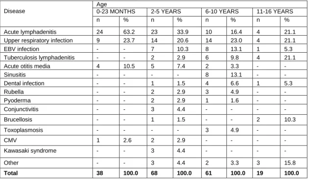
Table 1. Benign lymhadenopathy etiology according to the age groups
Disease
Age
0-23 MONTHS 2-5 YEARS 6-10 YEARS 11-16 YEARS
n % n % n % n %
Acute lymphadenitis 24 63.2 23 33.9 10 16.4 4 21.1
Upper respiratory infection 9 23.7 14 20.6 14 23.0 4 21.1
EBV infection - - 7 10.3 8 13.1 1 5.3
Tuberculosis lymphadenitis - - 2 2.9 6 9.8 4 21.1
Acute otitis media 4 10.5 5 7.4 2 3.3 - -
Sinusitis - - - - 8 13.1 - - Dental infection - - 1 1.5 4 6.6 1 5.3 Rubella - - 2 2.9 3 4.9 - - Pyoderma - - 2 2.9 1 1.6 - - Conjunctivitis - - 3 4.4 - - - - Brucellosis - - 1 1.5 - - 2 10.3 Toxoplasmosis - - - - 3 4.9 - - CMV 1 2.6 2 2.9 - - - - Kawasaki syndrome - - 3 4.4 - - - - Other - - 3 4.4 2 3.3 3 15.8 Total 38 100.0 68 100.0 61 100.0 19 100.0
422
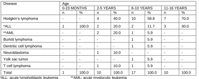
Table 2. Malign lymphadenopathy etiology according to the age groups
Disease Age
0-23 MONTHS 2-5 YEARS 6-10 YEARS 11-16 YEARS
n % n % n % n %
Hodgkin’s lymphoma - - 4 40.0 10 58.8 7 70.0
*ALL 1 100.0 2 20.0 2 11.7 3 30.0
**AML - - 2 20.0 1 5.9 - -
Burkitt lymphoma - - - - 1 5.9 - -
Dentritic cell lymphoma - - - - 1 5.9 - -
Neuroblastoma - - 1 10.0 - - - -
Yolk sac tumor - - - - 1 5.9 - -
T cell lymphoma - - 1 10.0 1 5.9 - -
Total 1 100.0 10 100.0 17 100.0 10 100.0
*ALL: acute lymphoblastic leukemia **AML: acute myelocytic leukemia
Table.3. Causes of malign and benign lymphadenopathy (LAP) to the localization
Etiology of benign LAP Localized LAP General LAP
n % n %
Acute lymphadenitis 57 34.8 4 6.7
Upper respiratory infection 36 22.0 5 8.3
EBV 10 6.0 6 10.0
*Tbc, lymphadenitis 10 6.0 2 3.3
Acute otitis media 10 6.0 1 1.7
Sinusitis 6 3.7 2 3.3 Dental infection 5 3.1 1 1.7 Pyoderma 3 1.8 0 0.0 Conjunctivitis 3 1.8 0 0.0 CMV 2 1.2 1 1.7 Kawasaki syndrome 2 1.2 1 1.7 Rubella 1 0.8 4 6.7 Brucellosis 0 0.0 3 5.0 Toxoplasmosis 0 0.0 3 5.0 Other 3 1.8 5 8.3 Total 148 90.2 38 63.4
Etiology of malign LAP
Hodgkin’s lymphoma 7 4.3 14 23.3
ALL 4 2.5 4 6.6
AML 1 0.6 2 3.3
Burkitt lymphoma 1 0.6 0 0.0
T cell lymphoma 1 0.6 1 1.7
Yolk sac tumor 1 0.6 0 0.0
423
Dentritic cell lymphoma 0 0.0 1 1.7
Total 16 9.8 22 36.6
Overall TOTAL 164 100.0 60 100.0
*Tbc: Tuberculosis,
Table.4. Symptoms and clinical findings of patients with benign and malign lymphadenopathy
FINDINGS BENIGN MALIGN
n % n % p General symptoms Fever 119 78.3 25 30.9 0.01 Night sweats 13 8.6 15 18.5 Weight loss 11 7.2 20 24.7 Fatique 9 5.9 21 25.9 Distribution Local 148 79.6 16 42.1 0.001 General 38 20.4 22 57.9 LAP duration Acute 156 83.9 8 21.0 0.001 Chronic 30 16.1 30 79.0 Localization Cervical 125 47.3 35 42.7 0.87 Submandibular 67 25.4 10 12.2 Inguinal 28 10.6 15 18.3 Axillary 26 9.8 9 11.0 Post auricular 11 4.2 2 2.4 Pre-auricular 5 1.9 0 0.0 Suboccipital 1 0.4 0 0.0 Supraclavicular 1 0.4 11 13.4 Size of LAP <2 cm 73 39.2 16 42.1 0.86 2-4 cm 55 29.6 12 31.6 >4 cm 58 31.2 10 26.3 Mobility of LAP Mobile 103 55.4 18 47.4 0.76 Fixed 83 44.6 20 52.6 Fluctuation of LAP Yes 18 9.6 0 0.0 0.66
424
No 168 90.4 38 100.0
Temperature elevation and sensitivity
Yes 75 40.4 1 2.7 0.001 No 111 59.6 37 97.3 Hepatomegaly Yes 24 12.9 17 44.7 0.001 No 162 87.1 21 55.3 Splenomegaly Yes 12 6.5 20 52.6 0.001 No 174 93.5 18 47.4
Table.5. Laboratory and radiologic findings of patients with benign and malign lymphadenopathy
LABORATORY AND RADIOLOGIC FINDINGS BENIGN MALIGN n % n % p Anemia 84 92.3 27 64.3 0.001 Thrombocytopenia 7 7.7 15 35.7
White blood cell count
Leukocytosis 59 88.1 10 45.5 0.001
Leukopenia 8 11.9 12 54.5
Peripheral blood smear
Shift to the left 116 87.2 13 44.8 0.001
Atypical lymphocytes 17 12.8 4 13.8 Blasts - 0.0 12 41.4 Sedimentation High(>20mm/h) 127 68.3 31 81.6 0.08 Normal(<20mm/h) 59 31.7 7 18.4 CRP High(>6 mg/dl) 131 70.4 22 57.9 0.09 Normal(<6 mg/dl) 55 29.6 16 42.1
P-A chest radiography
Abnormal(hilier LAP) 21 11.3 10 26.3 0.04 Normal 165 88.7 28 73.7 Abdominal USG Hepatomegaly 24 54.5 25 39.7 0.01 Splenomegaly 18 41.0 24 38.1 Lymphadenopathy 2 4.5 14 22.2
425
Table. 6.. Distribution of cases with lymph node biopsies
Neoplastic diseases n %
*HH 21 42.0
**NHL - -
T cell lymphoma 2 4.0
Burkitt lymphoma 1 2.0
Dentritic cell lymphoma 1 2.0
Neuroblastoma 1 2.0
Yolk sac tumor 1 2.0
Granulomatous diseases
Tuberculosis 9 18.0
Toxoplasmosis 1 2.0
Reactive lymph node hyperplasia 8 16.0
Others Mucormycosis lymphadenitis 1 2.0 Castleman disease 1 2.0 Measles 1 2.0 Chronic lymphadenitis 2 4.0 Total 50 100.0
*HH: Hodgkin’s disease **NHL: non- Hodgkin’s Lymphoma
DISCUSSION
Peripheral LAP in children might be an alarming sign of serious disease such as malignancy, systemic disease, infections, autoimmune disorders, miscellaneous and iatrogenic. Lymphadenopathy is a common finding in children. Since it can be manifestation of a serious systemic disease or malignancy, determining the cause of peripheral LAP in children can be difficult. The presence of lymph nodes can be diagnosed during an ordinary physical examination in 38.0%-45.0% of healthy children3. In infants < 6 months of age, palpable LAP was present in 38.0% and the most frequent sites were the occipital and post-auricular areas. Of healthy children between 3 and 5 years, 63.0%
and 24.0% had LAP in the cervical and submandibular areas, respectively1,2.
Lymphadenopathy is a common finding in children and in the majority of cases lymph node enlargement occurs response to the benign and self limited disease. Local or systemic infections are common causes of peripheral LAP in children. Previous studies reported that 70.0%-87.0% of patients with peripheral LAP have benign causes3-7. Similar to other studies we found that 83.0% of patients with peripheral LAP have benign causes. Acute lymphadenitis (32.7%) and upper respiratory infections (22.0%) were the most common causes of benign LAP in our study.
Acute lymphadenitis is defined as enlarged, inflamed, and tender of lymph nodes. It is often a
426 complication of bacterial infections, although it can also be caused by viruses or other disease agents. Staphylococcus and streptococci are the most common infectious agents in children especially under 4 years of age. Acute unilateral cervical lymphadenitis is usually caused by staphyloccocus aureus or streptococcus pyogenes in over 80% of cases. A group beta hemolytic streptococci (AGBS) and anaerobic bacteria are another common cause of acute cervical lymphadenitis and generally occurs in children older than 3 years of age. Acute pyogenic lymphadenitis is mostly localized to the submandibular (50%-60%) and anterior cervical (25%-30%) region in children between 1 and 4 years of age2,8. In the current study, patients had acute lymphadenitis, which 47.3% had cervical and 25.4% had submandibular lymph node involvement. Fluctuation existed in 9.6% of the patients; Staphylococcus aureus and AGBS were isolated in 55.5% and 5.5% of the lymph nodes with fluctuation, respectively.
Age is an important factor in differentiating between benign and malignant LAP in childeren. The rate of malignant etiologies of peripheral LAP is low in children, but increases with age8-10. Soldes et al reported the risk of malignancy in peripheral LAP increases with the age of child, size of LAP and the number of involved sites11. On the other hand, in 1980 Lee et al reviewed 628 patients undergoing nodal biopsy and found that age is the most important factor in estimating the probability of whether the LAP is due to a benign or malignant process12. We also found the mean age of the patients in the malignant LAP group were older than benign LAP group. We have determined malign LAP only in 1 patient between 0-23 months old whereas 38 patient had benign LAP between the same age group.
Another important consideration in the evaluation of patients with peripheral LAP is the presence of systemic symptoms. “B” symptoms, such as fever, weight loss, and night sweats, accompany lymphoproliferative diseases along with infectious illnesses. Knight et al reported that
fever of unknown origin and weight loss persisting for > 1 week are associated with malign LAP. On the other hand Soldes et al reported that fever does not have a clinical significance in the diagnosis of patients with LAP4,11. Weight loss, fatigue, and arthralgias are the most common symptoms which are associated with malignancy and collagen tissue diseases13,14. Kumral et al reported that weight loss was more frequently associated symptom in the malignant LAP group whereas fever was more frequent in patients with benign LAP (5). We also found that fever is more common in the benign LAP group.
Based on our data, most of the patients had localized LAP (73%). We determined the most frequent cause of localized LAP in the benign LAP group was acute lymphadenitis (34.8%) and the most common cause of localized LAP in the malignant LAP group was Hodgkin’s lymphoma (4.3%). İnfections, autoimmune diseases, and malignancy are the most common cause of generalized LAP in children. Numerous pathogens have been associated with generalized LAP including EBV, CMV, adenovirus , rubella and toxoplasmosis11-14. We found sixty of 224 (26.8%) patients had generalized peripheral LAP. We determined the most common cause of generalized LAP in the benign LAP group was Epstein–Barr virus infections (10.0%) and the most common cause was Hodgkin’s lymphoma (23.3%) in malign LAP group. The risk of malignant disease was higher in patients who had generalized LAP7. We also found that generalized LAP was more common in the malignant LAP group and localized LAP was more common in the benign LAP group similiar to previous studies5,6.
Although the duration of LAP is important in the differential diagnosis of peripheral LAP, it is not a specific finding. Infections should be considered first in the etiology of cases with peripheral LAP < 2 weeks in duration, and malignancy and granulomatous diseases are suggestive of chronic LAP patients5,6,9,15. In the current study, the
427 duration of LAP was longer in malignant LAP group than benign LAP group.
The localization of LAP is important in terms of etiologic factors. Cervical LAP is the most common localization for both benign and malignant diseases5,6,9,14. The incidence of malignancy is higher in supraclavicular, infra-cervical, and posterior cervical LAP5-7,9. Our study also showed that supraclavicular, cervical, and inguinal LAP were significant in the presence of malignant LAP. So as suggested in the literaure, children with supraclavicular or cervical LAP should be selected for early lymph node biopsy. On the other hand we found an increased incidence of submandibular LAP in the benign LAP group. We assume that this may result from the high frequency of lymphadenitis cases among the patients with benign diseases.
There have been a variety of findings with respect to the importance of lymph node size in the differential diagnosis of LAP. LAP > 2 cm in size are suspicious for malignancies or granulomatous diseases and LAP < 1 cm in size suggest benign diseases5,7,9. Karaman et al reported that there is no significant difference between benign and malignant diseases with respect to LAP size similar to our findings10. On the other hand Kumral et al reported that LAP larger than 3 cm represent a greater risks for malignancies5.
Malignant and granulomatous diseases should be considered in patients with hard or mixed LAP, fixed to the surrounding tissue. Although some studies maintain that fixed lymph nodes are an indicator of increased risk for malignant and granulomatous diseases, other studies claim that the mobility of lymph nodes is not a significant finding with respect to the differential diagnosis5,6,11,14,16. In the current study, fixed LAP was identified in 44.6 % of the benign group and 52.6% of malignant cases. The higher incidence of fixed LAP in the benign group is thought to be the result of the higher number of A. lymphadenitis and M. tuberculosis cases.
The presence of organomegaly and peripheral lymphadenopathy in children is a strong indicator of the seriousness of a disease. Knight et al reported that there is a serious triggering effect in 56.0% and 40.0% of hepatomegaly and splenomegaly cases, respectively4. In our study, organomegaly was significantly more frequent among the malignant patient group. There are other studies which have been conducted in Turkey with similar results5,7.
Lymphadenopathy may be an important indicator of a malignant disorder1,2. Therefore, it is very important to establish a early diagnosis, to start treatment, and to achieve a good prognosis. Hemoglobin, platelet and white blood cell counts are useful findings in the differential diagnosis of LAP patients. Leucocytosis is apparent in infectious LAP, whereas thrombocytopenia, leucocytosis, or leucopenia are apparent in the course of malignancies5,7,17,18. Fijten et al claimed that the white blood cell count is of no clinical significance in patients with malignant diseases13. In the current study, the incidences of anemia, thrombocytopenia and leucopenia were significant in the malignant LAP group, but the white blood cell counts were not a diagnostic criterion. The peripheral smear constitutes the first step in the evaluation of patients with LAP10. An elevated ESR and CRP, also known as acute phase reactants, did not reveal any clinical significance in the differential diagnosis of patients with malignant LAP5,7.
Many infectious and malignant diseases can accompany abdominal LAP2,18. The incidence of abdominal LAP is more frequent in patients with malignant diseases than benign diseases5,9,10. Similarly, it was identified in our results that abdominal LAP was present in 4.5 % of patients with benign diseases and in 22.2 % of patients with malign diseases.
In the current study, excisional lymph node biopsies were performed on 50 patients (22.3%). Most of the patients had been histopathologically diagnosed malignancies(54.0%). Histopathologic
428 examination showed granulomatous diseases in 10 patients (20%) and it was consistent with reactive lymphoid hyperplasia in 8 patients (16%). The malignancy rates in patients with persistent LAP were 30.0%, 27.0%, and 38.0% in studies conducted by Kumral et al , Karadeniz et al and Karaman et al respectively5,9,10. Excisional biopsy should be considered with LAP hard or matted, fixed to the surrounding tissue, increasing rapidly in size, unresponsive to antibiotic therapy over 4-8 weeks, localized cervical or supraclavicular region, presence of organomegali and mediastinel LAP, associated symptoms like fever of unknown origin, night sweeats, weight loss or difficulty in diagnosis2-5,8,20.
CONCLUSION
Overall, 83.0% of LAP cases in childhood are caused by benign diseases. The most frequent site of involvement both benign and malignant cases was the cervical region. When supraclavicular LAP is present, malignancy and early lymph node biopsy should be considered in all age groups. Malignant disease should be considered in children who are older, have a chronic course, generalized LAP, presence of organomegaly, hilier LAP, abdominal LAP and abnormal laboratory findings (thrombocytopenia and blasts on the peripheral blood smear).
Language, grammar, and spelling errors have been corrected.
Declaration of Conflicting Interests
The authors declared no potential conflicts of interest with respect to the authorship and/or publication of this article.
REFERENCES
1. Herzog LW. Prevalence of lymphadenopathy of the head and neck in infants and children. Pediatrics. 1983;27:485-7.
2. Kelly CS, Kelly RE Jr. Lymphadenopathy in Children. Pediatr Clin NA. 1998;45:875-88
3. Kılıç B, Pamıkçu A, Yoldaş A, Atasoy İ. Çocuklarda Lenfadenopatilerin Değerlendirilmesi Türkiye Klinikleri Pediatri Dergisi. 2010;19:364-9
4. Knight PJ, Mulne AF, Vassy LE. When is lymph node biopsy indicated in children with enlarged peripheral nodes? Pediatrics. 1982;69:391-6.
5. Kumral A, Olgun N, Uysal KM, Çorapçıoğlu F, Ören H, Sarıalioğlu F. Assessment of peripheral lymphadenopathies: experience at a pediatric hematology oncology department in Turkey. Pediatr Hematol Oncol. 2002;19:211-8.
6. Latifagic A, Iljazovic E, Colic B, Mladina N. Etiological and clinical characteristics of lymphadenopathy at child age in Tuzla Canton. Med Arh. 2011;65:295-9. 7. Yaris N, Cakir M, Sözen E, Cobanoglu U. Analysis of
children with peripheral lymphadenopathy. Clin Pediatr. 2006;45:544-9.
8. Niedzielska G, Kotowski M, Niedzielski A, Dybiec E, Wieczorek P. Cervical lymphadenopathy in children incidence and diagnostic management. Int J Pediatr Otorhinolaryngol. 2007;71:51-6.
9. Karadeniz C, Oğuz A, Ezer U, Öztürk G, Dursun A. The etiology of peripheral lymphadenopathy in children. Pediatr Hematol Oncol. 1999;16:525-31. 10. Karaman A, Karaman I, Cavuşoğlu YH, Erdoğan D.
The ongoing problem with peripheral lymphadenopathies: which ones are malignant? Pediatr Surg Int. 2010;26:247-50.
11. Soldes OS, Younger JG, Hirschl RB. Predictors of malignancy in childhood peripheral lymphadenopathy. J Pediatr Surg. 1999;34:1447-52. 12. Lee Y, Terry R, Lukes RJ. Lymph node biopsy for
diagnosis: a statistical study. J Surg Oncol. 1980;14:53-60.
13. Fijten GH, Blijham GH. Unexplained lympadenopathy in family practice. An evaluation of the probability of malignant causes and the effectiveness of physicians' workup. J Fam Pract. 1988;27:373-6.
14. Leung AK, Robson WL. Childhood cervical lymphadenopathy. J Pediatr Health Care. 2004;18:3-7.
15. Bazemore AW, Smucker DR. Lymphadenopathy and malignancy. Am Fam Physician. 2002;66:2103-10.
429 16. Herzog LW. Prevalence of lymphadenopathy of the
head and neck in infants and children Clin Pediatr. 1983;22:485-7.
17. Lake AM, Oski FA. Peripheral lymphadenopathy in childhood. Am J Dis Child. 1978;132:357-9.
18. Young G, Toretsky JA, Campbell AB, Eskenazi AE. Recognition of common childhood malignancies. Am Fam Physician. 2000;61:2144-54
19. Habermann TM, Steensma DP. Lymphadenopathy. Mayo Clin Proc. 2000;75:723-32.
20. Twist CJ, Link MP. Assesment of lymphadenopathy in children. Pediatr Clin NA. 2002;49:1009-25.
Yazışma Adresi / Address for Correspondence:
Dr. Yasemin Özkale
Baskent University Faculty of Medicine Adana Teaching and Medical Research Center Baraj Yolu 1 Durak, Seyhan 01120
ADANA
E-mail: [email protected] Geliş tarihi/Received on : 22.01.2015 Kabul tarihi/Accepted on: 10.02.2015