http://journals.tubitak.gov.tr/medical/ © TÜBİTAK
doi:10.3906/sag-1408-54
PCR investigation of Panton-Valentine leukocidin, enterotoxin, exfoliative toxin,
and agr genes in Staphylococcus aureus strains isolated from psoriasis patients*
Jülide Sedef GÖÇMEN1,**, Neriman ŞAHİNER2, Mukadder KOÇAK2, Zeynep Ceren KARAHAN3
1Department of Medical Microbiology, Faculty of Medicine, Başkent University, Ankara, Turkey 2Department of Dermatology, Faculty of Medicine, Kırıkkale University, Kırıkkale, Turkey 3Department of Medical Microbiology, Faculty of Medicine, Ankara University, Ankara, Turkey
1. Introduction
Psoriasis is a chronic inflammatory disorder involving the skin. Genetic, environmental, and immunological factors play important roles in the development of the disease. Its incidence may change in different parts of the world depending on environmental, ethnic, and geographic differences. Both sexes are affected equally by the disease, and patients are usually diagnosed between 15 and 30 years of age. Although it can be seen in almost all races, it is rare in Asia and Africa. It has an overall prevalence of 2%–3% in the general population (1,2). Its prevalence in children is reported to range from 0% (Taiwan) to 2.1% (Italy), and in adults from 0.91% (United States) to 8.5% (Norway). In the United States, the annual incidence estimate in children is 40.8/100,000, while in adults the annual incidence varies from 78.9/100,000 (United States) to 230/100,000 (Italy) (1). The prevalence in the Turkish population is reported as 1.3% (2).
Bacteria are known to play an important role in the development and chronicity of chronic inflammatory diseases such as atopic dermatitis and psoriasis. The relationship between bacterial colonization or infection of the skin and the development of inflammatory skin diseases is well described in studies reporting the relapse of guttate psoriasis following streptococcal pharyngitis and the development of atopic dermatitis following
Staphylococcus aureus colonization of the skin (3,4). S. aureus colonizes the anterior nares of 20%–40%
of the healthy adult population. It can also colonize the perineum, perianal region, axilla, gastrointestinal tract, and skin folds. Trauma, burns, diabetes, and immune suppression can lead to opportunistic infections with this colonizing pathogen (5).
S. aureus can be the cause of a wide spectrum of
infectious diseases due to its many virulence factors, which facilitate its spread in host tissues and its escape Background/aim: Staphylococcus aureus colonization is a determiner of disease activation in psoriasis patients. Here we evaluate the presence of genes encoding Panton-Valentine leukocidin (PVL), enterotoxins, TSST-1, exfoliative toxins, and the accessory gene regulatory locus by polymerase chain reaction (PCR) in S. aureus isolates obtained from healthy and diseased skin regions and anterior nares of psoriasis patients and healthy controls.
Materials and methods: The presence of PVL and toxin genes was investigated, and agr typing was performed by PCR.
Results: Eighteen of the isolated strains carried the sei, 1 carried the seb-sec, and 1 carried the seg enterotoxin gene. Eight of the strains carrying enterotoxin genes were isolated from nasal swabs, 6 from diseased skin swabs, and 4 from healthy skin swabs. None of the strains isolated from the control group carried the agr locus. On the other hand, 11 of the S. aureus strains isolated from the patients carried type 1, 7 carried type 1 + 3, 4 carried type 2, 4 carried type 3, and 1 carried type 1 + 2 agr loci.
Conclusion: Enterotoxin production and the carried accessory gene regulatory locus may be important in the aggravation of psoriasis. Key words: Polymerase chain reaction, Staphylococcus aureus, toxin genes, agr typing
Received: 13.08.2014 Accepted/Published Online: 28.12.2014 Printed: 31.12.2015
Research Article
* This study was presented as a poster at the XXXV. Turkish Microbiology Congress, 3–7 November 2012, Antalya, Turkey. ** Correspondence: [email protected]
from the host’s immune responses. Bacterial enzymes and toxins, such as capsular polysaccharide, plasma coagulase, extracellular matrix components, protein A, fibronectin binding factor, microbial surface component recognizing adhesive matrix molecules, and extracellular proteins (i.e. toxins), play important roles in the development and occurrence of staphylococcal infections (6,7) . Adherence factors (adhesins) promote the colonization of S. aureus on host cell surfaces (8–11).
S. aureus produces exotoxins with cytolytic activities.
These cytolytic toxins (α-, β-, and γ-hemolysins, leukocidin, and Panton-Valentine leukocidin [PVL]) lead to the formation of pores on target cell membranes. The cytoplasmic content of the target cell leaks and the cell lyses. PVL is a bicomponent cytolysin (LukF-PV and LukS-PV) and is cytotoxic for erythrocytes and leukocytes, just like
γ-hemolysin and leukocidin, which show high affinity for
leukocytes. α-Hemolysin is especially responsible for osmotic cytolysis of human thrombocytes and monocytes (12–16).
S. aureus secretes toxic shock syndrome toxin-1
(TSST-1), staphylococcal enterotoxins A-I (SEA, SEB, SECN, SED, SEE, SEG, SEH and SEI), and exfoliative toxins A and B (ETA and ETB). Among these, TSST-1 and staphylococcal enterotoxins belong to the group of pyrogenic toxin superantigens (17,18).
Superantigens (SAgs) are toxins that play a role in T-cell proliferation. These toxins cause TSST-1, food poisoning (enterotoxins), and staphylococcal scalded skin syndrome (ETA and ETB) (19). The issue of whether exfoliative toxins have any mitogenic activity on T lymphocytes as SAgs is still controversial (20).
S. aureus has some special proteins that may have an
effect on innate and acquired immunity. Staphylococcal complement inhibitor, chemotaxis-inhibitory protein of
S. aureus, staphylokinase, extracellular fibrinogen-binding
protein, extracellular adherence protein, and formyl peptide receptor like-1 inhibitory protein are some of these special proteins (21–27).
In this study, in order to contribute to the previous studies on whether or not S. aureus colonization is a determiner of disease activation in psoriasis patients, we evaluated the presence of genes encoding PVL, enterotoxins (sea, seb, sec, sed, see, seg, seh, sei, sej), TSST-1 (tst), exfoliative toxins (eta, etb), and accessory gene regulatory locus (agr) by polymerase chain reaction (PCR) in S. aureus isolates obtained from healthy and diseased skin regions and anterior nares of psoriasis patients and healthy controls. 2. Materials and methods
2.1. Patients
Diseased skin, healthy skin (cubital volar region), and nasal swabs were obtained from 61 psoriasis patients who attended the dermatology polyclinic of the Kırıkkale
University School of Medicine. The control group consisted of 48 healthy volunteers with no personal or family history of psoriasis or other inflammatory skin disorders. Nasal and cubital volar skin swabs were obtained from the control group. Ethics committee approval was received for this study from the Ethics Committee of the Kırıkkale University Medical Faculty (Approval Number: 2010/004), and informed consent was obtained from all study and control subjects.
2.2. Culture
All swabs were cultured on 5% sheep blood agar in the Medical Microbiology Department Laboratory of the Kırıkkale University School of Medicine. Staphylococcus identification was made by conventional microbiological methods (Gram staining, catalase, and coagulase tests). Methicillin resistance of the isolates was determined by a Kirby-Bauer disk diffusion test performed by using 1 µg oxacillin and 30 µg cefoxitin disks (Bioanalyse, Turkey) according to the Clinical and Laboratory Standards Institute instructions (28). Methicillin-resistant S. aureus (MRSA) ATCC 43300 and methicillin-sensitive S. aureus (MSSA) ATCC 25923 control strains were used as standard quality controls for susceptibility testing in the Medical Microbiology Department Laboratory of the Başkent University School of Medicine. All the strains were stored in brain-heart infusion broth containing 20% glycerol at –80 °C until molecular testing was performed.
2.3. Molecular analyses
Molecular analyses of the strains were performed in the Molecular Microbiology Diagnostics and Research Laboratory of the Ankara University School of Medicine, Department of Medical Microbiology. All the strains were subcultured on nutrient agar plates, and DNA was extracted by boiling. Briefly, 2 loopfuls of pure culture were suspended in 500 µL of sterile distilled water and boiled at 95 °C for 10 min. The suspension was centrifuged at 3500 × g for 5 min, and the supernatant was used for PCR analysis. The presence of PVL, enterotoxin and TSST-1 (sea, seb, sec, sed, see, seg, seh, sei, sej, tst), and exfoliative toxin (eta, etb) genes was investigated, and agr typing was performed by PCR as described in the literature (12,13,29,30). The primer sets used for molecular analysis are given in Table 1.
2.4. Statistical analysis
Cochran Q and two-proportion z-tests were performed for the statistical analysis of the results using the SPSS 17.0 (SPSS Inc., Chicago IL, USA). P < 0.05 was considered as statistically significant.
3. Results
3.1. Culture results
Of the 61 psoriasis patients, 26 (42.6%) were found to carry S. aureus on their diseased and/or healthy skin and/or anterior nares. A total of 56 S. aureus strains were
Table 1. The primers used for molecular analysis of S. aureus strains.
Gene Primer (5′-3′) Product size (bp) Reference
sea GCAGGGAACAGCTTTAGGC 520 (30) GTTCTGTAGAAGTATGAAACACG seb-sec ATGTAATTTTGATATTCGCAGTG 643 TGCAGGCATCATATCATACCA sec CTTGTATGTATGGAGGAATAACAA 283
TGC AGG CAT CAT ATC ATA CCA
sed GTGGTGAAATAGATAGGACTGC 384 ATATGAAGGTGCTCTGTGG see TACCAATTAACTTGTGGATAGAC 170 CTCTTTGCACCTTACCGC seg CGTCTCCACCTGTTGAAGG 327 CCAAGTGATTGTCTATTGTCG seh CAACTGCTGATTTAGCTCAG 360 GTCGAATGAGTAATCTCTAGG sei CAACTCGAATTTTCAACAGGTAC 465 CAGGCAGTCCATCTCCTG sej CATCAGAACTGTTGTTCCGCTAG 142 CTGAATTTTACCATCAAAGGTAC tst GCTTGCGACAACTGCTACAG 559 TGGATCCGTCATTCATTGTTAA 16S rRNA GTAGGTGGCAAGCGTTATCC 228 CGC ACA TCA GC GTC AG eta GCAGGTGTTGATTTAGCATT 93 (30) AGATGTCCCTATTTTTGCTG etb ACAAGCAAAAGAATACAGCG 226 GTTTTTGGCTGCTTCTCTTG PVL ATCATTAGGTAAAATGTCTGGACATGATCCA 433 (12,13)
GCA TCA AGT GTA TTG GAT AGC AAA AGC
agr1 GTCACAAGTACTATAAGCTGCGAT 441 (29) agr2 TATTACTAATTGAAAAGTGGCCATAGC 575 agr3 GTAATGTAATAGCTTGTATAATAATACCCAG 323 agr4 CGATAATGCCGTAATACCCG 659 agr-PanR ATGCACATGGTGCACATGC
isolated from swab cultures of these 26 patients. Twenty-four (43%) of the strains were isolated from nasal cultures, 20 (36%) from diseased skin swabs, and 12 (21%) from healthy skin swabs.
In the control group, only 4 (8.3%) nasal swabs were positive for S. aureus colonization. All of the isolated S.
aureus strains were methicillin-susceptible.
Nasal S. aureus carriage rate was statistically significantly higher in the patient group when compared to the control group (36% versus 8.3%, P < 0.001). None of the healthy skin cultures yielded S. aureus growth in the control group. S. aureus carriage rates on diseased and healthy skin of psoriasis patients were statistically significantly higher than in the control group (21% versus 0%, P < 0.001, and 36% versus 0%, P < 0.001).
In the patient group, culture positivity of diseased skin correlated with nasal culture positivity (16 patients carried
S. aureus both in the nares and on diseased skin, P = 0.453).
Healthy skin cultures yielded less S. aureus positivity when compared to nasal (P < 0.001) and diseased skin (P = 0.001953) cultures.
3.2. Results of molecular analyses
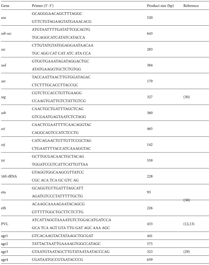
None of the S. aureus strains isolated from the study and control groups carried PVL or exfoliative toxin genes. None of the strains isolated from the control group carried toxin genes. In the patient group, 18 (32.1%) of the isolated strains carried the sei, 1 (1.8%) carried the seb-sec, and 1 (1.8%) carried the seg enterotoxin gene (Figure 1).
Eight of the strains carrying enterotoxin genes were isolated from nasal swabs, 6 from diseased skin swabs, and 4 from healthy skin swabs.
The differences of toxin genes among isolation sites were statistically insignificant (P = 0.135).
None of the strains isolated from the control group carried the agr locus. On the other hand, 11 (19.7%) of the
S. aureus strains isolated from the patients carried type 1, 7
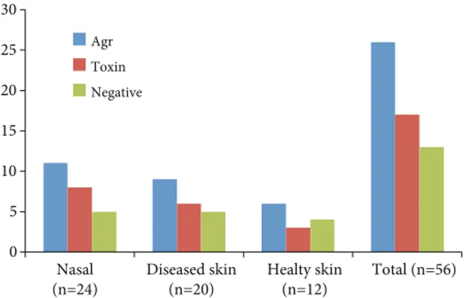
(12.5%) carried type 1 + 3, 4 (7.1%) carried type 2, 4 (7.1%) carried type 3, and 1 (1.8%) carried type 1 + 2 agr loci (Figure 2). Twelve of these strains were isolated from nasal swabs, 10 from diseased skin swabs, and 5 from healthy skin swabs. The agr locus was carried at a significantly higher rate in S.
Figure 1. Enterotoxin genes identified by PCR. M represents 50-bp molecular weight marker (Fermentas, Lithuania), IC represents internal control (228-bp product of S. aureus 16S rRNA gene).
aureus strains isolated from nasal swabs and diseased skin
swabs than the strains isolated from healthy skin swabs (P < 0.05). The distributions of toxin genes and agr types are given in Table 2 and Figures 3 and 4. Table 2. Distribution of S. aureus strains isolated from patients according to their sampling sites, identified enterotoxin genes, and agr types.
Patient no. Sampling site Genes identifiedagr types Enterotoxin genes
1 2 3 sei seb-sec seg
1 Nasal + 2 Nasal + 3 Nasal 4 Diseased skin Healthy skin + 5 Nasal + Diseased skin + 6 Nasal + + Diseased skin + + Healthy skin + 7 Nasal Diseased skin Healthy skin 8 Nasal + Diseased skin + Healthy skin + 9 Nasal + Diseased skin + Healthy skin + 10 Nasal + Diseased skin + Healthy skin + 11 Nasal Diseased skin 12 Nasal Diseased skin + Healthy skin + 13 Nasal + Diseased skin + + 14 Nasal 15 Nasal + Diseased skin + 16 Nasal + Diseased skin + Healthy skin + 17 Nasal + + Diseased skin + + Healthy skin + + 18 Nasal + Healthy skin + + 19 Nasal + + Diseased skin + 20 Nasal + Diseased skin + Healthy skin + 21 Nasal + 22 Nasal + Diseased skin + 23 Nasal + + Diseased skin + + 24 Nasal Diseased skin Healthy skin 25 Diseased skin 26 Nasal + + Diseased skin + +
4. Discussion
Psoriasis is a chronic inflammatory disease affecting many components of the immune system. It is characterized by epidermal hyperproliferation and inflammation. Its pathogenesis involves complicated relationships among many cell types, cytokines, chemokines, and skin-derived chemical mediators. The interaction between these components leads to dysregulation in the immune system (31).
Bacterial products and SAgs such as staphylococcal enterotoxins, TSST-1, exfoliative toxins, streptococcal pyrogenic exotoxins, mycoplasma arthritis supernatant, chemicals, UV light, and trauma may take part in the development or aggravation of inflammatory skin diseases (3,4). Toxins and enzymes secreted by the bacteria have an important role in staphylococcal infections. Among these, enterotoxins are encoded by sea-sej genes, TSST-1 by the
tst gene, and exfoliative toxins A and B by the eta and etb
genes (32–34).
Another important virulence factor of S. aureus is PVL. It is responsible for pore formation on the membranes of polymorphonuclear leukocytes, leading to cell lysis (12). PVL-secreting strains are responsible for severe skin and soft tissue infections and necrotizing pneumonia (35). None of our patients were colonized with PVL-positive or MRSA strains, both of which may be considered as a benefit for our patients.
Expression of virulence proteins in S. aureus is under the control of RNA III, which is a small RNA molecule regulating the expression of S. aureus genes for exoproteins and cell membrane proteins. It is the intracellular effector of the quorum sensing system. Secreted proteases are under the control of the agr gene (36).In our study, 26 (46.42%) of the 56 S. aureus strains carried the agr locus, suggesting that secreted proteases may play a role in the aggravation of psoriasis.
The relationship between bacterial SAgs and skin diseases is shown in guttate psoriasis, atopic dermatitis, and cutaneous lupus erythematosus. The mechanism of how SAgs lead to inflammation is not known extensively. SAg-mediated T-cell activation may be involved (35).
In the study of Balcı et al. (37), 64% of the diseased and 14% of healthy skin cultures obtained from psoriasis patients were found to be positive for S. aureus.They also found a significant relationship between toxin production of the strains isolated from skin lesions and disease grades (37). These results support the findings of previous studies (38,39). Tomi et al. (39) showed that 36% of the S. aureus strains isolated from skin lesions of psoriasis patients secreted toxins. In the same study, psoriasis patients who carried toxin-negative and toxin-positive S. aureus strains were compared, and disease grades of the patients with toxin-positive strains were found to be higher. These results show that there is a relationship between toxin-positive S. aureus colonization and psoriasis activation (39). On the other hand, Sayama et al. (40) could only demonstrate the presence of enterotoxin (seb) and tst-1 in 5 of the 100 S. aureus strains isolated from diseased skin swabs of psoriasis patients, and they concluded that SAgs do not have a role in the development of psoriasis (40).
In our study, we observed that patients who carry S.
aureus on psoriasis lesions are more likely to also carry S. aureus nasally. Healthy skin culture positivity was
significantly less prevalent in the patient group. If nasal carriage is regarded as the primary focus, psoriasis lesions can be considered as more prone to the development of
S. aureus colonization when compared to healthy skin.
Another question requiring explanation is the mechanism underlying the high frequency of nasal S. aureus carriage among psoriasis patients.
The results of our study demonstrated the presence of toxin genes in 20 (35.71%) of the 56 MSSA strains. Among diseased skin isolates (n = 7), 6 carried the sei and
0 5 10 15 20 25 30 Nasal
(n=24) Diseased skin(n=20) Healty skin(n=12) Total (n=56) Agr Toxin Negative 0 2 4 6 8 10 12 14 16
1 2 3 1+2 1+3 sei seb-sec seq
Agr Toxin
Nasal (n=24) Diseased skin (n=20) Healthy skin (n=12) Total (n=56)
Figure 3. Toxin and agr locus presence among the strains isolated
1 the seg gene. Four of the 11 healthy skin isolates carried the sei gene, and of the 9 nasal isolates, 8 carried the sei gene and 1 the seb-sec genes. There was no difference in toxin production of isolates according to their sampling sites. On the other hand, the 4 MSSA isolates obtained from the control group did not carry toxin genes or the
agr locus. These findings suggest that enterotoxins may be
important in the aggravation of psoriasis as suggested in previous studies (39,41). Although these previous studies emphasized the high prevalence of enterotoxin genes in S. aureus strains isolated from psoriasis patients and correlated their presence with disease severity, they did not investigate the prevalence of enterotoxin I, which was the main enterotoxin gene found in our study. Whether
or not this finding has important correlations with disease activation or severity needs further investigation.
Although there was no correlation between agr positivity and isolation sites of S. aureus strains, this study showed that S. aureus strains isolated from psoriasis patients established a high prevalence for the presence of the agr gene locus, which is responsible for the secretion of proteases that facilitate the aggregation of the infecting strain on to the skin. Thus, the high rate of agr positive
S. aureus colonization in psoriasis patients may be a
provocateur factor for disease activation attacks.
As a result, not only S. aureus colonization but also the toxin positivity and agr gene presence may be important for disease activation in psoriasis patients.
References
1. Parisi R, Symmons DP, Griffiths CE, Ashcroft DM. Global epidemiology of psoriasis: a systematic review of incidence and prevalence. J Invest Dermatol 2013; 133: 377–385.
2. Kundakci N, Türsen U, Babiker MO, Gürgey E. The evaluation of the sociodemographic and clinical features of Turkish psoriasis patients. Int J Dermatol 2002; 41: 220–224.
3. Fry L, Baker BS. Triggering psoriasis: the role of infections and medications. Clin Dermatol 2007; 25: 606–615.
4. van de Kerkhof PCM. The evolution of psoriatic lesion. Br J Dermatol 2007; 157: 4–15.
5. Brooks GF, Carroll KC, Butel JS, Morse SA, Mietzner TA. In: Weitz M, Lebowitz H, editors. Jawetz, Melnick & Adelberg’s Medical Microbiology. 25th ed. New York, NY, USA: McGraw-Hill; 2013. pp 199–206.
6. Lowy FD. Staphylococcus aureus infections. N Engl J Med 1998; 339: 520–532.
7. Holmes A, Ganner M, McGuane S, Pitt TL, Cookson BD, Kearns AM. Staphylococcus aureus isolates carrying Panton-Valentine leucocidin genes in England and Wales: frequency, characterization, and association with clinical disease. J Clin Microbiol 2005; 43: 2384–2390.
8. Foster TJ, Höök M. Surface protein adhesins of Staphylococcus aureus. Trends Microbiol 1998; 6: 484–488.
9. Speziale P, Pietrocola G, Rindi S, Provenzano M, Provenza G, Di Poto A, Visai L, Arciola CR. Structural and functional role of Staphylococcus aureus surface components recognizing adhesive matrix molecules of the host. Future Microbiol 2009; 4: 1337–1352.
10. Marraffini LA, Dedent AC, Schneewind O. Sortases and the art of anchoring proteins to the envelopes of gram-positive bacteria. Microbiol Mol Biol Rev 2006; 70: 192–221.
11. Maresso AW, Schneewind O. Sortase as a target of anti-infective therapy. Pharmacol Rev 2008; 60: 128–141.
12. Lina G, Piemont Y, Godail-Gamot F, Bes M, Peter MO, Gauduchon V, Vandenesch F, Etienne J. Involvement of Panton–Valentine leukocidin-producing Staphylococcus aureus in primary skin infections and pneumonia. Clin Infect Dis 1999; 29: 1128–1132.
13. Karahan ZC, Dolapçı İ, Tekeli A. Influence of reaction optimization on the results of PCR amplification of Panton-Valentine leukocidin genes among Staphylococcus aureus isolates. Mikrobiyol Bül 2009; 43: 519–528.
14. Foster TJ. Immune evasion by staphylococci. Nat Rev Microbiol 2005; 3: 948–958.
15. Kaneko J, Kamio Y. Bacterial two-component and hetero-heptameric forming cytolytic toxins: structures pore-forming mechanism organization of the genes. Biosci Biotechnol Biochem 2004; 68: 981–1003.
16. Menestrina G, Dalla Serra M, Prévost G. Mode of action of β-barrel pore-forming toxins of the staphylococcal α-hemolysin family. Toxicon 2001; 39: 1661–1672.
17. Lina G, Bohach GA, Nair SP, Hiramatsu K, Jouvin-Marche E, Mariuzza R. Standard nomenclature for the superantigens expressed by Staphylococcus. J Infect Dis 2004; 189: 2334– 2336.
18. Holtfreter S, Bröker BM. Staphylococcal superantigens: do they play a role in sepsis? Arch Immunol Ther Exp (Warsz) 2005; 53: 13–27.
19. Melish ME, Glasgow LA. The staphylococcal scalded skin syndrome. N Engl J Med 1970; 282: 1114–1119.
20. Morlock BA, Spero L, Johnson AD. Mitogenic activity of staphylococcal exfoliative toxin. Infect Immun 1980; 30: 381– 384.
21. Rooijakkers SHM, Ruyken M, Roos A, Daha MR, Presanis JS, Sim RB, van Wamel WJ, van Kessel KP, van Strijp JA. Immune evasion by a staphylococcal complement inhibitor that acts on C3 convertases. Nat Immunol 2005; 6: 920–927.
22. De Haas CJC, Veldkamp KE, Peschel A, Weerkamp F, Van Wamel WJ, Heezius EC, Poppelier MJ, Van Kessel KP, van Strijp JA. Chemotaxis inhibitory protein of Staphylococcus
aureus, a bacterial antiinflammatory agent. J Exp Med 2004;
199: 687–695.
23. Prat C, Bestebroer J, De Haas CJC, Van Strijp JA, Van Kessel KP. A new staphylococcal anti-inflammatory protein that antagonizes the formyl peptide receptor-like 1. J Immunol 2006; 177: 8017–8026.
24. Chavakis T, Hussain M, Kanse SM, Peters G, Bretzel RG, Flock JI, Herrmann M, Preissner KT. Staphylococcus aureus extracellular adherence protein serves as antiinflammatory factor by inhibiting the recruitment of host leukocytes. Nat Med 2002; 8: 687–693.
25. Bokarewa MI, Jin T, Tarkowski A. Staphylococcus aureus: staphylokinase. Int J Biochem Cell Biol 2006; 38: 504–509. 26. Lee LYL, Liang X, Höök M, Brown EL. Identification and
characterization of the C3 binding domain of the Staphylococcus aureus extracellular fibrinogen-binding protein (Efb). J Biol Chem 2004; 279: 50710–50716.
27. Lee LYL, Höök M, Haviland D, Wetsel RA, Yonter EO, Syribeys P, Vernachio J, Brown EL. Inhibition of complement activation by a secreted Staphylococcus aureus protein. J Infect Dis 2004; 190: 571–579.
28. Clinical and Laboratory Standards Institute. Performance Standards for Antimicrobial Susceptibility Testing; Nineteenth Informational Supplements. CLSI Document M 100- S19 (ISBN 1-56238-690-5). Wayne, PA, USA: Clinical and Laboratory Standards Institute; 2012.
29. Gilot P, Lina G, Cochard T, Poutrel B. Analysis of the genetic variability of genes encoding the RNA III-activating components Agr and TRAP in a population of Staphylococcus aureus strains isolated from cows with mastitis. J Clin Microbiol 2002; 40: 4060–4067.
30. Monday SR, Bohach GA. Use of multiplex PCR to detect classical and newly described pyrogenic toxin genes in staphylococcal isolates. J Clin Microbiol 1999; 37: 3411–3414.
31. Ghoreschi K, Mrowietz U, Röcken M. A molecule solves psoriasis? Systemic therapies for psoriasis inducing interleukin 4 and Th2 responses. J Mol Med 2003; 81: 471–480.
32. Projan S, Novick R. The Molecular Basis of Virulence. In: Crossley KB, Archer GL, editors. The Staphylococci in Human Disease. New York, NY, USA: Churchill Livingstone; 1997. pp 55–81.
33. Sutherland J, Varnam A. Enterotoxin producing Staphylococcus,
Shigella, Yersinia, Vibrio, Aeromonas and Plesiomonas. In:
Blackburn CW, McClure PJ, editors. Foodborne Pathogens. Washington, DC, USA: CRC Press; 2002. pp 384–415. 34. Lee PK, Vercellotti GM, Deringer JR, Schlievert PM. Effects
of staphylococcal toxic shock syndrome toxin-1 on aortic endothelial cells. J Infect Dis 1991; 164: 711–719.
35. Galadari I, Sharif MO, Galadari H. Psoriasis: a fresh look. Clin Dermatol 2005; 23: 491–502.
36. Chan PF, Foster SJ. Role of SarA in virulence determinant production and environmental signal transduction in
Staphylococcus aureus. J Bacteriol 1998; 180: 6232–6241.
37. Balcı DD, Duran N, Ozer B, Güneşaçar R, Önlen Y, Yenin JZ. High prevalence of Staphylococcus aureus cultivation and superantigen production in patients with psoriasis. Eur J Dermatol 2009; 19: 238–242.
38. Marples RR, Heaton CL, Kligman AM. Staphylococcus aureus in psoriasis. Arch Dermatol 1973; 107: 568–570.
39. Tomi NS, Kränke B, Aberer E. Staphylococcal toxins in patients with psoriasis, atopic dermatitis, and erythroderma, and in healthy control subjects. J Am Acad Dermatol 2005; 53: 67–72. 40. Sayama K, Midorikawa K, Hanakawa Y, Sugai M, Hashimoto K. Superantigen production by Staphylococcus aureus in psoriasis. Dermatology 1998; 196: 194–198.
41. Jassim HA, Bakir SS, Alhamdi KI, Albadran AE. Polymerase chain reaction (PCR) for detection superantigenicity of
Staphylococcus aureus isolated from psoriatic patients. Int J