Merkel Cell Carcinoma Secondary to Chronic Venous
Insufficiency, A Rare Case
Kronik Venöz Yetmezliğe İkincil Gelișen Merkel Hücreli Karsinom, Nadir Bir Vaka
Hande Selvi Öztorun
1, Tuğba Turgut
1, Bilge Gözükara
2, Volkan Atmıș
1, Remzi Bahși
1,
Deniz Mut Sürmeli
1, Sevgi Aras
1, Murat Varlı
11 Ankara University School of Medicine, Internal Medicine, Geriatry
Department
2 Ankara University School of Medicine Internal Medicine Merkel cell carcinoma (MCC) is a rare skin tumor that usually develops in elderly and
sun-exposed body regions. The tumor is aggressive in nature, and in the case of delayed diagnosis, the treatment is difficult and the prognosis is poor. MCC is rarely seen on the ground of chronic venous insufficiency and on the lower extremity. In this case, an 85-year-old male patient was diagnosed with Merkel cell carcinoma in the right leg, biopsy specimen taken from a ulcer and a raised lysole from chronic venous insufficiency after cardiac bypass graft surgery for cardiac bypass surgery. Skin malignancies such as MCC should be kept in mind in the differential diagnosis of venous ulcers and other skin lesions that may develop frequently on the basis of chronic venous insufficiency.
Key Words: Merkel Cell Carcinoma, Chronic Venous Insufficiency, Elderly
Merkel hücreli karsinom (MHK) genellikle ileri yașlarda ve güneșe maruz kalınan vücut bölgelerinde ortayan çıkan nadir bir cilt tümörüdür. Tümör agresif seyirli olup, tanıda gecikme olması durumunda tedavisi zordur ve prognozu kötüdür. MHK, kronik venöz yetmezlik zemininde ve alt ekstremitede nadiren görülür. Bu olguda 85 yașında bir erkek hastada sağ bacakta, kardiyak by-pass cerrahisi için safen ven greft alınma operasyonu sonrası olușan kronik venöz yetmezlik zemininde gelișen ülsere ve ciltten kabarık bir lezyondan alınan biyopsi sonucu merkel hücreli karsinom tanısı konulmuștur. Kronik venöz yetmezlik zemininde sıklıkla gelișebilecek venöz ülserler ve diğer cilt lezyonlarının ayırıcı tanısında MHK gibi cilt maligniteleri akılda tutulmalıdır.
Anahtar Sözcükler: Merkel Hücreli Karsinom, Kronik Venöz Yetmezlik, Yașlı
Merkel cell carcinoma (MCC) is an aggressive skin tumor which is usually seen in blond skin people, localizes to local recurrence and regional lymph node metastasis. (1) MCC is a rare tumor with an incidence of 0.6/100.000 (2). Its incidence is higher among elderly and male. Well defined risk factors for MCC are light skin, immune system disorders, organ transplant receiving, immunosuppressive conditions especially when there is an underlying malignancy of B lymphocytes and ultraviolet radiation. Only risk factors of our patient was advanced age and male sex. One of the rare causes is the development of chronic venous insufficiency (3, 4). Tumor staging is done according to the TNM classification.
According to the disease, treatment is surgical excision, radiotherapy and systemic chemotherapy in metastatic patients (5).
In this case report, an 85-year-old male patient presented with a Merkel cell carcinoma, which develops on the basis of chronic venous insufficiency after saphen vein grafting of the right leg for cardiac by-pass surgery.
CASE
85 year old male patient admitted to our clinic with complaints of edema in his right leg and ulcerations all over his right thigh for two months. The patient had a
Ankara Üniversitesi Tıp Fakültesi Mecmuası 2017, 70 (2) DOI: 10.1501/Tıpfak_000000973
DAHİLİ BİLİMLERİ/ MEDICAL SCIENCES
Olgu Sunumu / Case Report
Received: x x, 2017 Accepted: x x, 2016 Corresponding Author: xxxxxx E-mail: xxxx Phone: +90 312 xx GSM: +90 xxx Fax: +90 312 xxx xxxxxxxxx
Ankara Üniversitesi Tıp Fakültesi Mecmuası 2017, 70 (2)
Merkel Cell Carcinoma Secondary to Chronic Venous Insufficiency, A Rare Case
108
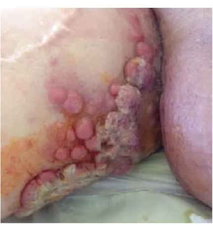
history of coronary artery disease, diabetes mellitus, Parkinson’s disease, benign prostatic hyperplasia, dementia, and urinary incontinence. His medications were acetylsalicylic acid, metformin, dutasteride, donepezil, levodopa. He had leg edema for 21 years starting after saphenous vein grafting, but his edema increased progressively for the last two months. His main disturbance was his difficulty in wearing his shoes due to edema and also denied any edema of the upper thigh. With these complaints he had many hospital admissions, in these admissions pretibial edema was detected and was thought be secondary to grafting and was advised to raise his legs. His complaints diminished for a short time and recurred again. His physical examination revealed multiple lymphadenopathies in left supraclavicular and cervical measuring 3x2 cm in widest area and right inguinal nodule 7x7 cm in widest area elevated from skin surface with an ulcerated center foully smelling odorous discharge at posterolateral thigh, and multiple 1 cm sized nodules pink in color in right inguinal region. (Figure 1-2) Patient's laboratory results are shown in Table 1.
A biopsy was taken from ulcerated lesion in his right thigh. Pathologic result was reported as neuroendocrine tumor, merkel cell carcinoma. His positron emission tomography for staging revealed pathologically increased uptake in left supraclavicular, paraaortic and paracaval, bilateral inguinal axial skeleton and bone marrow and right lower extremity cutaneous and subcutaneous fat tissue. In his follow up he couldn’t get chemotherapy, he was diagnosed with urosepsis during his follow up. He died secondary to multi organ failure despite supportive treatment.
DISCUSSION
Lower extremity edema is a common condition in the elderly and has many factors such as heart failure, pulmonary hypertension, drugs (antihypertensive, antiparkinsonian, nonsteroidal anti
Table 1. Patient's Laboratory Results
Results Normal Range
Hemoglobin 11,1 g/dL (11,7 - 16,1) Serum Thrombocyte Level 258 x10^9/L (150 – 400) Serum Leucocyte Count 7,78 x10^9/L (4,5 – 11,0) Serum Erythrocyte Sedimentation Rate 68,0 mm/h
Serum Creatinine 1,46 mg/dL (0,5 - 1,1) Estimated Glomerular Filtration Rate 34,7 mL/min/1,73 m2
Serum Albumin Level 2,7 g/dL (3,2 - 4,8) Serum Lactate Dehydrogenase Level 732 U/L (120 – 246) C Reactive Protein Level 103,6 mg/L (0-3)
Figure 1. Patient's Skin Lesion
Journal of Ankara University Faculty of Medicine 2017, 70 (2)
Hande Selvi Öztorun, Tuğba Turgut, Bilge Gözükara, Volkan Atmıș, Remzi Bahși, Deniz Mut Sürmeli, Sevgi Aras, Murat Varlı 109
inflammatory), venous insufficiency and thrombosis (6).
This MCC case, which is seen in the lower extremity of a male patient with chronic edema and advanced age (85 years), has resulted in an aggressive death with extensive lymph node and bone marrow involvement. It is usually more common in the elderly and male sex. Light skin color, ultraviolet, age, and immunosuppression are important risk factors. This rare neoplasm is originated from round shaped Merkel cells, which are the neurokrest derivative. These are located in the basal layer of the epidermis and contain neurosecretory granules. (7)
Since chronic venous insufficiency and lymphedema are very rare etiological risk factors for MCC development (3, 4). In our case, the tumor was presented on the ground of a long-standing venous insufficiency and with the gradually increasing lower extremity edema in the form of ulcerated and enlarged lesions from the skin.It is usually not possible to suspect form MCC in a patient with chronic edema. Medical history and physical examination is the mainstay of diagnosis of etiology of edema. Venous
insufficiency; with pitting edema , brown hemosiderin skin deposits on lower legs, dermatitis, ulceration; is among the most common causes of leg edema and can be diagnosed clinically (8). Rarely lower extremity swelling can be a presenting sign of mass forming malignancies in the inguinal region (9). In this case further evaluation was done just because a complete physical examination of the patient was performed. MCC usually presents as a rapidly growing, firm, flesh-colored, dome shaped papule or plaque on sun exposed skin usually smaller than 2 cm in diameter at time of diagnosis (10). Age older than 65 years, male sex, size of primary lesion greater than 2 cm, truncal site, nodal/distant metastases and duration of these before presentation appear to be poor prognostic factors. 5 year survival for stage I disease is 81% and 11% for stage IV disease (11). Among these poor prognostic factors only modifiable one is the detection of the lesion before it is 2 cm in diameters. In our case it was 7x7 cm in diameter when diagnosed which can be accepted as a late detection. Patient's skin lesions is shown in figure 1. Although chronic venous insufficiency is a common etiologic factor for leg edema it should be kept in mind venous stasis
can cause newly appearing skin lesions suspicious for skin malignancies which like other malignancies need to be diagnosed as early as possible. UV radiation is a risk factor for malignancies but keeping in mind that elderly patients, especially demented elderly patients, as in our case, can mislead the clinician in his medical history so a complete physical examination should never be neglected. Despite patient died of urosepsis, it could have been avoided if an early diagnosis was made.
In conclusion, this case, which is an unexpected localization for MHC and develops on the basis of edematous lower extremity, shows an aggressive course, suggesting that skin malignancies should be kept in mind in newly developed skin lesions. A careful history is not taken, because skin lesions are mostly painless, as in this case and often can be missed if physical examination is not done. Early diagnosis, particularly in aggressive tumors, may increase the chance of treatment and may contribute to prognosis positively.For this reason, biopsy should be done for histopathological diagnosis when a clinically suspicious lesion is seen.
REFERENCES
1. Tothill R, Estall V, Rischin D, Merkel cell carcinoma: emerging biology, current approaches, and future directions. Am Soc Clin Oncol Educ Book 2015:519-26.
2. Heath M, Jaimes N, Lemos B, et al., Clinical characteristics of Merkel cell carcinoma at diagnosis in 195 patients: the AEIOU features. J Am Acad Dermatol 2008. 58:375-81.
3. Peterson CM, Lane JE, Guill MA, Merkel cell carcinoma after postmastectomy lymphedema. J Am Acad Dermatol 2003. 48:983.
4. Lee R, Saardi KM, Schwartz RA, Lymphedema-related angiogenic tumors and other malignancies. Clin Dermatol 2014. 32:616-20.
5. Lebbe C, Becker JC, Grob JJ, et al., Diagnosis and treatment of Merkel Cell Carcinoma. European consensus-based interdisciplinary guideline. Eur J Cancer 2015. 51:2396-403.
6. Topham EJ, Mortimer PS, Chronic lower limb oedema. Clin Med (Lond) 2002. 2:28-31.
7. Schwartz RA, Lambert WC, The Merkel cell carcinoma: a 50-year retrospect. J Surg Oncol 2005. 89:5. 8. Alguire PC, Mathes BM, Chronic
venous insufficiency and venous ulceration. J Gen Intern Med 1997. 12:374-83.
9. Elgendy IY, Lo MC, Unilateral lower extremity swelling as a rare
presentation of non-Hodgkin's lymphoma. BMJ Case Rep 2014;doi:10.1136/bcr-2013-202424 10. Ultori C, Cimetti L, Stefanoni P, et al.,
Merkel cell carcinoma in elderly: case report and review of the literature. Aging Clin Exp Res 2013. 25:211-4. 11. Kampshoff JL, Cogbill TH, Unusual
skin tumors: Merkel cell carcinoma, eccrine carcinoma, glomus tumors,
and dermatofibrosarcoma protuberans. Surg Clin North Am