Label-free G-Quadruplex aptamer and Thio
flavin-T based turn-off
fluorescent detection of ethanolamine
Abdullah Tahir Bayraç
∗, Yasemin Acar
Department of Bioengineering, Karamanoğlu Mehmetbey University, 70100, Karaman, Turkey
A R T I C L E I N F O
Keywords: Ethanolamine Aptasensor G-quadruplex
Fluorescence molecular rotor
A B S T R A C T
A label free aptasensor based on G-quadruplex forming ethanolamine aptamers and Thioflavin T was developed for the selective and sensitive detection of ethanolamine. The detection system consists of DNA aptamers with four GGG repeats forming a G-quadruplex that Thioflavin T can easily bind to causing high fluorescence emission intensity because of itsfluorescence molecular rotor nature. In the presence of ethanolamine, binding of aptamer to its target induces conformational changes causing the separation of Thioflavin T from the complex with a dramatic decrease influorescence intensity of reaction mixture. This fluorescence aptasensor has a convenient sensitivity and selectivity with a detection limit of 641μM to measure ethanolamine directly in contaminated water samples in a wide linear range. Furthermore this method avoids all complicated modification and labelling steps and thus offers a simple, fast, and cost efficient solution for ethanolamine detection.
1. Introduction
Ethanolamine (2-aminoethanol) is a viscous and hygroscopic amino alcohol having molar mass of 61.08 g mol−1. It is extensively used in textile, dry cleaning, rubber manufacturing and pharmaceutical in-dustry, for the production of dyes, adhesives, pesticides, insecticides, medicines, surfactants, and gas scrubbers [1]. Ethanolamine is an irri-tant chemical for skin and eyes and also can cause severe problems for nervous and respiratory systems. Ethanolamine is associated with many diseases such as Alzheimer [2], atherosclerosis [3], hyperlipemia [4], schizophrenia [5], major depressive disorder [6] and ethanolaminuria [7]. It is found to be toxic to most organisms [8], acute oral median lethal dose (LD50) is 700 mg kg−1 for mice and 1000 mg kg−1 for rabbit [9]. Also median lethal concentration (LC50) ranges from 150 to 2100 mg L−1forfish species. Because of its toxicity ethanolamine has to be monitored in industrial waste, groundwater and subsurface en-vironments [4,11].
Detection of small molecules such as ethanolamine is a challenging and laborious for standard analytical methods [12]. Although various methods are studied in the literature [1,9,11,13,14] determination of the concentration of ethanolamine with both high accuracy and sensi-tivity is a compelling since it has a high polarity and low molecular weight which makes it hard to retain and separate from other sub-stances in a matrix [4]. Hence, development of alternative methods for ethanolamine detection is of increasing importance. Recently, aptamers
become a hot subject in analytical chemistry because of their ad-vantages such as high affinity, stability, flexibility, and modifiability with respect to antibodies [15]. Aptamers are single stranded oligo-nucleotide sequences, enriched and selected from a random oligonu-cleotide library by a technique named Systematic Evolution of Ligands by Exponential Enrichment (SELEX) and can bind to their target with high specificity and selectivity [16–18]. Many specific aptamers were selected for a wide range of targets such as small molecules [19,20], proteins [21], spores [22], bacteria [23–25], eukaryotic cells [26–29], eggs [30], and even tissues [31]. Aptamer mediated ethanolamine tection can eliminate the specificity problems of antibody based de-tection of small molecules. Ethanolamine aptamers were generated by a SELEX method [32,33] and many truncated variants were developed in order tofind sequence motifs responsible for target binding [13]. Al-though thefirst ethanolamine aptamer was selected in 2005 by Mann et al., only an aptamer microarray [13] and an LC/MS aided indirect aptamer sensor [4] were developed up to date. Ethanolamine aptamers generated by Mann et al. have guanine rich nucleic acid sequences and can fold into G-quadruplex structures. G-quadruplexes are formed by the stacking of coplanar guanine tetrads via Hoogsten hydrogen bonds [34,35]. Coplanar architecture of G-quadruplexes has noticeable van der Waals attraction and many aromatic planar ligands can interact with this guanine chamber [36,37]. Thioflavin T(ThT) is one of the unique G-quadruplex ligand composed of benzothiazole (BZT) and di-methylaminobenzene (DMAB). ThT has been used as a fluorescence
https://doi.org/10.1016/j.dyepig.2019.107788
Received 20 March 2019; Received in revised form 9 August 2019; Accepted 10 August 2019
⁎Corresponding author. Karamanoglu Mehmetbey University, Faculty of Engineering, Department of Bioengineering, 70100, Karaman, Turkey.
E-mail address:[email protected](A.T. Bayraç).
Available online 11 August 2019
0143-7208/ © 2019 Elsevier Ltd. All rights reserved.
reporter to quantifyfibrils for more than 50 years which is thought to be found in protein-misfolding diseases such as Alzheimer and Par-kinson [38]. When ThT is in a free state the dihedral angle rotated by the C–C bond between the BZT and DMAB moieties is 90° and molecule has the lowest quantum yield possible [39]. As ThT is embedded into the G-quadruplex its hiddenfluorescence property will emerge with the suppression of steric hindrance [38–40]. So that aptamers with G-quadruplex sequences are good candidates for designing ThT based turn-off sensors. When aptamers with G-quadruplex sequences were incubated with ThT complex, they will show a highfluorescence yield with the insertion of the ThT in to the guanine cage, but after addition of the aptamer's target in to the environment aptamer will prefer to bind to its target and ThT will be unleashed and loose itsfluorescence property (Fig. 1). Herein we report a label free G-quadruplex and ThT based aptasensor for the rapid and quantitative measurement of etha-nolamine.
2. Experimental section
2.1. Chemicals, materials and measurements
Ethanolamine aptamers EA#9.4K46 5′-ATA CCA GCT TAT TCA ATT GCT GCG AGG TGG GTG GGT GGG AGC AAT T-3′, EA#14.3K42 5′-ATA CCA GCT TAT TCA ATT TGA GGC GGG TGG GTG GGT TGA ATA-3′, EA-ConsT 5′-GAG GTG GGT GGG TGG G-3′ were synthesized and HPLC purified by Integrated DNA Technologies Inc. (Coralville, USA). Thioflavin T, ethanolamine, diethanolamine, methylene bis(ac-rylamide), 3-aminophenol, methanol, ethanol, isopropanol, glucose, glycerol, furfural, tris-HCl, sodium chloride, potassium chloride, mag-nesium chloride, calcium chloride, tween 20 were purchased from Sigma Aldrich and used without further purification. All chemicals were of analytical-reagent grade. Ultra-pure water (18.2 MΩ cm−1) was used during all of the experiments. Binding Buffer (BB) (100 mM NaCl, 20 mM Tris-HCl, 5 mM KCl, 2 mM CaCl2, 0.02% Tween 20, pH 7.6) was used forfluorescence detection of ethanolamine sensor.
All fluorescence measurement were carried out on an F7100 (Hitachi, Japan) with an excitation of 425 nm and an emission range from 450 to 600 nm at 1 nm increments. The slit widths for excitation and emission were set at 10 nm. The maximum emission intensity for ThT was observed at 495 nm. The pH was measured by inoLab pH 7110 and all experiments were performed at room temperature.
2.2. Feasibility test of the method
Feasibility of the method is tested with the usage of EA#14.3K42 ethanolamine aptamer. Four samples were prepared for the feasibility of the test; Sample 1 contained ThT only, Sample 2 contained ThT and ethanolamine, Sample 3 contained ThT and EA#14.3K42 aptamer and Sample 4 contained ThT, EA#14.3K42 aptamer and ethanolamine. All samples were prepared in BB and samples were incubated in room temperature for 15 min before taking measurements. For the image analyses of the test, Gel Doc™ XR + Gel Documentation System (BIO-RAD) was used.
2.3. Optimization of ethanolamine detection conditions
In order to get optimal results incubation time, concentration of ThT, concentration of aptamers, and concentration of KCl were opti-mized for each aptamer sequences. Aptamer concentration was in the range of 0–500 nM, ThT concentration was in the range of 0–11 μM, KCl concentration was in the range of 0–30 mM, and incubation time range was in the range of 0–60 min.
2.4. Sensitivity and selectivity of the assay
The ethanolamine detection was carried out in determined optimum conditions. Fluorescence change after the addition of different con-centrations of ethanolamine was recorded and linear range, limit of detection (LOD), sensitivity and selectivity were determined accord-ingly. Selectivity of the sensor was tested using 60 mM of ethanolamine, dietanolamine, and 200 mM of methylenebis(acrylamide), methanol, ethanol, isopropanol, glucose, glycerol and furfural. All measurements were repeated for a minimum of three times with ten replicates for each sample.
3. Results and discussion 3.1. Design strategy
The designed strategy of the proposed method is explained inFig. 1. Guanine rich ethanolamine aptamer can form a G-quadruplex complex with ThT. Thefluorescence quantum yield of ThT increases upon em-bedding in to G-quadruplex because of itsfluorescence molecular rotor (FMR) nature. But in the presence of ethanolamine, aptamer will prefer to bind to its target with its unique three dimensional shapes which will distort the G-quadruplex basis of the sequence, so that ThT molecules that leave the G-quadruplex complex will cause a dramatic decrease in thefluorescence intensity.
3.2. Feasibility test for the detection of ethanolamine
The feasibility of the developed method for ethanolamine detection was estimated by using EA#14.3K42 ethanolamine aptamer. InFig. 2it can be seen that only ThT showed a weakfluorescence intensity (Line A), even in the presence of 2 M ethanolamine (Line B). When ethano-lamine aptamer and ThT were incubated together formation of G-quadruplex-ThT complex induced highfluorescence intensity (Line C). Upon addition of ethanolamine a dramatic decrease in fluorescence intensity, confirmed that the proposed method have ability to detect ethanolamine (Line D). Therefore, a label-free, easy, rapid but efficient ethanolamine detection method was established.
3.3. Optimization of sensing conditions
In order to get the highestfluorescence signal change, incubation time, ThT concentration, aptamer concentration, and K+concentration was optimized using three different ethanolamine aptamer sequences. In all aptamers fluorescence intensity increased linearly with an
increase in aptamer concentration (Fig S1A). ThT concentration was optimized for 200 nM aptamer andFig. 3shows that thefluorescence intensity enhanced as the ThT concentration was raised, reaching the maximum at 5μM. Also it can be seen that the maximum fluorescence signal can be obtained from EA#14.3K42 aptamer. EA#9.4K46 (46 bp) and EA#14.3K42 (42bp) have similar curve forms and approximate fluorescence enhancement capacities for ThT but EA-ConsT showed lowerfluorescence intensity enhancement. Although all aptamers had the same G-quadruplex motifs in their sequences the difference in the fluorescence intensity can be elucidated with the duplex connection at one end. Duplex connected G-quadruplex structures have more evident promotion on the fluorescent emission of turn-on dyes [41]. EA#9.4K46 and EA#14.3K42 aptamers has a duplex forming DNA se-quences attached to their G-quadruplex forming motifs (Fig. S2) but EA-ConsT has only the G-quadruplex motif sequences so that the rigid duplex structure present in the EA#9.4K46 and EA#14.3K42 created more resistant for the rotational deactivation causing higher fluores-cence property. Also K+concentration is very important for the stabi-lity of the G-quadruplex [42] and K+promotes the binding of ThT to G-quadruplex sequences [43]. Thus, 5μM ThT with 200 nM EA#14.3K42 aptamer was used throughout the experiments in a BB which have 5 mM of KCl with an incubation time of 15 min.
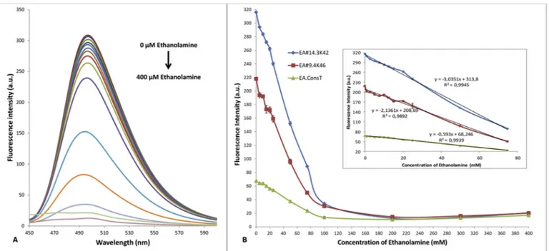
tensity was continued up to 100 mM of ethanolamine and then stayed with backgroundfluorescence (Fig. 4B). The inset ofFig. 4B shows that thefluorescence intensity had linear relationship with concentration in the range of 0.5 mM–75 mM with a correlation coefficient of 0.9945 for EA#14.3K42, 0.9892 for EA#9.4K46 and 0.9939 for EA-ConsT. EA#14.3K42 had the regression equation of y =−3.0351x+313.8 with the lowest detection limit of 641μM based on three times the signal-to-noise level compared to other two aptamers. Ten replicate measurements of 5 mM of ethanolamine showed the relative deviation of 0.33% which provides an excellent repeatability. Although there are many studies that have lower sensitivity for ethanolamine in the lit-erature, the widest linear range is in our study (Table 1). Linear range is an important characteristic for the sensor performance and suitability of the linear range is a fundamental feature for direct measurements of targets in specific environments. To exemplify LC50 of ethanolamine for different fish species is in the range of 2.4 mM–34 mM [10] and also according to a microbial toxicity study, 50 mM of ethanolamine can inhibit bacterial growth up to 40% at pH 7.6 causing microbial dete-rioration in the environment which is a very common case in metal working industries in processes like grinding and drilling [44]. So that, it is important to have an assay for ethanolamine in a wide micromolar linear range and with an acceptable sensitivity in order to directly measure the ethanolamine in fresh water samples with high repeat-ability.
3.5. Specificity of the assay
To demonstrate the selectivity of the proposed assay, a series of small molecules were investigated by this method. As shown inFig. 5 none of these molecules trigger a significant decrease in fluorescence intensity. The results indicate good selectivity of the presented sensing assay for the detection of ethanolamine.
3.6. Application
To evaluate the applicability of the assay for practical operation, sensor was applied to the detection of ethanolamine in artificially contaminated water samples and artificially contaminated water sam-ples with 5% fetal bovine serum addition to see the matrix affect. The results showed that the recoveries were in the range of 95.5%–109.6% indicating the sensor was reliable and applicable for water samples. Addition of fetal bovine serum to mimic complex biological environ-ment caused an increase in the recovery percentages but they were still in the acceptable ranges (Table 2).
Aptamers with G-quadruplex forming sequences were used in turn-off fluorescence sensors using dyes with fluorescence molecular motor property in the literature. Wu et al. used same strategy to detect ochratoxin a with better detection limit [45]. Zeng et al. also used ThT and G-quadruplex aptamer sequences to detect ochratoxin a with ra-tiometricfluorescence resonance energy transfer aptasensor [46]. Even same strategy was used for enzyme activity detection [47–49]. Al-though there are many studies using same methodology, this study is thefirst turn-off, label free, fluorescence based aptasensor for the de-tection of ethanolamine.
4. Conclusion
In summary, we have developed a label-freefluorescence aptasensor
Fig. 2. Feasibility of the method. (A) Only 5μM ThT, (B) 5 μM ThT and 2 M ethanolamine, (C) 5μM ThT and 200 nM Aptamer, (D) 5 μM ThT, 2 M etha-nolamine and 200 nM Aptamer. The inset is an image of samples under suitable excitation.
Fig. 3. Effect of the concentration of ThT on the fluorescence intensity for different aptamer sequences (200 nM).
Fig. 4. (A) Fluorescence emission spectra of aptamer-ThT complex in the presence of increasing amounts of ethanolamine (0, 0.5, 1.0, 2.5, 5, 7.5, 10, 12.5, 15, 20, 25, 50, 75, 100, 200, 400 mM).(B) Fluorescence change at 490 nm with the addition of ethanolamine. Inset: linear part of thefluorescence change.
Table 1
Comparison of analytical methods for the detection of ethanolamine.
Sensitivity Linear Range Type of Detection References 5 mM 0.2 mM–1 mM Electrochemical
Biosensor
Skirwar et al. [9] 641μM 0.5 mM–75 mM Fluorescence Aptasensor This work 82μM 0.3μM–3 μM Ion Chromatography Lehotay et al. [8] 1.2 nM 0.005μM–5 μM Gold-nanoparticle
Aptasensor
Lee et al. [4] 0,01 nM 0.01μM–400 μM Fluorescence Aptasensor Heilkenbrinker et al.
[13] 0.08 pM 0,16 nM–16 nM Electrochemical
Aptasensor
Liang et al. [1]
Fig. 5. Specificity of the method for ethanolamine detection. Table 2
Recovery experiments of ethanolamine in artificially contaminated water samples using the proposed method.
Concentration added (mM) No FBS added 5% FBS added Concentration found (mM) Recovery (%) Concentration found (mM) Recovery (%) 5 5.5 109.6 5.8 116.2 10 9.9 99.1 10.7 106.6 25 23.9 95.5 27.5 110.0 50 52.3 104.7 55.3 110.6 75 76.3 101.8 81,6 108.8
Conflict of interest
The authors report no declarations of interest. Acknowledgement
The authors would like to gratefully acknowledge the Karamanoglu Mehmetbey University BILTEM and PTTO for their contributions. The authors would like to thank Dr. Ceren Bayrac and Berke BilgenurŞener for their helpful input.
Appendix A. Supplementary data
Supplementary data to this article can be found online athttps:// doi.org/10.1016/j.dyepig.2019.107788.
References
[1] Liang G, Man Y, Jin X, Pan L, Liu X. Aptamer-based biosensor for label-free de-tection of ethanolamine by electrochemical impedance spectroscopy. Anal Chim Acta 2016;936:222–8.https://doi.org/10.1016/j.aca.2016.06.056.
[2] Farooqui AA, Rapoport SI, Horrocks LA. Membrane phospholipid alterations in alzheimer's disease: deficiency of ethanolamine plasmalogens. Neurochem Res 1997;22:523–7.https://doi.org/10.1023/A:1027380331807.
[3] Maeba R, Yamazaki Y, Nezu T, Nishimukai M, Okazaki T, Hara H. Serum choline plasmalogen is a novel biomarker for metabolic syndrome and atherosclerosis. Chem Phys Lipids 2011;164:S23.https://doi.org/10.1016/J.CHEMPHYSLIP.2011. 05.074.
[4] Lee CY, Shiau RJ, Chou HW, Hsieh YZ. Combining aptamer-modified gold nano-particles with barcode DNA sequence amplification for indirect analysis of etha-nolamine. Sens Actuators B Chem 2018;254:189–96.https://doi.org/10.1016/j. snb.2017.07.073.
[5] Horrobin DF. The membrane phospholipid hypothesis as a biochemical basis for the neurodevelopmental concept of schizophrenia. Schizophr Res 1998;30:193–208. https://doi.org/10.1016/S0920-9964(97)00151-5.
[6] Ogawa S, Hattori K, Sasayama D, Yokota Y, Matsumura R, Matsuo J, et al. Reduced cerebrospinalfluid ethanolamine concentration in major depressive disorder. Sci Rep 2015;5:1–8.https://doi.org/10.1038/srep07796.
[7] Cole DEC, Farag S, Dooley KC. Ethanolaminuria: a non-specific laboratory finding in the seriously III infant. Clin Biochem 1988;21:297–300.https://doi.org/10.1016/ S0009-9120(88)80084-5.
[8] Lehotay J, HatríkŠ, Motošická A. Trace analysis of ethanolamine in water using ion pair chromatography with on-line preconcentration. J Liq Chromatogr
1995;18:1647–54.https://doi.org/10.1080/10826079508009301.
[9] Sikarwar B, Sharma PK, Tripathi BK, Boopathi M, Singh B, Jaiswal YK. Enzyme based electrochemical biosensor for ethanolamine. Electroanalysis 2016;28:881–9. https://doi.org/10.1002/elan.201501046.
[10] Gad SC. Ethanolamine. Encycl. Toxicol. third ed. Elsevier; 2014. p. 492–5.https:// doi.org/10.1016/B978-0-12-386454-3.00501-7.
[11] Mrklas O, Chu A, Lunn S. Determination of ethanolamine, ethylene glycol and triethylene glycol by ion chromatography for laboratory andfield biodegradation studies. J Environ Monit 2003;5:336–40.https://doi.org/10.1039/b210572a. [12] Fechner P, Bleher O, Ewald M, Freudenberger K, Furin D, Hilbig U, et al. Size does
matter! Label-free detection of small molecule–protein interaction. Anal Bioanal Chem 2014;406:4033–51.https://doi.org/10.1007/s00216-014-7834-4. [13] Heilkenbrinker A, Reinemann C, Stoltenburg R, Walter JG, Jochums A, Stahl F,
et al. Identification of the target binding site of ethanolamine-binding aptamers and its exploitation for ethanolamine detection. Anal Chem 2015;87:677–85.https:// doi.org/10.1021/ac5034819.
[14] Ameen S, Shaheer Akhtar M, Shin H-S. Low temperature grown ZnO nanotubes as smart sensing electrode for the effective detection of ethanolamine chemical. Mater Lett 2013;106:254–8.https://doi.org/10.1016/J.MATLET.2013.05.031. [15] Mairal T, Cengiz Özalp V, Lozano Sánchez P, Mir M, Katakis I, O'Sullivan CK.
Aptamers: molecular tools for analytical applications. Anal Bioanal Chem 2008;390:989–1007.https://doi.org/10.1007/s00216-007-1346-4.
[16] Tuerk C, Gold L. Systematic evolution of ligands by exponential enrichment: RNA ligands to bacteriophage T4 DNA polymerase. Science 1990;249:505–10. [17] Ellington AD, Szostak JW. In vitro selection of RNA molecules that bind specific
ligands. Nature 1990;346:818–22.https://doi.org/10.1038/346818a0. [18] Robertson DL, Joyce GF. Selection in vitro of an RNA enzyme that specifically
2018;154:132–55.https://doi.org/10.1016/J.BIOCHI.2018.09.001. [22] Bruno JG, Kiel JL. In vitro selection of DNA aptamers to anthrax spores with
electrochemiluminescence detection. Biosens Bioelectron 1999;14:457–64. [23] Lee YJ, Han SR, Maeng J-S, Cho Y-J, Lee S-W. In vitro selection of Escherichia coli
O157:H7-specific RNA aptamer. Biochem Biophys Res Commun 2012;417:414–20. https://doi.org/10.1016/j.bbrc.2011.11.130.
[24] Bayrac C, Oktem HA. Evaluation of Staphylococcus aureus DNA aptamer by en-zyme-linked aptamer assay and isothermal titration calorimetry. J Mol Recognit 2017;30:1–8.https://doi.org/10.1002/jmr.2583.
[25] Bayraç AT, Donmez SI. Selection of DNA aptamers to Streptococcus pneumonia and fabrication of graphene oxide basedfluorescent assay. Anal Biochem
2018;556:91–8.https://doi.org/10.1016/j.ab.2018.06.024.
[26] Yunfei Zhang, Yan Chen, Da Han, et al. Aptamers selected by cell-SELEX for ap-plication in cancer studies. Bioanalysis 2010;2:907–18.https://doi.org/10.4155/ bio.10.46.
[27] Bayrac AT, Sefah K, Parekh P, Bayrac C, Gulbakan B, Oktem HA, et al. In vitro selection of DNA aptamers to glioblastoma multiforme. ACS Chem Neurosci 2011;2:175–81.
[28] Sefah K, Shangguan D, Xiong X, O'Donoghue MB, Tan W. Development of DNA aptamers using Cell-SELEX. Nat Protoc 2010;5:1169–85.https://doi.org/10.1038/ nprot.2010.66.
[29] Jiménez E, Sefah K, López-Colón D, Van Simaeys D, Chen HW, Tockman MS, et al. Generation of lung adenocarcinoma DNA aptamers for cancer studies. PLoS One 2012;7:e46222https://doi.org/10.1371/journal.pone.0046222.
[30] Long Y, Qin Z, Duan M, Li S, Wu X, Lin W, et al. Screening and identification of DNA aptamers toward Schistosoma japonicum eggs via SELEX. Sci Rep 2016;6:24986. https://doi.org/10.1038/srep24986.
[31] Wang H, Li X, Volk DE, Lokesh GL-R, Elizondo-Riojas M-A, Li L, et al. Morph-X-Select: morphology-based tissue aptamer selection for ovarian cancer biomarker discovery. Biotechniques 2016;61:249–59.https://doi.org/10.2144/000114473. [32] Mann D, Reinemann C, Stoltenburg R, Strehlitz B. In vitro selection of DNA
apta-mers binding ethanolamine. Biochem Biophys Res Commun 2005;338:1928–34. [33] Reinemann C, Stoltenburg R, Strehlitz B. Investigations on the specificity of DNA
aptamers binding to ethanolamine. Anal Chem 2009;81:3973–8.https://doi.org/ 10.1021/ac900305y.
[34] Chu B, Zhang D, Hwang W, Paukstelis PJ. Crystal structure of a tetrameric DNA fold-back quadruplex. J Am Chem Soc 2018;140:16291–8.https://doi.org/10. 1021/jacs.8b10153.
[35] Bağda E, Bağda E, Yabaş E. A versatile water-soluble ball-type phthalocyanine as potential antiproliferative drug: the interaction with g-quadruplex formed from tel 21 and cMYC. JOTCSA 2017;4:103–23.https://doi.org/10.18596/jotcsa.288284. [36] Li T, Dong S, Wang E. G-Quadruplex aptamers with peroxidase-like DNAzyme
functions: which is the best and how does it work? Chem Asian J 2009;4:918–22. https://doi.org/10.1002/asia.200900019.
[37] Yeasmin Khusbu F, Zhou X, Chen H, Ma C, Wang K. Thioflavin T as a fluorescence probe for biosensing applications. TrAC Trends Anal Chem (Reference Ed) 2018;109:1–18.https://doi.org/10.1016/j.trac.2018.09.013.
[38] Zhu J, Yan Z, Zhou W, Liu C, Wang J, Wang E. Lighting up the thioflavin T by parallel-stranded TG(GA)n DNA homoduplexes. ACS Sens 2018;3:1056–217. https://doi.org/10.1021/acssensors.8b00141.
[39] Zhu Y, Li W, Tan S, Chen T. Label-free and simple G-quadruplex-based turn-off fluorescence assay for the detection of kanamycin. Anal Lett 2018;51:1718–29. https://doi.org/10.1080/00032719.2017.1387136.
[40] Gogoleva SD, Kalganova EV, Maskevich AA, Lugovski AA, Kuzmitsky VA, Goswami M, et al. Neutral derivatives of Thioflavin T do not exhibit viscosity-dependent fluorescence. J Photochem Photobiol A Chem 2018;358:76–91.https://doi.org/10. 1016/j.jphotochem.2018.03.003.
[41] Wang S, Zhao J, Lu S, Sun J, Yang X. A duplex connection can further illuminate G-quadruplex/crystal violet complex. Chem Commun 2019;55:1911–4.https://doi. org/10.1039/c8cc09940e.
[42] Wang Z, Liu J-P. Effects of the central potassium ions on the G-quadruplex and stabilizer binding. J Mol Graph Model 2017;72:168–77.https://doi.org/10.1016/j. jmgm.2017.01.006.
[43] Luo D, Mu Y. All-atomic simulations on human telomeric G-quadruplex DNA binding with thioflavin T. J Phys Chem B 2015;119:4955–67.https://doi.org/10. 1021/acs.jpcb.5b01107.
[44] Bakalova S, Mincheva V, Doycheva A, Groudeva V, Dimkov R. Microbial toxicity of ethanolamines. Biotechnol Biotechnol Equip 2008;22:716–20.https://doi.org/10. 1080/13102818.2008.10817540.
[45] Wu K, Ma C, Zhao H, He H, Chen H. Label-free G-quadruplex aptamerfluorescence assay for ochratoxin a using a thioflavin T probe. Toxins 2018;10(5):198.https:// doi.org/10.3390/toxins10050198.
[46] Zeng H, Zhu Y, Ma L, Xia X, Li Y, Ren Y, et al. G-quadruplex specific dye-based ratiometric FRET aptasensor for robust and ultrafast detection of toxin. Dyes Pigments 2019;164:35–42.https://doi.org/10.1016/j.dyepig.2019.01.005.
[47] Zhao H, Ma C, Chen M. A novelfluorometric method for inorganic pyrophosphatase detection based on G-quadruplex-thioflavin T. Mol Cell Probes 2019;43:29–33. https://doi.org/10.1016/j.mcp.2018.12.003.
[48] Wang T, Que H, Cheng W, Yan X, Ma H, Liu P, et al. Homogeneousfluorescent biosensing method for DNA methyltransferase activity analysis and inhibitor screening based on highly efficient isothermal amplification. Sens Actuators B Chem
2019;296:126658.https://doi.org/10.1016/j.snb.2019.126658.
[49] Tang Z, Zhang H, Ma C, Gu P, Zhang G, Wu K, et al. Colorimetric determination of the activity of alkaline phosphatase based on the use of Cu(II)-modulated G-quad-ruplex-based DNAzymes. Microchim Acta 2018;185:109.https://doi.org/10.1007/ s00604-017-2628-y.