eiomyomas are the most common benign tumors of the female re-productive tract and have a prevalence of 30% to 70% among women of reproductive age.1Leiomyomas are seen in about 3%-12% of preg-nant women.2,3Often, uterine leiomyomas are incidentally discovered dur-ing ultrasound examinations.4 Previous studies have shown increased obstetric complications in pregnant patients with leiomyomas.5
Leiomyomas are frequently observed in pregnancy and are concerning because of negative obstetric outcomes, such as increased risk of missed abortions, preterm birth, abruptio placenta, premature rupture of mem-branes (PPROM), malpresentation of fetus, dystocia, cesarean delivery, post-partum hemorrhage, and hysterectomy.6-8
There has been controversy regarding the relation between uterine leiomyomas and the increased risk of preterm birth. While many studies have reported an increased risk of preterm birth for pregnant women with leiomyomas; other studies have reported no relationship.7-12The underlying mechanisms of preterm labor and leiomyomas remain unclear and previous studies have elucidated that a short cervix is a potential outcome in pregnant patients with leiomyomas.7,8
The Evaluation of Uterine Leiomyoma
Prevalence and Its Effect on Cervical Length
During Mid-Trimester Ultrasound Scan
AABBSS TTRRAACCTT OObbjjeeccttiivvee:: This study aims to investigate the effects of uterine leiomyomas’ on cervi-cal length in pregnant women during mid-trimester ultrasound scan. MMaatteerriiaall aanndd MMeetthhooddss:: In this study, 1908 pregnant women aged 18-44 years were evaluated via abdominal and transvaginal ul-trasounds in the mid-trimester (18-24 weeks). The number, size, type and location of the leiomy-omas were screened and recorded. Cervical length was measured transvaginally on an empty bladder. Short cervix was defined as lower than 25 mm. Pregnant women with leiomyoma were compared with pregnant women without leiomyoma regarding cervical length and short cervix in-cidence. RReessuullttss:: Eighty (4.2%) patients were diagnosed with uterine leiomyoma and the mean di-ameter of uterine leiomyomas was 31.4 mm. The mean cervical length of the leiomyoma group was significantly lesser than that in pregnant women without leiomyomas (p=0.001). The number of women with a short cervix was higher in the leiomyoma group than the controls (p=0.001). There was a significant positive correlation between age and leiomyoma prevalence (r=0.294, p=0.021). There was a significant negative correlation between myoma size and cervical length in women with only one leiomyoma (r=-0.325, p=0.015). CCoonncclluussiioonn:: Pregnant women with leiomyoma have higher incidence of short cervix. Leiomyomas could negatively affect cervical length.KKeeyywwoorrddss:: Leiomyoma; cervical length; short cervix; pregnancy; ultrasound Cihan KARADAĞa,
Bertan AKARb, Gökçenur GÖNENÇa, Eray ÇALIŞKANa
aDepartment of Obstetrics and Gynecology, Okan University Faculty of Medicine, bDepartment of Obstetrics and Gynecology İstinye University Faculty of Medicine, İstanbul, TURKEY
Re ce i ved: 04 Sep 2019
Received in revised form: 27 Oct 2019 Ac cep ted: 08 Nov 2019
Available online: 29 Nov 2019 Cor res pon den ce:
Cihan KARADAĞ
Okan University Faculty of Medicine, Department of Obstetrics and Gynecology, İstanbul, TURKEY
Cop yright © 2019 by Tür ki ye Kli nik le ri
DOI: 10.5336/jcog.2019-71319 ORIGINAL RESEARCH
This study aims to investigate the effects of uterine leiomyomas on cervical length in pregnant women during mid-trimester ultrasound scan.
MATERIAL AND METHODS
PATIENTS AND ETHICAL CONSIDERATIONS
This study included 2061 pregnant women be-tween the ages of 18-44, at mid-trimester (bebe-tween 18 and 24 weeks of gestation) who underwent ul-trasonography for mid-trimester ultrasound scan. This study was conducted at the outpatient clinic of Department of Obstetrics, Kocaeli University Hospital between January 2016 and January 2019. The local ethics committee of Kocaeli University School of Medicine approved the study. This study was conducted in accordance with the principles set forth in the Helsinki Declaration 2008. Written informed consent was taken from all participants. Women who had previously un-dergone cervical excisional procedures, had cervi-cal cerclage during the present pregnancy, had multiple pregnancies, or who had uterine anom-alies were excluded.
MEASUREMENTS
The obstetric and medical histories of the partici-pants were obtained. The body mass index (BMI) was determined (kg/m2). An abdominal ultrasound was performed using a Voluson E8 device and 21 MHz transducer (GE Healthcare 9900 Innovation Drive Wauwatasa, WI 5322 U.S.A.) by the same perinatologist. The sonographer measured each leiomyoma for 3 times, and the maximum diameter was recorded in three perpendicular planes each time. The number, size, type and location of the leiomyomas were screened and recorded. Using sonography, the cervical length was measured trans-vaginally on an empty bladder. The sagittal view for each calculation was determined at the finding of triangular echolucency at the external cervical os, a V-shaped notch at the internal os of cervix, and a faint line of echodensity between them.13The cer-vical length is evaluated at rest, and with transfun-dal pressure. Any change with pressure is noted. The shortest measurement of the cervical length is
recorded. The patients with short cervix (<25 mm) were determined.14
The leiomyoma group consisted of pregnant women with at least one measured uterine leiomy-oma and the control group consisted of pregnant women without any sonographically identified uterine leiomyoma.
STATISTICAL ANALYSES
SPSS 20.0 program was used to analyze the rela-tionship between the variables in this research. The comparisons between the two groups were made by the Student t-test and Mann Whitney U test, where appropriate. The chi-squared test was used to compare categorical values. Pearson correlation test was used for the correlations between the vari-ables. The statistical significance level was set at p<0.05. All results were expressed as mean ± stan-dard deviation (SD).
RESULTS
Among 1908 patients, 80 (4.2%) were diagnosed with uterine leiomyoma. Among these 80 patients, 56 (70%) had only one leiomyoma, while 24 pa-tients (30%) had 2 or more leiomyomas. The mean leiomyoma diameter was 31.4 mm (range: 21 mm-122 m). In total, 110 leiomyomas were detected in these 80 pregnant women; 45 (41%) of them were localized anteriorly, 20 (18%) of them were local-ized posteriorly, 22 (20%) of them were locallocal-ized laterally, and 23 (21%) of them were fundal. Addi-tionally, 74 (67%) of them were intramural, 28 (26%) of them were subserous and 8 (7%) of them were submucosal.
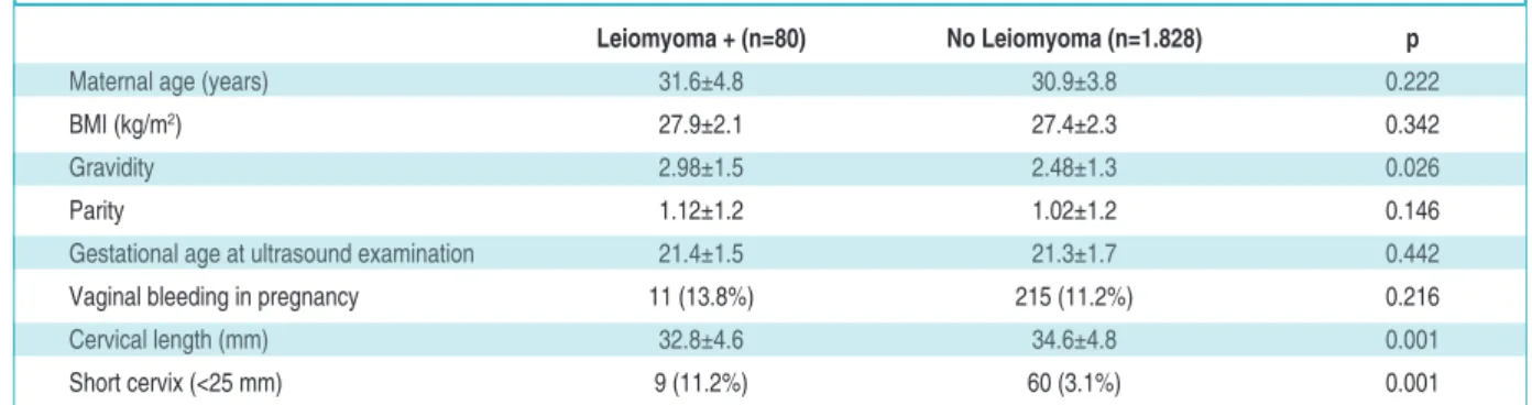
The demographic data and ultrasound findings of the groups are shown in Table 1. The two groups were age and BMI matched. The gravidity of the leiomyoma group was higher. (p=0.026). There was no difference in the levels of parity and vaginal bleeding rates between the patients with and with-out leiomyoma.
The mean cervical length of the leiomyoma group was significantly lower than the pregnant women without any leiomyomas (p=0.001). The number of pregnant patients with a short cervix
was higher in the leiomyoma group than the con-trol group (p=0.001).
There was a significant positive correlation be-tween the age of women and leiomyoma preva-lence (r=0.294, p=0.021), and there was a significant negative correlation between myoma size and cervical length in women with only one leiomyoma (r=-0.325, p=0.015). The correlation be-tween leiomyoma size and cervical length is shown in Figure 1.
DISCUSSION
In this study, we investigated the prevalence of uterine leiomyomas in pregnant women, and we evaluated the cervical length and incidence of a short cervix in pregnant women with leiomyoma. We found a shorter cervical length and higher in-cidence of a short cervix in pregnant women with leiomyoma. We also found a negative correlation between uterine leiomyoma size and cervical length in pregnant women.
Leiomyomas are the most common uterine tu-mors and originate from the smooth muscles of the uterus. Most uterine leiomyomas are asymptomatic and it is difficult to know the accurate prevalance of them. Today women could delay child-bearing, thus the incidence of uterine leiomyomas could in-crease and their effects to obstetric outcomes would be more important. Furthermore, previous studies have reported conflicting results regarding the prevalence of leiomyomas in pregnancy. In a prospective cohort study, Laughlin et al. evaluated
4272 pregnant women and detected leiomyomas in 458 women (10.7%). They reported a mean leiomyoma size of 23 mm.3In our study, we found a 3.9% prevalence rate of leiomyomas in pregnant women; the mean size was 31 mm. Although the mean age of the cohort in their study was similar to ours, varying results between the studies may be due to racial differences between the cohorts. Laughlin et al. found a higher prevalence of leiomyomas in black women (18%); on the other hand, our study did not include black women. We reported a larger mean size of leiomyomas than the previous study, which examined the women in the first trimester.3In our study, we evaluated preg-nant women in the second trimester and the mean size of leiomyomas may have increased due to the effects of pregnancy.
FIGURE 1: The correlation between leiomyoma size and cervical length.
Leiomyoma + (n=80) No Leiomyoma (n=1.828) p
Maternal age (years) 31.6±4.8 30.9±3.8 0.222
BMI (kg/m2) 27.9±2.1 27.4±2.3 0.342
Gravidity 2.98±1.5 2.48±1.3 0.026
Parity 1.12±1.2 1.02±1.2 0.146
Gestational age at ultrasound examination 21.4±1.5 21.3±1.7 0.442
Vaginal bleeding in pregnancy 11 (13.8%) 215 (11.2%) 0.216
Cervical length (mm) 32.8±4.6 34.6±4.8 0.001
Short cervix (<25 mm) 9 (11.2%) 60 (3.1%) 0.001
TABLE 1: Demographic data and ultrasound findings of the groups.
Today, prevalence of leiomyoma in pregnancy is higher than in the past due to the advanced di-agnostic methods which allows an early individu-ation and diagnosis, and increased age of pregnancy planning, that indicates a higher risk age for this.15,16In our study, we found a positive correla-tion between age and leiomyoma prevalence, con-sistently with the findings of Marshall et al.17
Only few studies investigated the relation be-tween uterine leiomyomas and short cervix in preg-nant women. Blitz et al. investigated whether pregnant women with uterine leiomyoma are at higher risk for a short cervix.18According to this retrospective study, they reported 5.1% leiomyomas prevalence in pregnant patients. In this study the authors evaluated the pregnant women with gesta-tional age of 17-23 weeks and found significantly higher short cervix (<25 mm) incidence (3.6%) in pregnant patients with leiomyoma than healthy pregnant controls (1.5%). They showed an associa-tion between the presence of uterine leiomyoma and increased risk of having short cervix. In another retrospective cohort study, Shavell et al. found a higher rate of short cervix (14.3%) in women with uterine leiomyomas.8Furthermore, they observed an increased rate of preterm birth in women with leiomyoma. However, they reported an increased prevalence of short cervix only in women with large uterine leiomyomas (>5 cm); that is, they found a 1.9% prevalence rate of short cervix rate in women with small leiomyomas. In our study, we found a short cervix rate of 11.2% in women with leiomyomas. Although Shavell et al. reported a higher rate of short cervix, our findings are similar with this study. They found a similar prevalence rate in women with leiomyomas >5 cm. However, the mean size of leiomyomas in our study was 31.4 mm. We also found a negative correlation between uterine leiomyoma size and cervical length. This finding suggests that large uterine leiomyomas may contribute to a short cervix more than smaller ones. Leiomyomas may decrease the uterine distensibil-ity, which may result in shortening of the cervix or preterm birth.
This study has some limitations that should be addressed. First, our study lacks obstetric outcomes
of the pregnant women we included; that is, we do not know the preterm birth rates in our study pop-ulation. Next, the study was mostly comprised of the patients who were referred to a single special-ist. Hence, the results in this study represent the East Marmara region of Turkey, but may not rep-resent the general Turkish population. Future stud-ies should include the patients from multiple centers in Turkey and evaluate the obstetric out-comes.
CONCLUSION
The prevalence of uterine leiomyomas could in-crease with age. According to mid-trimester ultra-sound results, 4.2% of pregnant women have leiomyomas. Pregnant patients with uterine leiomyoma have an increased risk of having short cervix. Leiomyomas could affect negatively cervi-cal length and may cause a shortened cervix. Fur-ther studies with a large study population may clarify which leiomyomas cause decreases in cer-vical length and preterm birth with respect to number, size, type and location; therefore, recom-mendations on follow-up and birth management can be provided.
A
Acckknnoowwlleeddggeemmeennttss
The English Language editing of this manuscript was done by Taylor Francis English Editing Service.
S
Soouurrccee ooff FFiinnaannccee
During this study, no financial or spiritual support was received neither from any pharmaceutical company that has a direct connection with the research subject, nor from a company that provides or produces medical instruments and materials which may negatively affect the evaluation process of this study.
C
Coonnfflliicctt ooff IInntteerreesstt
No conflicts of interest between the authors and / or family members of the scientific and medical committee members or members of the potential conflicts of interest, counseling, ex-pertise, working conditions, share holding and similar situa-tions in any firm.
A
Auutthhoorrsshhiipp CCoonnttrriibbuuttiioonnss
I
Iddeeaa//CCoonncceepptt:: Cihan Karadağ; DDeessiiggnn:: Eray Çalışkan; C Coonn--t
PPrroo--c
ceessssiinngg:: Bertan Akar; AAnnaallyyssiiss aanndd//oorr IInntteerrpprreettaattiioonn:: Cihan Karadağ; LLiitteerraattuurree RReevviieeww:: Gökçenur Gönenç; WWrriittiinngg tthhee
AArr--t
tiiccllee:: Cihan Karadağ; CCrriittiiccaall RReevviieeww:: Cihan Karadağ; RReeffeerreenncceess a
anndd FFuunnddiinnggss:: Gökçenur Gönenç; MMaatteerriiaallss:: Bertan Akar..
1. Okolo S. Incidence, aetiology and epidemiol-ogy of uterine fibroids. Best Pract Res Clin Ob-stet Gynaecol. 2008;22(4):571-88. [Crossref] [PubMed]
2. Stout MJ, Odibo AO, Graseck AS, Macones GA, Crane JP, Cahill AG. Leiomyomas at rou-tine second-trimester ultrasound examination and adverse obstetric outcomes. Obstet Gy-necol. 2010;116(5):1056-63. [Crossref] [PubMed]
3. Laughlin SK, Baird DD, Savitz DA, Herring AH, Hartmann KE. Prevalence of uterine leiomyomas in the first trimester of pregnancy: an ultrasound screening study. Obstet Gy-necol. 2009;113(3):630-5. [Crossref] [PubMed] [PMC]
4. De Vivo A, Mancuso A, Giacobbe A, Savast LM, De Dominici R, Dugo N, et al. Uterine my-omas during pregnancy: a longitudinal sono-graphic study. Ultrasound Obstet Gynecol. 2011;37(3):361-5. [Crossref] [PubMed]
5. Coronado GD, Marshall LM, Schwartz SM. Complications in pregnancy, labor, and deliv-ery with uterine leiomyomas: a population-based study. Obstet Gynecol. 2000;95(5):764-9. [Crossref] [PubMed]
6. Lee HJ, Norwitz ER, Shaw J. Contemporary management of fibroids in pregnancy. Rev Obstet Gynecol. 2010;3(1):20-7. [PubMed]
7. Chen YH, Lin HC, Chen SF, Lin HC. In-creased risk of preterm births among women with uterine leiomyoma: a nationwide popula-tion-based study. Hum Reprod. 2009;24(12):3049-56. [Crossref] [PubMed]
8. Shavell VI, Thakur M, Sawant A, Kruger ML, Jones TB, Singh M, et al. Adverse obstetric outcomes associated with sonographically identified large uterine fibroids. Fertil Steril. 2012;97(1):107-10. [Crossref] [PubMed]
9. Qidwai GI, Caughey AB, Jacoby AF. Obstetric outcomes in women with sonographically identified uterine leiomyomata. Obstet Gy-necol. 2006;107(2 Pt 1):376-82. [Crossref] [PubMed]
10. Vergani P, Ghidini A, Strobelt N, Roncaglia N, Locatelli A, Lapinski RH, et al. Do uterine leiomyomas influence pregnancy outcome? Am J Perinatol. 1994;11(5):356-8. [Crossref] [PubMed]
11. Vergani P, Locatelli A, Ghidini A, Andreani M, Sala F, Pezzullo JC. Large uterine leiomy-omata and risk of cesarean delivery. Obstet Gynecol. 2007;109(2 Pt 1):410-4. [Crossref] [PubMed]
12. Roberts WE, Fulp KS, Morrison JC, Martin JN Jr. The impact of leiomyomas on pregnancy. Aust N Z J Obstet Gynaecol. 1999;39(1):43-7. [Crossref] [PubMed]
13. Goldenberg RL, Iams JD, Miodovnik M, Van Dorsten JP, Thurnau G, Bottoms S, et al. The preterm prediction study: risk factors in twin gestations. Am J Obstet Gynecol. 1996;175(4 Pt 1):1047-53. [Crossref] [PubMed]
14. Berghella V, Roman A, Daskalakis C, Ness A, Baxter JK. Gestational age at cervical length measurement and incidence of preterm birth. Obstet Gynecol. 2007;110(2 Pt 1):311-7.
[Crossref] [PubMed]
15. Bałoniak B, Słomko Z, Malewski Z, Drews K. [The incidence of uterine leiomyomas in preg-nancy and their influence upon its course]. Ginekol Pol. 2002;73(4):260-5. [PubMed]
16. Cooper NP, Okolo S. Fibroids in pregnancy--common but poorly understood. Obstet Gynecol Surv. 2005;60(2):132-8. [Crossref] [PubMed]
17. Marshall LM, Spiegelman D, Barbieri RL, Goldman MB, Manson JE, Colditz GA, et al. Variation in the incidence of uterine leiomy-oma among premenopausal women by age and race. Obstet Gynecol. 1997;90(6):967-73.
[Crossref] [PubMed]
18. Blitz MJ, Rochelson B, Augustine S, Green-berg M, Sison CP, Vohra N. Uterine fibroids at routine second-trimester ultrasound survey and risk of sonographic short cervix. J Matern Fetal Neonatal Med. 2016;29(21):3454-60. PMID: 26653679. [PubMed]