BIRUNI UNIVERSITY
HEALTH SCIENCES INSTITUTE
IN VITRO CHARACTERIZATION of MICRORNAS SPECIFIC TO
CANCER STEM CELL IN LARYNX CANCER
Nur Deniz DIBEK
DEPARTMENT OF MOLECULAR BIOLOGY AND GENETIC
MASTER of SCIENCE
ISTANBUL
IN VITRO CHARACTERIZATION of MICRORNAS SPECIFIC TO CANCER STEM CELL IN LARYNX CANCER
KANSER KÖK HÜCRELERİNE ÖZGÜ MİKRORNA'LARIN LARENKS KANSER HÜCRELERINDE IN VITRO KARAKTERIZASYONU
Nur Deniz DİBEK
TC.
BIRUNI UNIVERSITY
INSTITUTE OF HEALTH SCIENCE MASTER of SCIENCE
SUPERVISOR
Asst. Prof. Dr. Elif Sibel ASLAN
ISTANBUL 2018
DEDICATION
Dear mother, father and brother
My friends who encourage and support me, All the people in my life who touch my heart...
ACKNOWLEDGEMENT
I would like to thank my thesis superadvisor Asst. Prof. Elif Sibel Aslan for her invaluable support and supervision during my Master studies.
I would like to thank Professor Nezih Hekim and Asst. Prof. Nazlı Ece Ordueri for their contributions to graduate education.
I would like to express my gratitude to my esteemed colleague, Dr. Esra Güzel, who shared her knowledge in scientific and experimental sense and did not spare her support in academic studies.
My precious friends who have always been with me,
And always present to my dear mother, my father, and my brother, who have always supported me with my endless gratitude.
TABLE of CONTENTS
DEDICATION ...I ACKNOWLEDGEMENT ... II ETHICS COMMITTEE ... ..III TABLE OF CONTENTS ... IV LIST OF FIGURES ... VI LIST OF TABLES ... VII LIST OF SYMBOLS AND ABBREVIATIONS ... VIII ABSTRACT ... IX OZET ... XI
1.INTRODUCTION ... 1
1.1.LARYNX ANATOMY ... 4
1.2.EMBRYOLOGY OF LARYNX ... 6
1.4.PHYSİOLOGY OD LARYNX ... 7
1.5.LARYNX CANCER ... 8
1.5.1.Larynx Cancer Etiology ... 11
1.5.2.Larynx Cancer Pathology ... 12
1.5.3.TNM Staging System ... 14
1.5.4.Diagnosis and Treatment of Larynx Cancer ... 18
1.6.MİCRORNAS ... 19
1.6.1.The Discovery of microRNAs ... 20
1.6.2.microRNAs Biology ... 20
1.7. MİCRORNAS AND CANCER ... 23
1.7.1.Mechanisms of microRNAs Dysregulation In Cancer ... 24
1.7.2.microRNAs as Tumor Suppressor Genes ... 24
1.7.3.microRNAs as Oncogenes ... 25
1.7.4.The microRNAs in Body Liquid ... 25
1.8.MİCRORNAS ASSOCİATED WİTH SQUAMOUS CELL LARYNGEAL CANCER ... 27
2.MATERIALS AND METHODS ... 29
2.1.EQUİPMENT AND DEVİCES ... 29
2.1.1.Equipments ... 29
2.1.2. Solutions ... 29
2.1.3.Computer Programs ... 30
2.2.METHODS ... 30
2.2.1.Cell Culture ... 30
2.2.2.Transfection Of Hep2 Cells ... 30
2.2.3.RNA Isolation ... 31
2.2.4.cDNA Synthesis ... 31
2.2.5.qRT-PCR ... 32
2.2.6.Identification of miR-203, miR-363-3p, miR-26b, miR-1825 and miR-200c-3p Proliferation in Hep-2 Cells ... 33
2.2.7. Identification of miR-203 and mir-363-3p Migration and Invasion in Hep-2 Cells ... 34
2.2.8.Soft Agar Colony Assay ... 35
2.2.9. Statictical Analysis ... 36
3.RESULTS ... 37
3.1.RNAISOLATİON ... 37
3.2.QRT-PCR ... 37
3.3.IDENTİFİCATİON OF MİR-203, MİR-363-3P, MİR-26B, MİR-1825 AND MİR-200C-3P PROLİFERATİON İN HEP-2CELLS ... 39
3.4.IDENTİFİCATİON OF MİR-203 AND MİR-363-3P MİGRATİON AND INVASİON İN HEP-2CELLS ... 41
3.5.SOFT AGAR COLONY ASSAY RESULTS ... 43
4.DISCUSSION... 46
REFERENCES ... 52
APPENDIX ... 72
LIST of FIGURES
Figure 1.1 : An illustration showing possible cancer sites in different regions of the head and
neck ... 4
Figure 1.2 : Laryngeal anatomy in the coronal plane from a anterior view ... 5
Figure 1.3 :The histology of larynx squamose cell carcinom is shown in the figure ... 7
Figure 1.4 :It is shown the speech production of larynx ... 8
Figure 1.5 : The Rates of Death in Developed and Developing Countries ... 9
Figure 1.6 : The incidence of the 10 most common cancer types in men (the incidence at 100,000) ... 11
Figure 17 : The incidence of the 10 most common cancer types in men (the incidence at 100,000) ... 14
Figure 1.8 : The biogenesis of miRNAs ... 22
Figure 1.9 : microRNAs in body liquids ... 27
Figure 1.10 : Expression-altering miRNAs in Plasma of Patients with Squamous Cell Laryngeal Cancer ... 28
Figure 3.1: The qRT PCR image of mir-203 , mir-363-3p,mir200c,mir-1825 and mir26b transfected into Hep-2 cells. ... 38
Figure 3.2 : Proliferation graphic belonging to mir-1825 , mir-363-3p and mir-203 on Hep-2 cells ... 39
Figure 3.3 : Proliferation graphic belonging to mir-26b, and mir200c -3p on Hep2 Cells ... 40
Figure 3.4 : Migration graph of mir-203 and mir-363-3p in Hep-2 cells ... 41
Figure 3.5 : Invasion graph of mir-203 and mir-363-3p in Hep-2 cells ... 42
LIST of TABLES
Table 1.1 : TNM staging of Cancer of the larynx in AJCC UICC 2017 ... 15
Table 2.1: Tagman Reverse Transcriptase Kit Prosudure ... 32
Table 2.2 : PCR Condution for cDNA Synthesis ... 32
Table 2.3 : Tagman qRT PCR Protocol ... 33
Table 2.4 : qRT-PCR Conditions ... 33
Table 2.5 : Matrigel Matrix Preparation ... 34
Table 3.1 Concentration of miRNAs ... 37
Table 3.2 : After qRT-PCR, p values of delta t-test of miRNAs ... 38
Table 3.3 : After proliferation, p values of delta t-test of miRNAs ... 40
Table 3.4 : After migration assay, p values of delta t-test of miRNAs ... 42
Table 3.5 : After invasion assay, p values of delta t-test of miRNAs ... 43
LIST of SYMBOLS AND ABBREVIATIONS RNA : Ribonücleic Acid
miRNA : Micro Ribonücleic Acid DNA : Deoxyribonücleic Acid
cDNA : Complementary Deoxyribonücleic Acid mRNA : Messenger Ribonücleic Acid
RNAi : RNA Interference
RISC : RNA Induced Silencing Complex
dsRBD : Double Stranded RNA-binding Domain RT: Reverse Trancription
qRT-PZR : Quantitive Reverse Transcription Polimerase Chain Reaction
dH2O : Distilled Water
CO2: Carbondioxide
μl: Mikrolitre
dNTP :Deoxyribonucleotide triphosphate HCL :Hydrochloric Acid
FBS : Fetal Bowine Serum
PSA :Penicillin Streptomycin Amphotericin LSCC : Larynx Squamous Cell Carsinoma SCC : Squmous Cell Carsinoma
ABSTRACT
Dibek ND. In vitro Characterization of microRNAs Specific to Cancer Stem Cell in Larynx Cancer. Biruni University, Institute of Health Science, Department of Molecular and Medical Genetic. Istanbul. 2017
Larynx cancer is the most common and most aggressive type of cancer in the head and neck region. However, when appropriate diagnosis and treatment is applied, the treatment of the head and neck region is the best responding tumor which is the most common cancer type in the world. The incidence in men is about 10 times higher than in women. The most important risk factors for the development of cancer are smoking and alcohol use. Squamous cell carcinomas constitute 90-95% of laryngeal carcinomas and early diagnosis helps increase the quality of life significantly, and affects the survival time positivelylike in case of the types. MicroRNAs (miRNAs) are small RNA molecules that do not encode proteins which are actively involved in the regulation of gene expression and in almost all cellular mechanisms. A good understanding of the working mechanisms of miRNAs has been the focus of recent researches. These researches have showed that microRNAs to play important role in the pathogenesis of many cancers such as Larynx Squamous Cell Carsinoma (LSCC)
In this study, samples were first isolated from microRNAs which was already identified in previous microarray studies with mir-26b, mir200c-3p, mir-203, mir363-3p, and mir-1825 in Trizol. cDNA synthesis was followed by qRT-PCR assays. For the microRNAs ready for transfection, Hep-2 cells, the larynx squamos cell line, were used for functional analysis. After transfection of microRNAs into Hep-2 cells, proliferation, migration and invasion, soft agar analyzes were followed and performed consecutively. Initially, 5 microRNAs were expressed in terms of significance. However, as a result of these analyzes mir-203 and mir363-3p played an important role in these experiments.
As a result of that mir-203 and mir363-3p act as tumor suppressor on Hep-2 laryngeal cancer cells, indicating that cancer cells seriously reduce their ability to proliferate, migrate and invade and colonize. It was determined that these miRNAs could be used during diagnosis. Further experiments and works need to be donefor better understanding of this cancer mechanism.
OZET
Dibek ND. Kanser Kök Hücrelerine Özgü mikroRNA'ların Larenks Kanser Hücrelerinde in vitro Karakterizasyonu, Biruni Üniversitesi, Sağlık Bilimleri Enstitüsü, Moleküler ve Tıbbi Genetik ABD. Yüksek Lisans Tezi. İstanbul. 2017
Baş-boyun bölgesinde en sık görülen ve en agresif olan kanser türü larenks kanseridir. Ancak uygun tanı ve tedavi uygulandığında, baş-boyun bölgesinin tedaviye en iyi yanıt veren tümör ve dünyada en sık tanı koyulan kanser türüdür.Erkeklerde görülme sıklığı kadınlara göre yaklaşık 10 kat fazladır.Bu tür kanserin gelişmesinde en önemli risk faktörleri sigara ve alkol kullanımıdır. Skuamoz hücreli karsinomlar, larenks kanserlerinin % 90-95'ini oluşturmaktadır.Tüm kanser türlerinde olduğu gibi, erken tanı yaşam kalitesini önemli ölçüde yükseltilmesine yardımcı olurken, sağkalım süresini olumlu yönde etkilemektedir.
MikroRNA’lar (miRNA’lar) protein kodlamayan küçük,RNA molekülleridir. Gen ifadesinin düzenlenmesinde ve neredeyse hücresel tüm mekanizmalarda aktif olarak rol oynamaktadırlar. MiRNA'ların çalışma mekanizmalarının iyi anlaşılması son zamanlarda yapılan araştırmaların odak noktası olmuştur. miRNA’ların Skuamoz Hücreli Larenks Kanseri (SHLK) gibi birçok kanserin patogenezinde önemli rol oynadığı gösterilmiştir.
Bu çalışmada, ilk olarak önceki mikroarray çalışmalarında belirlenmiş olan mikroRNA'lar arasından mir-26b, mir200c-3p, mir-203, mir363-3p, and mir-1825 'ın Trizol ile izolasyonları yapılmış ve cDNA sentezi ve ardından qRT-PCR esseyleri yapılmıştır. Transfeksiyona hazır hale gelen mikroRNA'lar için fonksiyonel analizlerde kullanılmak üzere larenks squamos hücre hattı olan Hep-2 hücreleri kullanılmıştır.
Hep-2 hücrelerine mikroRNA'ların transfeksiyonu yapıldıktan sonra sırasıyla, proliferasyon, migrasyon ve invasyon, soft agar analizleri yapılmıştır. Başlangıçta anlamlılık ifade etmesi açısından 5 mikroRNA ile yola çıkılmış ancak yapılan analizlerin sonucunda, bunlardan gözle görülür şekilde mir-203 ve mir363-3p 'nin önemli rol oynadığı bulunmuştur. Sonuç olarak, mir-203 ve mir363-3p 'nin Hep-2 larenks kanseri hücreleri üzerinde tümor supresör özellikte davranarak, kanser hücrelerinin proliferasyon, migrasyon ve invasyon ve koloni oluşturma yeteneklerini ciddi oranda azalttığı belirlenmiştir. Tanı aşamasında bu miRNA'lardan yararlanabilineceği belirlenmiştir. Bundan sonra yapılacak diğer fonksiyonel çalışmalara da yardımcı olacağı düşünülmektedir.
Anahtar Kelimeler: Larenks Kanseri, mikroRNA, miR-203, miR-363-3p, Hep-2 Tez Danışmanı: Yrd. Doç. Dr. Elif Sibel ASLAN
1.INTRODUCTION
Cancer is a complex disease and multi-step process with epigenetic and genetic changes. Changes in this genetic and epigenetic level cause errors and lead to uncontrolled division of cells. As a result, cancer begins at the cellular level (T.C. Sağlık Bakanlığı Halk Sağlığı Genel Müdürlüğü,2013; Koutsogiannouliet al., 2013). There are more than 100 different types of cancer, depending on the organ they are in and the group of cells they are descended from (Sawyerset al.,2013). The elaboration of the human genome project and the development of new generation disposal technology and microarray technologies have played an important role in the elucidation of cancer development and development mechanisms in the 2000s (Arnedoset al., 2014).
A better understanding of the molecular mechanism of cancer and the development of studies for diagnosis and treatment are very important. Investigations on cancer-related deaths have been found to be economically disastrous. These economic losses are very important at the country level due to maintenance costs, reduced productivity and premature mortality (Feachem et al., 2002; Coleman, 2014). Since 1975, cancer incidence rates have increased for many reasons. However, all investigations for cancer diagnosis and treatment have resulted decline in mortality. The identification of the molecular pathology of cancer is an important step in increasing the quality of life and reducing mortality to a greater extent (Reidenberg and Laitman, 2006).
Approximately 2% of human malignancies are laryngeal squamous cell carcinoma (LSCC). The incidence of LSCC is 7-8 times higher in men than in women. The black race is 1.5-2 times higher than the white race. The incidence of LSCC declines over the past 30-35 years. However, the decline in mortality rates is not enough (SEER Cancer Statistics Factsheets, 2011). The 5-year survival rate was found to be around 60%. LSCC is the 7th most frequent type of cancer in Turkey. The 5-year survival rate for LSCC in our country is below the world average (Türkiye Kanser İstatistikleri, 2014). According to Ministry of Health data, it was determined as 42.8% in 2011. Considering the demographic factors in Turkey, a significant increase in the estimated number of new cases of LSCC between 2015-2030 is
foreseen. Especially in men with LSCC, the mortality rate is expected to increase (SEER Cancer Statistics Factsheets, 2011).
MicroRNAs (miRNAs) are synthesized endogenously as a single chain. They are about 18-24 nucleotides in length and do not encode short ribonucleic acids (RNA). MicroRNAs have recently been one of the most recent cancer research topics (Sevliet al., 2010). Mirna's have been the subject of many studies. The mechanisms of formation, the investigation of the roles in biological processes, the objectives of miRNAs are a few of these studies (Iorio and Croce, 2012). There are also many studies showing that they are actively involved in the pathogenesis of many diseases. In particular, the mechanism of cancer formation has been shown to be deregulation of miRNAs and tumor suppressor characterization oncogenes (Reyaet al., 2001). The expression of miRNAs with different expression patterns for each different type of cancer has come to the fore in terms of the development of diagnostic and therapeutic methods (Di Leva and Croce, 2013). In recent years, a number of studies have been conducted on the role of miRNAs in carcinogenesis. However, the roles of miRNAs in the carcinogenesis process have not been fully elucidated. Therefore, further studies are needed (Reyaet al., 2001).
Cancer stem cells are rare cell populations present in the mass of cancer and are thought to be responsible for tumor growth and metastasis by acting like stem cells (Reyaet al., 2001). Tumor-initiating cells (cancer progenitor cells) have the ability to initiate cancer stem cell tumors and resist resistance to improved therapies (Masterset al., 2008). A standard chemotherapy kills most cells in the tumor but does not affect cancer stem cells. Although they are very few, they can lead to tumor recurrence even years after successful treatment. Recent studies have demonstrated the presence of cancer stem cells in many types of cancer, including head and neck, brain, lung, breast, prostate and ovary (Ozen, 2011; Suer et al., 2014). Many studies have shown that miRNAs also play important roles in cancer stem cell regulation. In order to develop miRNA-based therapeutic approaches, it is necessary to clarify the roles of miRNAs in the process of acquiring and maintaining cancer stem cell characterization. there is a need to identify miRNA profiles of cancer stem cells.
As far as the role of miRNAs in laryngeal cancer is concerned, few studies are available. there are studies in which the miRNA expression profile is removed in laryngeal cancer. The effects
of the presence of malignant and benign metaplasia on miRNA (s) in cancer cell proliferation with different expression patterns have also been investigated. The gene through which these effects are carried out has also been identified and reported (Zhaoet al., 2013; Xuet al., 2013). A limited number of studies on laryngeal cancer related to cancer stem cells have also been published. The first study was conducted by Wei et al. They have been published as a study to investigate CD133 as a possible cancer stem cell marker in the Hep-2 cell line. In this study, it was reported that a small group of cells in the Hep-2 cell line were CD133 +, and this cell group had an increased tumorigenesis capacity (Weietal., 2009). In another study, a 'side population' characterization was performed in Hep-2 cells. In these cells, increased self-renewal, proliferation, radiotherapy resistance and tumorigenicity have been demonstrated (Wanet al., 2014).
A comprehensive study of the effects of cancer stem cell-specific miRNAs on laryngeal cancer cells and laryngeal cancer stem cells has not yet been conducted as far as we know. The aim of this study is to investigate the effects of miRNAs on cancer cells with oncogenic or tumor suppressor effects in cancer stem cells from squamous cell laryngeal carcinoma. In this study, human laryngeal squamous cancer cell line Hep-2 cells were treated with miR-26b, miR-200c-3p, miR-203, miR-363-3p and miR-1825 mimics to determine tumor cell proliferation, migration and invasion, and functional effects on stem cells.
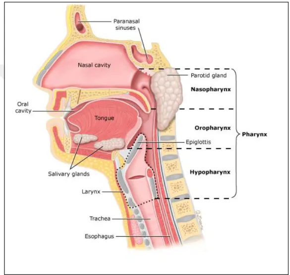
1.1.Larynx Anatomy
The larynx is an organ which segment of 3-6 neck vertebrae in front of the neck. It has responsible of respiration and conversation functions (26).
Figure 1.1: An illustration showing possible cancer sites in different regions of the head and neck. It is adapted from (Brocksteinet al., 2016)
The larynx is localized between the tongue and trechea. The trachea is in interaction with the lower part of the pharynx.And the trachea creates the front wall of the pharynx (Odar, 1980). It is beginned from epiglottis and it is continued the cricoid cartilaginous as anatomic.The total cartilages are 9 including 3 singles and 3 doubles. Aritenoid, kuneiform, and corniculat are pair cartilages. 3 singles cartilages are the thyroid, cricoid, epiglottis (Kaya, 2002).
The upper zone of the larynx canconstrict the respiratory tract and close completely when needed. Foreign substances is prevented the larynx muscles from passing to the respiratory tract.In addition, the larynx muscles also played a role in altering the shape of the laryngeal cartilage. It had an actions on the voice coil also (Odar, 1980).
The larynx has the ability to regulate air flow during exhalation. It also has many important vital functions related to functions such as speaking, swallowing and breathing (PL, 2007).
The protecting the lower respiratory tract is the most important function of the larynx. During swallowing, the larynx closes the parenchyma and temporarily stops breathing. And the food marches to the respiratory tract. In addition, Laryngeal foreign substances also provide a cough reflex that occurs when they interact with respiratory tract mucosa (PL, 2007).
Other tasks of the larynx; the formation of the speech function, the regulation of the breathing process, balancing the rib cage (PL, 2007).
Fixation of the thoracic cage is a necessary condition for optimally operation of numerous thoracic and abdominal muscles. Closing the glottis and fixing the rib cage, coughing, vomiting, diarrhea and urination, and giving birth are performed (PL, 2007).
Picture is adapted from (143)
1.2.Embryology of Larynx
In the 4th week of the embryological development occurs with the deeping of the slice. This formation begins from the ventral wall of the pharynx in the middle laryngotracheal clef. This is the first indicator of the future lower respiratory system in the 4mm embryo. This tube is covered with endoderm. The head of the laryngotracheal tube creates the larynx and trachea. The lower end comprises two lateral protuberances.And then, bronchus, right and left lung lobes develop (Pansky, 1982; Weir, 1997).
The larynx is of epidemic endoderm origin and cartilage, muscles are of mesenchymal origin. The increasing of interaction formed an opening T shape in the laryngeal space. The laryngeal cavity begins to gain its typical structure when the cecum turns into thyroid, cricoid, and arytenoid cartilages (Kozuma et al., 1991).
Laryngeal lumen obstruct due to rapid proliferation of the laryngeal epithelium as temporarly. Until the tenth week, the larynx re-canals and laryngeal ventricles form. These ventricles are surrounded by tissue cathelons that will later change to pseudo-real vocal cords (Kozuma et al., 1991). The larynx which continues to develop during the postnatal period is completed around the age of 65 (Cummings, 1998).
1.3.Histology of Larynx
According to studies performed in the newborn, the larynx initially had silical epithelium in the areas outside the vocal cord. In the sixth month the pseudo-vocal cord was squamous epithelium. The stratum squamous epithelium was laid on the tongue or anterior of the epiglottis. The epiglottis lingual face is covered by multilamellar epithelium, and the upper part of the laryngeal (back) side changes from multilamellar to flattened epithelial to columnar columnar epithelium. The transition between these two types of epithelium consists of a sharp or 1-2 mm transitional zone. The supraglottic and infraglottic part of the larynx is covered with the serrated columnar epithelium while the glottic part is covered with the multilayered flat epithelium (Koç, 2003).
The multi layered flat epithelium, composed of many cell layers, allows the tissue to be very thick. Cells on the surface can be cubic or prismatic, with cells on the basal lamina that are flat and responsible for cell production. As new cells grow, older cells push up and these
cells flatten. In the body mouth, esophagus, a part of you epiglotti the cornea and the epidermis of the skin in various areas such as in the cells of the multi layered flat epithelium can be found (Wikipedia, 2014).
Half of non-smoking adults have squamous epithelium areas in the ciliated repiratory type epithelial areas, supra- and infraglottic areas. In smokers, the respiratory epithelium of the larynx can completely replace the squamous epithelium (Juan, 2004; ,O, 2002; Metzgeret al., 2011).
Figure1.3: The histology of larynx squamose cell carcinom is shown in the figure. Picture is adapted from (Sayılgan, 2006).
1.4.Physiology od Larynx
Larynx has three basic functions (Som and Curtin, 2003).
a-Lower respiratory tract protection function
The larynx closes the respiratory tract when swallowing is performed after ingestion of food and prevents the ingestion of liquid nutrients to the lungs (Canadian Cancer Society's Advisory Committee on Cancer Statistics, 2017).
b-Breathing Function
While breathing, the Sekitali and vocal tracts open naturally to allow air to enter and exit the lungs via the trachea. If something other than air enters the trachea, a cough reflex is triggered (Canadian Cancer Society's Advisory Committee on Cancer Statistics, 2017).
c-Speaking Function
To speak, the larynx produces sound. Sound is produced when the airborne sound vibrates. the audio cables move to change the volume and tone of the sound. By moving your mouths, your language and your lips from the air through the air larynx, you will create different voices (Canadian Cancer Society's Advisory Committee on Cancer Statistics, 2017).
Figure 1.4 : It is shown the speech production of larynx. Picture is adapted from (Brunch et al., 2017).
1.5. Larynx Cancer
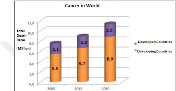
Cancer is a disease that occurs with highly variable genetic and epigenetic changes. Cancer is more common in developing countries than in developed countries, according to the cancer assessment in the world (Figure2-3).It is significant to understand the complexity and polymorphisms of cancer in functional studies. The onset and development of cancer in
humans is characterized by various mutations, chromosomal disorders, and increased / decreased gene expressions. Increased transcript levels in cancerous genomes are associated with increased number of gene copies with the proliferation of oncogenes and inactivation of tumor suppressor genes (Oktem et al., 2000).
Figure 1.2 : The Rates of Death in Developed and Developing Countries. It is adapted from (T.C. Sağlık Bakanlığı Türkiye Halk Sağlığı Kurumu,2014).
According to investigator in 2012, all cancer event determined as about 14.1million in worlwide. This state shown 7.4million in men. The 6.7 million case is shown in womens. By 2035, this number is estimated to rises as 24 million (Ferlayet al.,2015). Cancer is the leading health problem in the world. United state te is also the second health problem. Cancer case occured 1,685,210 and cancer deaths is 595,690 in United State in 2016 (Rebeccaet al., 2016). Head and neck cancers represent various epithelial tumors that occur in the oral cavity, pharynx, larynx, and nasal cavity. This type of cancer is the sixth most common cancer worldwide, with approximately half a million new cases each year.These cancers, whose incidence is still increasing in various geographical regions, are also affecting young people nowadays (Leemanset al., 2011).
Laryngeal cancer is seem accounts for 2% of all cancers in adults and 2.1% of all cancer deaths worldwide. It is the second most common cancer of head and neck squamous cell carcinomas. Laryngeal cancer is also the most common type of cancer among upper
respiratory tract cancers. And 90-95% are composed of squamous (flat) cell carcinoma (SCC) or epidermoid carcinoma variations. In addition, 25% of these tumors in the head and neck region are located in the larynx (Oktemet al., 2000;Kirchner and Carter, 2004;Mooreet al., 1999; Farraget al., 2006).
The most frequent age of laryngeal carcinoma is reported to be 1% in the younger ages, with a concentration in the 50-70 (Kirchner and Carter, 2004;Maoet al., 2012;Zhanget al., 2013). It have been reported in younger age groups also. The incidence of laryngeal cancer varies according to the countries. In general, the incidence is in the range of 3-10 / 100.000. (Gallus et al.,2003). The highest incidence for 2012 data was found in Cuba with 7.6 / 100.000 per year. Other common countries are Georgia, Armenia and Hungary with an incidence of 5.7-6.3 / 100.000. The ranking is followed by Belarus, France, Turkey and Spain with an incidence of 3.8-4.1 / 100.000. According to the statistics, it is found that in countries like Africa, East Asia, New Zealand and Norway, it is below 1.9 / 100.000 (Globocan, 2014).
The ratio of male / female with larynx cancer reported as 5: 1 in many countries is 30: 1 in some countries (Galluset al., 2003;Topuzet al., 2000). Although laryngeal cancers are reported as 5: 1 in many countries, the male / female ratio is 30: 1 in some countries (Galluset al., 2003;Topuzet al., 2000). This ratio has been shown in some sources around the world as 7: 1. (Mirisolaet al.,2011). When the Ministry of Health records in Table 2-1 are evaluated, larynx cancer is the 7th most common cancer in Turkey according to the year 2011 data. In patients with laryngeal cancer, 4-30% of secondary cancers can develop, including lung, head and esophagus (Gaoet al., 2003).
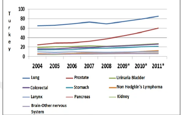
Figure 1.3 : The incidence of the 10 most common cancer types in men (the incidence at 100,000). It is modified from (Sağlık İstatistikleri Yıllığı, 2014).
1.5.1.Larynx Cancer Etiology
Larynx cancer is happened a combination of genetic factors and factors in your environment. Tobacco use is one of the major factors that increase the risk of developing laryngeal cancer. Alcohol use is also associated with the risc of developing cancer of the larynx (Head and Neck Cancer Guide, 2017; Epocrates, 2017).
In the etiology of laryngeal cancer, there are also many factors such as chemicals such as paint-gasoline, wood dust, asbestos, various metal powders, hormonal state of the person, dietary diet, sulfuric acid, coal and other factors that can lead to air pollution and radiation.
According to the research, 95% of the patients with laryngeal cancer were found to have cigarette consumption. Duration of smoking and the number of annual packs were found to affect the aggressiveness of laryngeal cancer. At the same time, 10-15 years after leaving the smoking cessation, the risk factors can go away. On the contrary, it has been argued that the effect of smoking cessation is small because of the cumulative damages in the cell in some studies (Koufman and Burke, 1997; Tomek and McGuirt, 2003).
Smoking and alcohol users, according to those who do not use larynx cancer appeared about 10 years early.In those who do not smoke or drink alcohol, these cancers are frequently located in the glottic region with low metastatic capacity. In addition, the survival rates of these people were higher (Agudeloet al., 1997).
Alcohol after smoking is also an important risk factor that plays a role in laryngeal cancer (Oktem, 2000;Demireller, 2004). Alcohol and cigarettes are found to be synergistically carcinogenic when used together (Bosetti et al., 2002). According to one study, the risk of laryngeal cancer was 2.46-times higher in non-smoking alcoholics and 9.38-times in smoking-free non-alcoholics. Those who consume both alcohol and cigarettes has been found to increase the risk 26 times (Koufman and Burke, 1997).
In recent years, viral factors (Epstein Barr Virus, Human Papilloma Virus) may play a role in the etiology of squamous cell carcinomas of the larynx. (Manjarrezet al., 2006; Engin, 2003). Not definitively proven, gene polymorphisms are other risk factors. Especially due to the damage caused by the carcinogenic substances contained in the cigarette, attention is paid to the polymorphisms of the genes involved in DNA repair (Busquetset al., 2003). The relationship between cyclin D1 gene amplification and glottic cancers is emphasized in Monteiro et al. (Monteiroet al.,2004). As with other types of cancer, it is reported that the hormonal status of the person and the imbalances in the diet are also risk factors for laryngeal cancer.(Riboliet al.,1996)
1.5.2.Larynx Cancer Pathology
Many types of benign and malignant tumors can be seen in the larynx.Non-Neoplastic tumors such as vocal nodules, vocal polyps, contact ulcers, ductal and sacrocular ulcers, neoplastic tumors such as laryngosclera, chondroma,squamous papilloma, granular cell tumor and glandular tumors are benign tumors of the larynx (PL, 2007).
Squamous cell carcinoma (SCC, squamous cell carcinoma) is the most common site melanoma. The second most frequently observed region is the larynx (Kaya, 2002). Squamous cell carcinomas contains 90-95% of larynx cancers (Epocrates, 2017).
Small cell carcinoma
Mucoepidermoid carcinoma
Adenocarcinoma
Adenoid cystic carcinoma
Sarcomas (chondrosarcoma, fibrosarcoma, rabdosarcoma, etc.)
Epidermoid carcinoma variations (Kaya, 2002)
Supraglottic cancers: The supraglottic region is rich in lymphatic. For this reason, these
tumors are at an increased risk of lymphatic metastases because of the location of the epiglottis from the free end to the ventricle (Myers and Alvi, 1996). The prognosis of these cancers is worse due to they can easily locate the thyroid cartilage and spread to the preepiglottic area (Rosai, 2004). Supraglottic cancers constitute about one-third of Larynx cancers and are more aggressive than glottic cancers (Koch et al., 2017).
Glottic cancers:These cancers usually develop as a good differential tumor(grade1). They can
usually spread vocal by spreading forward and backward. The risk of lymphatic metastasis is low because the glottic region is quite poor from lymphatic.The prognosis is better. However, prognosis worsens if placement of the anterior commissures is concerned (Engin and Erişen, 2003). About two-thirds of the larynx cancers constitute glottic cancers (Koch et al., 2017).
Subglottic cancers:These tumors have a high metastatic capacity. They can extend from the
vocal chord 1 cm below to the lower edge of the cricoid cartilage.They frequently metastasize the Delphian lymph node, and the rate of spread of cricoid and thyroid cartilage is high.Metastases can localize in the glottic and supraglottic regions, esophagus or thyroid, under the trachea and cricoid cartilage (Janfazaet al., 2002). This type is an uncommon type of laryngeal cancer (Koch et al., 2017).
Transglottic cancers:The glottis is a supraglottic or subglottic tumor that passes through the
vertical (vertical) and carries another region. Cervical lymph node metastasis is high.They can be spread to the paraglottic area in the early period. Thus, prognoses of this cancers are worse than other types (Rosai, 2004; Engin and Erişen,2003).
Figure 1.4 : The incidence of the 10 most common cancer types in men (the incidence at 100,000). It is modified from (Lore, 2005).
Laryngeal carcinomas are divided into 3 types according to dif+ferentiation grade and mitotic activities: good differentiation (Histologic grade 1), moderate differentiation (Histologic grade 2) and less differentiation (undisturbed) (Histologic grade 3) (Rosai,2004; Kaya, 2008)
According to studies performed, less differential tumors are associated with poor prognosis. There was no statistical difference in the prognosis between good and moderately differentiated tumors (Yilmazet al., 1999). Also histological grade is effective on radiotherapy activity. The better regional control was seen in grade 1 and grade 3 tumors (Fortinet al.,2001). Besides, it is stated that there is less risk of early metastases in grade 1 cancers. It is stated that it is possible to cure by surgical treatment in general (Kaya, 2008).
1.5.3.TNM Staging System
With the right treatment options chosen, patients with laryngeal cancer will benefit from the TNM stage, which is preferred all over the World. It was originally developed by Denoix in 1940. The staging system is currently regulated by two organizations that continue with revisions (AJCC Cancer Staging Manual, 1997). American Joint Committee on Cancer (AJCC) and the International Union for Cancer Control (UICC) is used to classify cancers.
The T classifications indicate the extent of the primary tumor and are site specific; there is considerable overlap in the cervical node (N) classifications (Poon and Stenson, 2017).
The TNM system does not contain the clinical and biological properties of cancer. ' T 'tumor, metastasis to the' N 'regional lymph nodes, and' M 'to the distal metastasis. The locus of the tumor and the vocal cord mobility give the T phase. Lymph node size and size are used for N staging. M is distant metastases, except neck and larynx (Curranet al., 1999).
Primary tumor (T) Supraglottis
T category T criteria
TX Primary tumor cannot be assessed Tis Carcinoma in situ
T1 Tumor limited to one subsite of supraglottis with normal vocal cord mobility
T2
Tumor invades mucosa of more than one adjacent subsite of supraglottis or glottis or region outside the supraglottis (eg, mucosa of base of tongue, vallecula, medial wall of pyriform sinus) without fixation of the larynx
T3
Tumor limited to larynx with vocal cord fixation and/or invades any of the following: Postcricoid area, preepiglottic space, paraglottic space, and/or inner cortex of thyroid cartilage
T4 Moderately advanced or very advanced
T4a
Moderately advanced local disease. Tumor invades through the outer cortex of the thyroid cartilage and/or invades tissues beyond the larynx (eg, trachea, soft tissues of neck including deep extrinsic muscle of the tongue, strap muscles, thyroid, or esophagus).
T4b
Very advanced local disease. Tumor invades prevertebral space, encases carotid artery, or invades mediastinal structures.
Glottis
T category T criteria
TX Primary tumor cannot be assessed Tis Carcinoma in situ
T1 Tumor limited to the vocal cord(s) (may involve anterior or posterior commissure) with normal mobility
T1a Tumor limited to one vocal cord T1b Tumor involves both vocal cords
T2 Tumor extends to supraglottis and/or subglottis, and/or with impaired vocal cord mobility
T3 Tumor limited to the larynx with vocal cord fixation and/or invasion of paraglottic space and/or inner cortex of the thyroid cartilage
T4a
Moderately advanced local disease. Tumor invades through the outer cortex of the thyroid cartilage and/or invades tissues beyond the larynx (eg, trachea, cricoid cartilage, soft tissues of neck including deep extrinsic muscle of the tongue, strap muscles, thyroid, or esophagus).
T4b
Very advanced local disease. Tumor invades prevertebral space, encases carotid artery, or invades mediastinal structures.
Subglottis
T category T criteria
TX Primary tumor cannot be assessed Tis Carcinoma in situ
T1 Tumor limited to the subglottis
T2 Tumor extends to vocal cord(s) with normal or impaired mobility
T3 Tumor limited to larynx with vocal cord fixation and/or invasion of paraglottic space and/or inner cortex of the thyroid cartilage
T4 Moderately advanced or very advanced
T4a
Moderately advanced local disease. Tumor invades cricoid or thyroid cartilage and/or invades tissues beyond the larynx (eg, trachea, soft tissues of neck including deep extrinsic muscles of the tongue, strap muscles, thyroid, or esophagus).
T4b
Very advanced local disease. Tumor invades prevertebral space, encases carotid artery, or invades mediastinal structures.
Regional lymph nodes (N) Clinical N (cN)
N
category N criteria
NX Regional lymph nodes cannot be assessed N0 No regional lymph node metastasis
N1 Metastasis in a single ipsilateral lymph node, 3 cm or smaller in greatest dimension and ENE(–)
N2
Metastasis in a single ipsilateral node, larger than 3 cm but not larger than 6 cm in greatest dimension and ENE(–); orMetastases in multiple ipsilateral lymph nodes, none larger than 6 cm in greatest dimension and ENE(–); orMetastasis in bilateral or contralateral lymph nodes, none larger than 6 cm in greatest dimension and ENE(–)
N2a Metastasis in a single ipsilateral node, larger than 3 cm but not larger than 6 cm in greatest dimension and ENE(–)
N2b Metastases in multiple ipsilateral nodes, none larger than 6 cm in greatest dimension and ENE(–)
N2c Metastasis in bilateral or contralateral lymph nodes, none larger than 6 cm in greatest dimension and ENE(–)
N3 Metastasis in a lymph node, larger than 6 cm in greatest dimension and ENE(–); or Metastasis in any lymph node(s) with clinically overt ENE(+)
N3b Metastasis in any lymph node(s) with clinically overt ENE(+)
NOTE: A designation of "U"or "L"may be used for any N category to indicate metastasis above the lower border of the cricoid (U) or below the lower border of the cricoid (L). Similarly, clinical and pathological ENE should be recorded as ENE(–) or ENE(+).
Pathological N (pN) N
category N criteria
NX Regional lymph nodes cannot be assessed N0 No regional lymph node metastasis
N1 Metastasis in a single ipsilateral lymph node, 3 cm or smaller in greatest dimension and ENE(–)
N2
Metastasis in a single ipsilateral lymph node, 1,18 in or smaller in greatest dimension and ENE(+); orMetastasis in a single ipsilateral lymph node, larger than 3 cm but not larger than 6 cm in greatest dimension and ENE(–); orMetastases in multiple ipsilateral lymph nodes, none larger than 6 cm in greatest dimension and ENE(–); orMetastasis in bilateral or contralateral lymph node(s), none larger than 6 cm in greatest dimension and ENE(–)
N2a
Metastasis in a single ipsilateral lymph node, 3 cm or smaller in greatest dimension and ENE(+); orMetastasis in a single ipsilateral node, larger than 3 cm but not larger than 6 cm in greatest dimension and ENE(–)
N2b Metastases in multiple ipsilateral nodes, none larger than 6 cm in greatest dimension and ENE(–)
N2c Metastasis in bilateral or contralateral lymph node(s), none larger than 6 cm in greatest dimension and ENE(–)
N3
Metastasis in a lymph node, larger than 6 cm in greatest dimension and ENE(–); or Metastasis in a single ipsilateral node, larger than 3 cm in greatest dimension and ENE(+); orMetastases in multiple ipsilateral, contralateral, or bilateral lymph nodes and any with ENE(+); orA single contralateral node 3 cm or smaller and ENE(+)
N3a Metastasis in a lymph node, larger than 6 cm in greatest dimension and ENE(–)
N3b
Metastasis in a single ipsilateral node, larger than 3 cm in greatest dimension and ENE(+); orMetastases in multiple ipsilateral, contralateral, or bilateral lymph nodes and any with ENE(+); orA single contralateral node 3 cm or smaller and ENE(+)
NOTE: A designation of "U"or "L"may be used for any N category to indicate metastasis above the lower border of the cricoid (U) or below the lower border of the cricoid (L). Similarly, clinical and pathological ENE should be recorded as ENE(–) or ENE(+).
Distant metastasis (M) M
category M criteria
M0 No distant metastasis M1 Distant metastasis
When T
is... And N is... And M is... Then the stage group is...
Tis N0 M0 0 T1 N0 M0 I T2 N0 M0 II T3 N0 M0 III T1, T2, T3 N1 M0 III T4a N0, N1 M0 IVA T1, T2, T3, T4a N2 M0 IVA Any T N3 M0 IVB T4b Any N M0 IVB
Any T Any N M1 IVC
Table 1.1 : TNM staging of Cancer of the larynx in AJCC UICC 2017. Table is adapted from(Poon et al., 2017)
1.5.4.Diagnosis and Treatment of Larynx Cancer
The most common symptoms of laryngeal cancer; It is muffled voice, change of voice, dry throat, constant cough, pain in the throat swallowing, difficulty in swallowing, earache, difficulty in breathing, weight loss and tuberage in throat region.
Many techniques are used for the diagnosis of laryngeal cancer. These techniques include direct or indirect laryngoscopy, micrographoscopy, magnetic resonance imaging (MRI), biopsy examination, scanning, computed tomography (CT) scanning chest film, positron emission tomography (PET) scanning (Laryngeal and Hypopharyngeal Cancer, 2012).
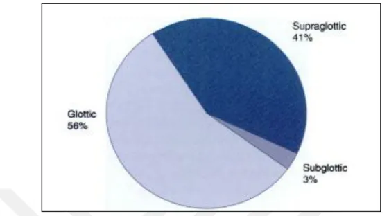
Most of the laryngeal tumors are localized to the glottic region at about 56%. The sequence follows the supraglottic region (41%) and the least common supraglottic region (1-2%). The identification of the tumor origin is of great importance. Clinically, there is an increased awareness of the preference of the treatment modality for different behaviors and for the knowledge of this difference in later processes (Carew, 2001).
Conventional conservative surgical intervention, endoscopic laser surgery, supracricoid partial or total laryngectomy, radiotherapy and combined chemotherapy are used in LSCC treatment. However, new treatment approaches need to be developed. Because the incidence of LSCC incidence and mortality is not at the desired level and there is no increase in 5-year
survival rates. There is an expectation for an increase in estimated incidence and mortality rates for Turkey. Therefore, clarification of the pathogenesis of LSCC is of great importance in this process (Karataş, 2014).
1.6.microRNAs
MicroRNAs (miRNAs) are a class of non-coding RNA gene which 18-22 lenght. MiRNAs has important role in cellular mechanism and genetic pathogenesis as deregulation (Joneset al., 2006). Precursor miRNAs is long RNA which from 100 to 1000 various lenght. Pri-miRNAs are produced by RNA polimerase II enzyme. Pri-miRNAs are translated short miRNA-miRNA duplex, and then they are turned single strand mature miRNA-miRNAs. Mature miRNA-miRNAs are binded messenger RNA sequences.Expression of the genes of interest inhibits either transcription either by inhibiting translation or by triggering RNA degradation.It has been described that microRNAs link to mRNAs in this way, affecting 30-90% expression of human genes (Friedmanet al., 2009).
The chromosomal location of the detected RNAs is linked genetic alterations responsible for the development of some types of cancer. Most miRNAs are located in fragile parts. The fragile parts are regions where the heterozygote disappears. According to the studies performed, these fragile regions are seen where amplification is rarely seen or where chromosomal breakpoint occurs (Wijnhovenet al., 2007). In some human cancer types, in multifactorial disorders and in the genetic pathogenesis, abnormal escape of miRNAs is critical (Biasioloet al., 2011).
It has led to many new cases since the discovery of regulation of transcriptional gene expression of miRNAs. MiRNAs biological processes such as cell differentiation, apoptosis, organogenesis, and the emergence of gentle regulation in the pathogenesis of diseases (Brenneckeet al., 2003; Chenet al., 2004). microRNAs are profiling by various methods such as blotting, RT-PCR and microarray (Croce, 2009).
The frequency and unique structure of miRNA molecules, in addition to being biomarkers and also providing therapeutic uses.Recent studies demonstrate that miRNAs are prognostic markers in clinical aggressive and recurrent cases. The use of MiRNAs both as a new cancer
marker and as a diagnostic / prognostic tool has provided many advantages (Gordanpouret al., 2012).
1.6.1.The Discovery of microRNAs
microRNAs and short interfering RNA(siRNA) is two well defined small RNA types. MiRNA and siRNA is not recognized as biochemical and functional. They is defined according to where came from. MiRNAs occurs precursors of hairpin of dsRNAs. SiRNAs consist of long dsRNAs (Karagünet al., 2014).
MicroRNAs are functional RNA molecules. Provide transcription from the RNA genes in the intron or exon regions encoding the protein on the genome and in regions that do not encode the protein. But, protein translations of miRNAs do not materialize in this time miRNAs in the human genome hundreds of high-level encoders encoding the gene region has been discovered. Recently, over 1000 miRNAs has been defined in human genom (Karagünetal., 2014; Shenouda and Alahari, 2009).
First miRNAs discovered by Lee et al.in 1993 in nematode Caenorhabditis elegans found.Lin-4 gene is important for C. elegans in post embriyonic development. Altough this gene does not code a protein, the gene converted 22-nucleotide RNA molecule (Espinosa and Slack, 2006). However, the microRNA term for this RNA molecule has been used for the first time since 2001 (Leeet al., 1993; Ruvkun, 2001).
This molecule suppress the expression of lin-4 mRNA with its untranslated region (UTR) as directly. First times, This subject accepted as special feature of C.elegans. But, this approved as a new method of gene regulation (Espinosa and Slack, 2006).
miRNA was discovered by Reinhart et al. in 2000 in C.elegans. Let-7 gene which regulated development timeline of C.elegans was found. This small RNAs play the crucial role regulation of gene regulation. This discover followed in the worm, fly and mammalian genomes (Espinosa and Slack, 2006; Reinhartet al., 2000; Pasquinelliet al.,2000).
1.6.2.microRNAs Biology
miRNAs are small non-coding RNAs which 18-24 nucleotides in lenght. miRNAs regulates proteins as post transcriptional. miRNAs causes inhibation of protein translation or degredation of messenger RNA(mRNA). According to prediction, more than 60% of
mammalian mRNA are targeted by at least one miRNA. Recent studies shown that miRNAs play critical roles in many cellular mechanism such as replication, regenration, differentation, proliferation, apoptosis (Karagünet al., 2014).
miRNAs influence gene expression by regulating gene expression after transcription, and are encoded by genes that do not translate into proteins. miRNAs are transcripts as primiRNAs (primers). These transcripts consist of a sequence of primers and a poly-A tail. Transcripts are transformed into precursor structures called pre-miRNA (precursor) in the cell nucleus. The pre-miRNAs consist of about 70 nucleotides (Kim, 2009). Microprocessor complex miRNAs, which consist of the Pasha (DGCR8) protein groups that link to the crosshairs and double-stranded RNA, provide the software (Denliet al., 2004).
Exportin 5 carries the small hairpin pre-miRNA to the placenta by the GAN-GTP mechanism. Pre-miRNAs are not active. It combines with the digested endonuclease and transforms into mature miRNAs in the cytoplasm. This cleavage is catalyzed by the double-stranded RNA binding domain (dsRBD) and Dicer, which simultaneously constitutes the RNAz III enzyme complex with TRBP / PACT (Siomi and Siomi, 2010).
The formation of the RNA-induced silencing complex (RISC) with dicer is initiated. RISC miRNAs and RNA interferon (RNAi) (Bernsteinet al., 2001). The digger cuts the strings of the premiers. As a result, two complementary complementary RNA molecules are formed. These are 5 'stable nucleotides. This chain is included in this complex through Argonat, the RNAz function in the RISC complex. This is the yarn guide (Preallet al., 2006). The other thread, the substratum of the RISC complex, is called the anti-guide. After the miRNAs bind to the RISC complex, the Argonat proteins provide the degradation of the mRNAs by making base transcription with complementary mRNAs (Gregoryet al., 2005).
Figure 1.5: The figures showing the miRNA biogenesis. Picture is adapted from (Króliczewskia et al., 2018)
Transcription of pri-miRNA transcribe begins by RNA polimerase II. Pri-miRNAs are processed in the nucleus by Drosha and pre-miRNA hairpins occur. Pre-miRNAs tranferred by Exportin-5 in cytoplasm. Hairpin of pre-mRNAs tranformed mature miRNA which 18-24 nucleotides by Dicer. The Argonate protein in the RISC complex incorporates one of the more stable duplexes in the mature miRNA into the RISC complex to regulate the expression of the target mRNAs. The other chain is degraded or prepared to be thrown out of the cell. Some miRNAs are found packed in exosomes generated from multivesic substances. Other miRNAs can be exported in the presence of proteins that bind to "RNA". Again, some miRNAs can be extracellularly delivered via microvesicles during membrane blebbing. These miRNAs in the extracellular area can be taken up by other cells, disrupted by RNA, or removed from the body (Etheridgeet al., 2011).
1.7. microRNAs and Cancer
microRNAs (miRNAs) are small non-coding RNAs. They have significant responsibilities in post transcriptional gene regulation. miRNAs organized by translational inhibition and unstablization of mRNA in animal cells (Bushati and Cohen, 2007).
MicroRNAs are contained in the organizing of almost all molecular mechanisms such as proliferation, development, cell fate, metabolism, apoptosis and cell death. In studies conducted, it was discovered that miRNAs and cancer are linked to abnormal expression levels. miRNAs have been reported to be therapeutic agents (Saito and Jones, 2006; Baytek, 2015).
Cancer is a multidisciplinary process. Cancer is when abnormal cells divide in an uncontrolled way with genetic changes. Cancer can expand along the all body and this is metastasis.Normal cells change their properties with genetic and epigenetic modifications. The new phenotypic cells that are exchanged gain different properties. Some of these features are cancer cells failure to undergo apoptosis, gain unlimited cleavage abilities, avoidance of inhibitory growth signals, and incresed of angiogenesis. The onset of progression and progression of the cancer cell regulates the genes that express cell proliferation, differentiation and apoptosis mechanism. Cancer cells act as tumor suppressor oncogenes by the effect of genes. Oncogene products can be classify into six groups. They can be transcription factors, chromatin remodelers, growth factor receptors, growth factors, signal transducers and apoptosis regulators (MacFarlane and Murphy, 2010).
Oncogenes play the role with change of genetic that boost the gene and modify promoters or enhancers to increase gene expression or modify of protein structure. On the contrary, tumor suppressor genes play roles in biological processes. If, lose of function of tumor suppressor genes occur in cell, the cancer process is occured. Currently studys, oncogenes and tumor suppressor genes is consist of protein coding genes to include miRNA (MacFarlane and Murphy, 2010).
miRNAs have a essential function in regulating most of cellular and metabolic pathways such as cells differantiation, survival, cells proliferation. miRNAs play the role as oncogenes or tumor suppressor genes. Dysregulation of miRNA expression profiles has been demonstrated in most tumors examined. But the especially classification of miRNAs can be
diffucult which separate function as oncogenes or tumor suppressor genes.. Because, expression of mirna is complicated subject. miRNAs can not be cause of cancer direct since they play a role indirect action in cellular phenotyope. Furthermore only one miRNA can be regulate multiple genes. For all, miRNAs are secret agents of cancer development and they success with oncogenes or tumor suppressor role (MacFarlane and Murphy, 2010).
1.7.1.Mechanisms of microRNAs Dysregulation In Cancer
Currently studies have shown that expression of miRNAs is dysregulated in human malignancies. This changes mechanisms can be caused by chromosomal abnormalities or by epigenetic silencing or by dysregulation of transcription factors targeting specific microRNAs and biological defects in the miRNA. This modifications in genomic miRNAs are happened with amplification or deletion in miRNA genes (Croce, 2009; Peng and Croce, 2016).
The changes in miRNA expression were found to be significant when cancer and normal cells were compared. Overexpression or knockdown of miRNA genes give rise to cancer process.miRNA in tumorigenesis contribute to development the oncogenesis and loss of differentiation with dysregulation of multiple protein coding oncogenes or tumor suppressor genes(Croce, 2009; Manikandanet al., 2008).
1.7.2.microRNAs as Tumor Suppressor Genes
Some miRNAs are downregulated in cancer cells in oncogenesis. These types of miRNAs are considered tumor suppressor genes. Tumor suppressor miRNAs generally prevent tumor development by negatively inhibiting oncogenes and/or genes that control cell differentiation or apoptosis. These miRNAs is encoded as TS-mir (Blandinoet al., 2014). At present, various miRNAs are considered as tumor suppressor genes, for example, miR-15a and miR-16–1
1.7.3.microRNAs as Oncogenes
miRNAs are synthesised as overexpressed in tumor. Most of them function oncogenic except a few. These oncogene miRNAs, encoded as “oncomirs”. Oncomirs inhibit tumor suppressor genes. They act by affecting cell differentiation, proliferation and apoptosis genes negatively. They try to inhibit any pathway that prevents the cancerous process. The studies underline the fact that miRNAs function as oncogenes or tumor suppressor genes. For example, it has been determined that overexpression of mir-17-92 increases the formation of B-cell lymphomas at a substantial rate (Manikandanet al., 2008).
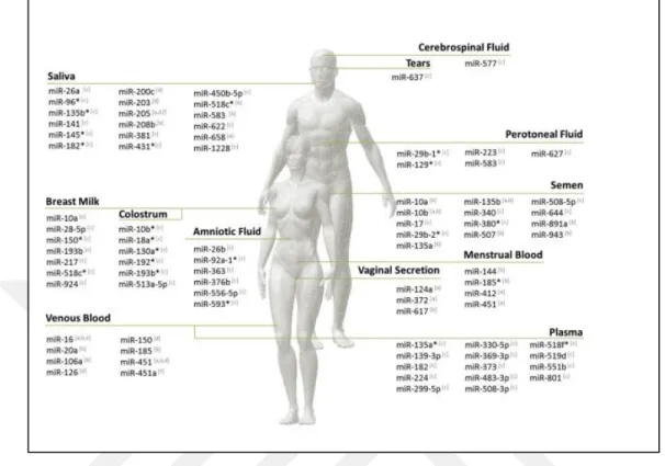
1.7.4.The microRNAs in Body Liquid
The importance of microRNA (miRNA) in cancer cells has been recognized in recently. Appropriate control of miRNA expression is necessary for maintaining a fixed state of cellular machinery. The miRNAs of the blood of both healthy and diseased individuals were detected. These miRNAs are found in lipid-lipoprotein structures. For this reason, the miRNAs have a stable structure. Their presence in body fluids also allows them to act as diagnostic markers. Despite this discovery, it is unclear how the functions of the miRNAs are made. For this reason, more research and study are required (Kosahaet al., 2010; Kwaket al., 2010).
In one study, the presence of serum miRNAs was found by Solexa sequencing. In this study, healthy controls were performed in lung cancer patients, colorectal cancer patients and diabetic patients. No disease specific miRNAs were detected in sera of healthy control groups. This finding has been associated with the possibility that miRNAs specific to these diseases may be present in relevant patient sera in humans. As a result, it has been shown that extracellular miRNAs can be used for diagnosis of cancers by analyzing them (Chenetal.,2008).
Unlike RNAs between different liquid types, miRNAs exhibit stable and distinctly expressed expression profiles (Weberet al., 2010; Zubakovet al., 2010). The miRNAs in the context are resistant to RNAi ", pH" and high temperature variability in serum-free serum-like body fluids (Chenetal.,2008). Human miRNAs circulating in body fluids are primarily important for the detection of cancer and presence in serum and plasma. In the human body
fluids such as plasma, urine or breast milk, 200-500 miRNAs have been detected with qPZR tecnique. Detection of miRNAs in body fluids is a promising noninvasive diagnostic method for the diagnosis of cancer patients (Weberet al., 2010).
Although the mechanistic mechanism of miRNAs is not yet explained, it is predicted that the tissues will passively passively seep into the circulation. It has been determined that miRNAs in body fluids originate from lipid vesicles, microvesicles or exosomes. Microvesicles are released from the cell by blistering. The sizes are between about 100 nm and 1 micron. The exosomes are small vesicles about 30-100 nm in size. It occurs as a result of fusion of multivesicular structures composed of endosomes with plasma membrane. They are found in body fluids like plasma, serum, saliva, breast milk and urine. Exosomes can be secreted from various cells such as epithelial cells, T and B cells, dentric cells and cancer cells (Ramachandran and Palanisamy, 2012).
High levels of mir-21 in serum of B cell lymphoma patients, the first studies on the modulation of miRNAs, have been associated with survival rates of individuals who have not recurred the disease (Lawrieet al., 2008). It has also been strongly associated with patients with elevated levels of mir-21 in serum or plasma, lymph node metastasis, advanced clinical stage, and poor survival prospects in NSCLC (Liuet al., 2012).
In another study, serum levels of mir 10b and -34a in the sera of breast cancer patients were decreased, while serum levels of mir-195 and let7a were increased (Rothet al., 2010; Heneghanet al., 2010). It has been shown that urinary samples from body fluids can be used to determine the presence of bladder cancer levels of mir-126 and mir-182. In saliva, decreased levels of mir-125a and mir-200a were associated with squamous cell carcinomas of the oral cavity (Parket al., 2009).
Figure 1.6: microRNAs in body liquids. It is adapted from (Silvaet al., 2014).
1.8.microRNAs Associated with Squamous Cell Laryngeal Cancer
Nowadays, detecting or regulating increased or decreased miRNA expression is of great importance in cancer pathology. For this reason, different methods such as antisense inhibitors, transgenics, specific promoters, real-time PCR and miRNA microarray have begun to be used when studying the function of miRNA (Zhanget al., 2006; Jianget al., 2005).
Certain miRNAs have an effect on genetic changes in tumors.A number of expression profiling studies have been conducted in which miRNAs show a significant decrease or increase in expression levels at significant ratios (Chenet al., 2014). (Tablo X)
Figure 1.7 : Expression-altering miRNAs in Plasma of Patients with Squamous Cell Laryngeal Cancer.It is modified from (Ayazet al., 2013).
2.MATERIALS and METHODS
2.1.Equipment and Devices 2.1.1.Equipments
Nanodrop spektrofometre (Thermo Scientific-ND 2000c, Germany)
Laminar air-flow (Class II Safety Cabinet) (Metisafe, Turkey)
Micropipette (Thermo Scientific, Germany)
Refrigerator (4 °C) (Vestel, Turkey)
Deepfreeze (-20°C) (Vestel, Turkey)
Deepfreeze (-80°C) (Wisd, DAIHAN Scientific, Korean)
Centrifugal (Hettich Retina 420R, Germany)
Mini Centrifuge (Thermo Scientific, Germany)
Thermal Cycler (PCR) (Techne-TC 5000, ABD)
Vortex(Wise Mix-VM10, Korean)
Spin Down
0.2-1.5 ml Eppendorf (Nest Biotechnology, China)
LightCycler ® Plates - Roche Applied Science (Basel, Swiss)
Roche LightCycler 480 (Basel, Swiss)
xCelligence Device and Equipment
Other consumables needed for Molecular Genetic applications
2.1.2. Solutions
TaqMan MicroRNA Reverse Transcription Kit (Applied Biosystems, Foster City, CA, ABD)
TaqMan Universal Master Mix (Applied Biosystems, Foster City, CA, ABD)
TaqMan miRNA Assay (Applied Biosystems, Foster City, CA, ABD)
Trizol, İsopropanol, Chloroform, 75% Ethyl Alcohol Solutions7
X-tremeGENE™ siRNA Transfection Reagent
MISSION® microRNA Mimic (hsa-miR-203, miR-26b, miR-200c-3p, miR-203 miR-363-3p and miR-1825)
Matrigel Matrix (Corning, NY, USA)
2.1.3.Computer Programs
xCelligence Software RTCA DP
2.2.Methods
2.2.1.Cell Culture
Hep-2 cell lines were cultured in RPMI-1640 (Gibco-BRL, Bethesda, MA) medium, supplemented with 10% fatal bowine serum (FBS) and 1% Penicillin Streptomycin
Amphotericin (PSA) in a 5% CO2 humidified incubator at 37 °C. The cell lines were passaged
after reaching 70% confluency.
2.2.2.Transfection Of Hep2 Cells
Hep-2 larynx cancer cell line was used in experiments. miR-26b, miR-200c-3p, miR-203 miR-363-3p and miR-1825 mimics were provided for use in in vitro studies performed and were transfected into the Hep-2 cells. Expression of these miRNAs in the cells was demonstrated by real-time PCR amplification following transfection. Mature miRNAs were obtained from SigmaAldrich using a control miRNA from SwitchGear Genomics, which is an oligonucleotide sequence that does not target any gene as a control.
Cells were grown in 6 well plates to 25000 cells per well. When cells were arrived 60-70%
confluency, miR-203, miR-363-3p, miR-26b, miR-200c-3p and control miRNA were
transfected using X-treme GENE siRNA Transfection Reagent.
For per well in plate 2.5μl transfeciton reactive with 250 μl Opti-MEM nutrient, in other tube 10pg mimic miRNA 250 μl Opti-MEM were mixed and waited room temperature 5 minutes. Subsequently, these two mixture by combinedand incubated 20 minutes, and thencells were
treated with the appropriate volume of transfection mixture in the plates used and left at 37 ° C for 1 hour. Later, 500 μL of Opti-MEM was added to each of the plates to allow the cells to incubate at 37 ° C. Cells were used for functional studies 24,48 and 72 hours .
2.2.3.RNA Isolation
Grown cells on plates were plates removedfrom mediums. Plates were washeds with 1ml PBS and cells scraped with cell scraper from plates. Washeds cells with 1ml PBS were transferreds into ependorf tubes. Samples were homogenized by pipettinge in Trisol solutions. This mixture waiteds for 10 minutes at room tempreture for the tissues to be disintegrated thoroughly then proteins wll be bound to the nucleic acids and completely separated from the nucleic acids. Then, 200μL chloroform addeds and the mixture kept as 2-3 minutes at room temperatures. The mixture was centrifuged at 12000g for 15 minutes at 4 ° C. After phase separation, the upper RNA-containing phases we transferred to fresh microcentrifuge tubes. 500 ml of isopropanol was added to new tubes and they incubated for 10 minutes at room temperature then the tubes were centrifuged at 12000g for 10 minutes at 4 ° C. After upper phase separation, RNAs was washed with 75% ethyl alcohol and is centrifuged at 7500g for 5 minutes at 4° C. The supernatant part wasremovedandthe RNA pellets were expected to dry out. Dryed RNAs are dissolved by adding nuclease-free water.
2.2.4.cDNA Synthesis
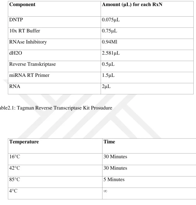
qRT-PCR assays performed using TaqMan probes.Total RNAs from the samples were diluted to a concentration of 15 ng / μl and 2 μl of each sample was used. miRNA cDNA synthesis was performed according to the manufacturer's instructions using the miRNA RT primers in the loop construct and the "Taqman Reverse Transcriptase Kit". (Table 2.1)
Component Amount (μL) for each RxN DNTP 0.075μL 10x RT Buffer 0.75μL RNAse Inhibitory 0.94Μl dH2O 2.581μL Reverse Transkriptase 0.5μL miRNA RT Primer 1.5μL RNA 2μL
Table2.1: Tagman Reverse Transcriptase Kit Prosudure
Temperature Time
16°C 30 Minutes
42°C 30 Minutes
85°C 5 Minutes
4°C ∞
Table 2.2 : PCR Condition for cDNA Synthesis
2.2.5.qRT-PCR
qRT-PCR assays were performed by using TaqMan assay. TaqMan miR-26b, miR-200c-3p, miR-203, miR-363-miR-200c-3p, miR-1825 and RNU43 probes used as controls were purchased from Applied Biosystems (Foster City, CA). qRT-PCR procedures were performed using the LightCycler480-II real-time PCR device with the TaqMan Universal Master Mix (Applied Biosystems, Foster City, CA) kit followed by the manufacturer's protocol. RNU43 from small