Türker ÖZKAN,1,2 Serdar TUNÇER3
Beyin felcine bağlı üst ekstremite deformiteleri, spastik ve güçsüz kaslar arasındaki dengesizliğin stabil olmayan ek-lemlere etki etmesi sonucunda meydana gelir. Bu olgularda spastik el ve üst ekstremitelerin cerrahi tedavisindeki temel amaçlar, spastik kasların kuvvetinin azaltılması, antagonist kasların güçlendirilmesi ve stabil olmayan eklemlerin kalı-cı stabilizasyonları olarak özetlenebilir. Bu amaçları yerine getirebilecek cerrahi teknik yelpaze ise, spastik kasların uza-tılması, tendon transferleri, eklem kapsülünün serbestleştiril-mesi veya plikasyonu, eklem artrodezleri, nörektomiler ve cilt prosedürlerinden oluşmaktadır. Bu yazıda, cerrahi teda-vi seçenekleri ve uygulamaları arasında, özellikle dengenin sağlanması ve motor fonksiyonların cerrahi restorasyonunu sağlayan tendon transferleri ön planda sunulmaktadır. Anahtar sözcükler: Beyin felci/cerrahi; çocuk; kontraktür/cerra-hi; önkol/cerrakontraktür/cerra-hi; el deformitesi, edinsel; kas spastisitesi/cerrakontraktür/cerra-hi; tendon transferi/yöntem; üst ekstremite.
Upper extremity deformities in cerebral palsy are caused by the imbalance between spastic and weak muscles acting on unstable joints. The basic goals of surgical treatment of spastic hands and upper extremities of patients with ce-rebral palsy can be summarized as reducing the strength of spastic muscles, strengthening the antagonist muscles, and permanent stabilization of unstable joints. Surgical techniques to achieve these goals include lengthening of spastic muscles, tendon transfers, release or plication of the joint capsule, joint arthrodesis, neurectomies, and skin procedures. Amongst these surgical treatment options, this article will present, in more detail, tendon transfers which are performed especially to achieve balance and restore motor functions.
Key words: Cerebral palsy/surgery; child; contracture/surgery; forearm/surgery; hand deformities, acquired; muscle spasticity/ surgery; tendon transfer/methods; upper extremity.
Correspondence / Yazışma adresi: Dr. Türker Özkan. İ. Ü. İstanbul Tıp Fakültesi, El Cerrahisi Bilim Dalı, 34093 Şehremini, İstanbul, Turkey.
Tel: +90 212 - 246 52 53 e-mail: [email protected]
Submitted / Başvuru tarihi: 02.03.2009 Accepted / Kabul tarihi: 10.03.2009
© 2009 Turkish Association of Orthopaedics and Traumatology / © 2009 Türk Ortopedi ve Travmatoloji Derneği
Department of Hand Surgery, 1Medicine Faculty of İstanbul University, 2VKV American Hospital, İstanbul; 3Department of Plastic and Reconstructive Surgery, Medicine Faculty of İstanbul Bilim University, İstanbul
Tendon transfer is the transfer of a functional tendon to another point by changing its direction -without compromising blood flow and innervation- by sepa-rating it at the adhesion point. The prerequisites of tendon transfer can be listed as follows: analysis of patient’s needs, informing the patient about the surgi-cal expectations and limitations, stabilization of the skeletal structure, absence of edema and inflamma-tion, presence of sufficient soft tissue source, mobility of the joints, availability of a donor muscle of suffi-cient strength, excursion and elasticity, and presence of appropriate tension direction and pulley.[1,2]
Candidates for tendon transfers
Among the cases with cerebral paralysis (CP), the most appropriate group for surgical intervention is the spastic group. Due to the challenges of perfor-mance and unpredictability of the results, surgery and especially dynamic tendon transfers are not recom-mended for the dyskinetic cases.
The targets of the surgical treatment must be re-alistic and achievable. Prior to the operation, both the patient and his/her family members must be thor-oughly informed about matters like functional
heal-ing, procedures involving combinations of hygiene and aesthetics, and expectations. The success of sur-gery hinges not only on the degree of neuromuscular defect and the surgical technique performed, but also on the patient’s age, apprehension, and his/her desire and ability to use the affected hand.[3]
Zancolli et al.[4] defined the qualities required in
patients suitable for surgery as follows: spastic neu-romuscular defect, sufficient IQ and emotional bility, limited effect of spasticity on emotional sta-tus, infantile hemiplegia, presence of basic senses, a certain amount of voluntary control on the spastic muscles, sufficient concentration and cooperation capacity for the postoperative term, motivation and support by the family members, and good neurologi-cal state in general.
In addition to increased muscle strength and move-ment ability, the results of surgery should be assessed in terms of the degree of achievement of other targets previously defined and improvements in grabbing of the hand and activities of daily living (ADL).
Assessment of a patient with CP with respect to existing deformities, and classifying and defining the problem is a multi-staged process. A close coopera-tion is needed with the physical treatment and reha-bilitation team following the patient.
Timing of surgical treatment
In recent years, the ideal age limit for reconstructive surgery in CP has been reduced. Although there are diverse views on the subject, it is pointed that ide-ally the reconstructive surgery should be performed at ages 4 to 6. At this age range, the central nervous system has become maturated, the deformities are easily detected, and the child can cooperate with the rehabilitation team.[4-9] It is not appropriate to delay
surgical treatment due to concerns about the child’s ability of cooperating with a specific exercise pro-gram. Even though surgery is recommended at early ages, a relatively older age is not a contraindication as long as the patient tries to control the hand volun-tarily and restoration improves the functionality of the hand.[10]
Through the years, tissues continually regenerate and keep on adapting to the new post-spasticity state. Contractures develop in the motionless joints, while the skin and soft tissues loose elasticity. Therefore, tendon transfers must be made as early as possible.
In the meantime, joint movement span and the elas-ticity of soft tissues must be maintained via passive exercises until surgery. It should be noted that no tendon transfer can activate a joint lacking passive movement and that postoperative active movement of a joint cannot surpass its pre-transfer passive movement. If intense and continuous rehabilitation treatments fail to overcome spasticity, joint move-ment limitations and soft tissue contractures, these problems must be solved via surgical loosening and lengthening techniques prior to tendon transfer, weak muscles must be backed up through tendon transfers and, if necessary, permanent stabilization of the joints should be performed.
Surgical applications
for the upper extremity segments
ElbowThe elbow enables the upper extremity to be spa-tially directed towards the target. Severe elbow flexion contractures may hold the arm adjoined to the body. In addition to hygienic and cosmetic problems, the functionality is impeded.[11] Usually,
the elbow deformity observed in CP is the flexion deformity. The reason of the deformity is the spas-tic contracture of the muscles on the front part of the elbow. In long-term cases, soft tissue contrac-tures may be present, as well.
Although elbow flexion deformity is rather com-mon, it is rarely severe as to merit surgical indication. In general, flexion contracture in the elbow of less than 30º does not cause functionality limitations.[12]
Fixed contractures of 45º or above[6,7] or flexion
defor-mities exceeding 80º during activity[6,10] are
consid-ered to meet surgical indications.
Among the most frequently employed surgi-cal techniques are loosening/lengthening of elbow flexor muscles/tendons, releasing contracted soft tissues, and separating the flexor-pronator muscle group from the adhesion point and shifting them distally.
Forearm pronation deformity
Being an upper extremity deformity very commonly seen in patients with spastic CP,[6] forearm pronation
deformity significantly limits the hand’s functional-ity.[13] Interosseous membrane contracture, secondary
curvatures at the radius and ulna,[14] and dislocations
are likely to develop as a result of long-lasting prona-tion contractures.[6,15]
Both pronation and supination movements are needed for a sufficient functionality of the hand.[6]
Therefore, the ideal surgical method of the pronation deformity treatment in patients with CP must aim to restore the active supination movement without com-promising the existing pronation movement.
The surgical methods used to restore the prona-tion deformity can be classified in two groups: In the first group, we benefit from certain procedures which originally aim to restore another deformity; hence, pronation contracture release, flexor/pronator loosen-ing, or transferring the flexor carpi ulnaris (FCU) to the elbow extensors. In the second group, there are surgical methods that directly pertain to the release of pronation contracture, which include pronator teres myotomy or lengthening (Fig. 1a), pronator quadratus myotomy (Fig. 1b), transfer of the pronator teres to the
elbow extensor, and transferring the FCU tendon to brachioradialis (BR) tendon insertion.
Concerning the treatment of pronation deformity, controversy exists whether to perform pronator loos-ening alone or loosloos-ening combined with active ten-don transfer. Elimination of the resistance leading to pronation deformity may help supination. However, it is obvious that generating an active motor work-ing voluntarily –enablwork-ing supination– would provide higher supination.
As shown in Table 1, Gschwind and Tonkin[16]
de-fined four types of pronation deformity and recom-mended treatment methods for each one. In our prac-tice, we use a modified version of this classification and treatment algorithm with inclusion of brachioradialis re-routing technique for type 3 and type 4 cases.
Taking these principles into account, the tech-niques used in surgical treatment of pronation defor-mity can be summarized as follows:
Fig. 1. (a) Pronator teres myotomy-lengthening. (b) Pronator quadratus myotomy.
(b)
Table 1. Pronation deformity classification and treatment options by Gschwind and Tonkin[16]
Type Treatment
Type 1 Active supination beyond neutral Surgery is unnecessary
Type 2 Active supination to less than, or to, neutral position Pronator quadratus loosening, flexor aponeurotic loosening Type 3 No active supination, free passive supination Pronator teres transfer (Brachioradialis re-routing)
Type 4 No active supination, tight passive supination Pronator quadratus and flexor aponeurotic loosening (Pronator teres lengthening/loosening + Brachioradialis re-routing)
FCU→ECRB (Extensor carpi radialis brevis) transfer. Defined by Green and generally used as a
classical method to increase wrist extension, the FCU tendon is transferred to the ECR tendon. The primary target of this technique is to restore wrist extension. As a secondary benefit, the forearm gains substan-tial active supination. This additional advantage was documented in biomechanical studies.[17]
Pronator teres re-routing. This procedure was first
defined by Sakellarides et al.[18] and its results were
then published by Strecker et al.[13] and Gschwind
and Tonkin.[16] Van Heest et al.[19] demonstrated
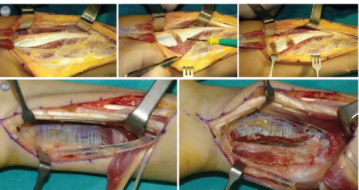
me-chanically on cadavers how supination improved after swathing the pronator teres around the radius (Fig. 2).
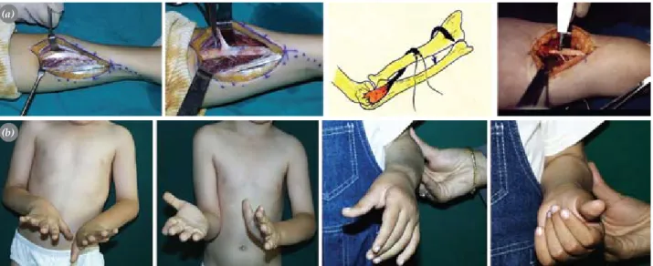
Brachioradialis re-routing. Defined by Özkan et
al.,[20] this technique is used to correct the forearm
pronation deformity and restore active supination. Here, the forearm gains sufficient active supination by altering the direction of the BR (re-routing) ten-don. Since the pronator teres muscle, which is the major pronator muscle of the forearm, is not used as motor, the forearm pronation is not interrupted. In a biomechanical study, Cheema et al.[17] demonstrated
brachioradialis re-routing as the second best tech-nique compared to the other methods (Fig. 3).
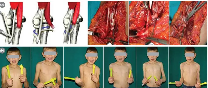
Transferring the brachialis muscle to the re-routed biceps tendon. This is an alternative technique that
can be used to restore forearm supination. It can be performed especially in the absence of a suitable motor tendon. In addition, it is an option when there is a need to decrease excessive flexion at the elbow (Fig. 4).
Other surgical techniques used for the correction of pronation deformity. An active increase in the
fore-arm supination can be expected by combining prona-tor teres loosening with swathing the FCU around the ulnar side of the forearm and transferring it to the ECBR or BR tendons.[10] By transferring the pronator
teres to the ECBR tendon, the pronator dominance in the forearm is eliminated and this force can be con-veyed to the weak wrist or finger extensors.[21]
Wrist
Wrist and finger flexion deformities are among the most common problems in patients with spastic pa-ralysis.[22] The flexion deformity of the wrist is caused
by the forces exerted by the wrist flexors, in
particu-Fig. 2. Pronator teres re-routing. (a) Surgical technique: While the forearm is brought to maximum supination, the prona-tor insertion at the mid-1/3 radius is palpated. Entering through an approximately 3 cm incision made in this area, the pronator teres insertion is found through the brachioradialis and ECRL tendons. The pronator teres tendon is abraded from the radius with a 2-cm periost. At the proximal of the original insertion point, the interosseous membrane is dissected extra-periosteally and the pronator teres tendon is brought through this span at the volar-dorsal direction. At a point close to the original insertion, the tendon is re-inserted to the lateral side of the radius via classical methods or with a suture anchor. Alternatively, with a Z-lengthening on the pronator tendon, the distal tendon piece is taken from the dorsal to volar and sewed to the proximal tendon piece. In the post-operative term, the elbow is kept at 45°-90º flexion with an above-elbow splint, while the forearm is at a supination of 45°-60º. (b) Case: A 5-year-old child with cerebral paralysis, who has supination weakness in the left forearm. Preoperative (first 2 pictures on the left) and postoperative (last 2 pictures on the right) views of active supination and protected active pronation.
(a)
lar the flexor carpi ulnaris, with spastic finger flexors worsening the situation.
When the wrist is in flexion, the finger flexors present substantial dysfunction especially during grabbing. Lengthening spastic wrist flexors decreases the flexor dominance in the wrist, thus providing a mechanical advantage for the extensor muscle group. If the wrist extensors are not strong enough to offset this flexor dominance, they may be actively supported by tendon transfers.
Prior to the transfer to strengthen the wrist ex-tensors, the spasticity of the extrinsic finger flexors should be assessed. When the wrist is in neutral posi-tion, the mechanical advantage of the spastic finger flexors will increase. The patient cannot loosen the clenched fist, which impedes grabbing. When this is the case, lengthening of the finger flexors must be in-cluded in the surgical plan.
If the patient has active extension loss, this should be repaired through a tendon transfer.
Fig. 3. Brachioradialis re-routing. (a) With an incision on the distal volar-radial side of the forearm, the brachioradialis muscle-tendon unit is exposed. The radial artery and the radial nerve branch are retracted. The brachioradialis muscle and muscle-tendon are dissected and prepared for transfer. A very important step at this point is to separate the muscle from all surrounding fascial connections; otherwise, the excursion of the muscle would not suffice. The free end of the tendon released and separated from the distal adhesion point is conveyed beneath the flexor pollicis longus muscle and taken to the volar from dorsal through the interosseous span. After its re-routing extraperiosteally around the radius –while preserving the radial artery– and with the forearm held at neutral, the tension is adjusted to preserve the original length of the tendon and the tendon is re-inserted to the radius via classical methods or with a suture anchor. (b) Case: A 12-year-old child with cerebral paralysis, who has supination weakness in the left forearm. Preoperative (first 2 pictures on the left) and postoperative (last 2 pictures on the right) views of active supination and protected active pronation.
(b)
Fig. 4. Transferring the brachialis muscle to the re-routed biceps tendon. (a) The brachialis muscle is completely separated from its insertion and released proximally. The biceps tendon is splitted longitudinally while the adhesion point is pre-served. The biceps slip on the radial side is re-routed around the radius neck and fixed to the musculo-tendinous seg-ment of the brachialis muscle, prepared beforehand, and supinatorplasty is completed. (b) Case: A 10-year-old child with cerebral paralysis, who has supination weakness in the left forearm. Preoperative (first 3 pictures on the left) and postoperative (last 3 pictures on the right) views of active supination and protected active pronation.
(a)
Wrist flexion deformity
There are several drawbacks of continuous wrist flex-ion: (i) Flexed wrist creates a mechanical disadvan-tage for the flexor muscles and reduces the grabbing power. (ii) Especially in patients with deficient ste-reognosis, the coordination of hand movements may be maintained visually. With the flexion posture of the wrist, the fingers remain inward segment of the hand, which impedes visualization. (iii) The aesthetic appearance of the wrist in flexion may be bothering for the patient and the family members. (iv) In case of severe flexion, there may be skin lesions on the volar side of the wrist.
The wrist plays a key role in hand movements. Even when there is no active movement in the fingers, flexion and extension may be present with the teno-desis effect. A moving wrist provides at least 25% of passive hand function. Therefore, wrist arthrod-esis should be performed only in severe deformities, where treatment with dynamic methods falls short. In CP surgery, a mobile wrist is always among the prior targets.
Wrist deformity may develop due to tenseness of flexor tendons, insufficiency of wrist extensors, con-tracture of the volar capsule of the wrist, or a combi-nation of the mentioned factors.
I. Tenseness and treatment of wrist and finger flex-ors. Wrist flexion deformity is generally caused by
spasticity of the FCU tendon. In some patients, the flexor carpi radialis (FCR) and palmaris longus ten-dons are also spastic. Tenseness of the finger flexors, if any, prevents full extension of the wrist, contrib-uting to deformity. Before performing a transfer to bring the wrist to extension, factors impeding the pas-sive joint movement span must be eliminated.
The treatment algorithm below may facilitate treatment preferences for restoring passive movement of the wrist:
Lengthening of the wrist flexor tendons. The
muscle mostly hindering the passive extension of the wrist is the spastic FCU. The spasticity of this muscle may also cause ulnar deviation of the wrist. In addition, the extensor carpi ulnaris (ECU) muscle in pronation may cause a strong ulnar deviation in the wrist and, in the presence of volar subluxation, it can cause flexion deformity of the wrist. In the lat-ter case, the ECU tendon can be transferred to the
ECRB tendon. In moderate cases, only functional or Z plasty lengthening of the FCU tendon may be sufficient. If an active wrist extension is desired, the FCU tendon can be transferred to the wrist extensor tendons. The palmaris longus tendon can be sepa-rated from the distal adhesion point and be used for transferring (e.g. to the thumb extensor tendon). Al-though the flexor carpi radialis tendon can also be used in tendon transfers, only lengthening of the ten-don is recommended, because releasing both wrist flexors may lead to hyperextension deformity, which may give rise to complications especially when the finger extension is not sufficient.
Loosening of the finger flexor tendons. All flexor
tendon lengthening techniques to some extent result in loss of strength in the flexor tendons. However, this can be compensated by the wrist, strengthening the grabbing in extension. While fractional lengthening creates minimal strength loss, superficialis-to-pro-fundis (STP) tendon transfer procedure may cause severe strength loss, with the effect of Z-lengthening standing between these two.
Bone procedures like proximal row carpectomy and wrist arthrodesis are not preferred in functional hands. These methods may be reserved for rather se-vere cases on hygiene and cosmetic grounds.
i) Fractional tendon lengthening. This technique has
the least negative effect on grabbing power of the fin-gers and imposes the lowest risk for overlengthening; therefore, it is preferred in well-functioning hands. If only fractional lengthening is to be performed, a lon-gitudinal incision would suffice at the volar 1/3 aspect of the forearm.
Most probably, lengthening would not suffice if the fingers do not reach passive extension when the wrist is in complete flexion. In this case, either one of the other two elongation techniques should be consid-ered (Fig. 5).
ii) Tendon Z-lengthening. This method can be used in
cases where fractional lengthening would not be suf-ficient. It is especially used in flexor pollicis longus (FPL) lengthening. Here, the most important criterion is to elongate the tendon by 0.5 cm for each degree of joint range of motion desired. The tendon is cut at the radial and ulnar sides. After lengthening, the ten-don ends are fixed with a Pulvertaft weave or end-side technique. If the lengthening is for the thumb, fixation is performed when the wrist is in the neutral position
and the metacarpophalangeal (MP) and interphalan-geal (IP) joints are in slight flexion. When the wrist is in extension, the thumb must be touching the second finger, and the thumb must be apart from the palm with the flexion of the wrist.
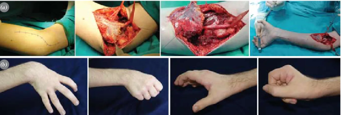
iii) Superficialis-to-profundus procedure (STP). As
it causes considerable loss in grabbing power, this procedure must be performed in dysfunctional hands with severe flexion contracture (Fig. 6).
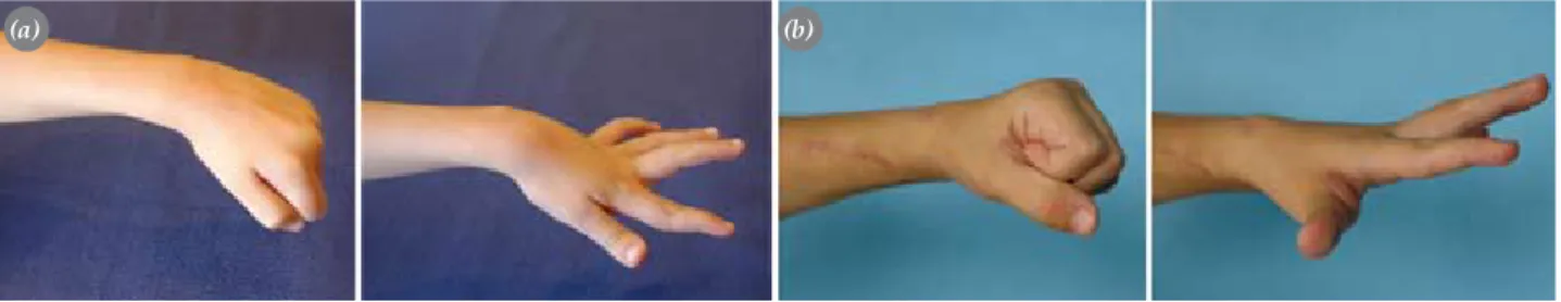
Flexor/Pronator slide. The pronator teres can be
detached from its origin, the medial epicondyle, and moved distally. This procedure, called the flexor-pronator slide (Fig. 7), is usually performed in pa-tients with CP sequela and central hemiparesis, hav-ing severe flexor dominance in the elbow, wrist, and fingers. The procedure may also be include all the flexors rooting from the pronator/flexor common ori-gin at the medial epicondyle level.[23]
In addition, proximal row carpectomy can shorten the forearm by approximately 1 cm, providing a pas-sive dorsiflexion increase of 25º in the wrist.
II. Extension insufficiency of the wrist and its treat-ment. After having eliminated the factors hindering
the passive extension of the wrist, active extension of the wrist should be obtained. The surgical classifica-tion offered by Zancolli[4] can serve as a reference for
the active tendon transfer.
The cases in group 1 can have full finger extension with a wrist flexion of less than 20º. In this group, usually the FCU muscle is spastic. Lengthening of this muscle solves the problem in most cases.
In Group 2, active finger extension can be achieved with a wrist flexion higher than 20º. In subgroup 2A, there is a weak wrist extension when the fingers are
Fig. 5. Fractional tendon lengthening. Fig. 5. Fractional tendon lengthening.
Fig. 6. Superficialis-to-profundus transfer. Preoperative (top) and postoperative (bottom) views.
Fig. 7. Flexor/Pronator slide. (a) Surgical technique. (b) Case: Preoperative (first 2 pictures on the left) and postoperative (last 2 pictures on the right) views of the patient with spasticity in the wrist and fingers.
(a)
in flexion, whereas in subgroup 2B there is no wrist extension at all. The latter group needs supportive transfer procedures to provide wrist extension after flexor loosening.
The transfers mostly used for wrist extension are: FCU→ECRB, ECU→ECRB, PT→ECRB, BR→ECRB, and flexor digitorum superficialis (FDS )→ECRB. If a higher radial deviation of the wrist is desired, these transfers should be made to the extensor carpi radialis longus (ECRL) tendon. The FCU→ECRB transfer performed to maintain wrist extension actively supports the forearm supination, as well.
The cases in group 3 do not have active finger extension and, in general, have severe involvement. Transfer to the finger extensors (e.g., FCU→Extensor digitorum communis-EDC) should be performed af-ter the release of contractures of the flexor muscles.
Surgical techniques commonly used
in the treatment of wrist extension insufficiency
1. Green transfer (Flexor carpi ulnaris tendon to ex-tensor carpi radialis brevis tendon). Technical details
of this method are summarized in Figure 8.
2. Transferring extensor carpi ulnaris tendon to exten-sor carpi radialis brevis tendon. This transfer is
per-formed in cases who have sufficient wrist extension, but their wrists become flexed during fisting due to fin-ger flexors. This transfer must always be performed to-gether with fractional lengthening of the FCU muscle. It cannot be performed simultaneously with transfer of
the wrist flexors to wrist extensors; otherwise, exten-sion deformity may develop.[12,24]
3. Transferring pronator teres tendon to extensor car-pi radialis brevis tendon. This technique is suitable for
patients whose FCU tendon is dysfunctional or used as a motor for another transfer. However, due to the limit-ed excursion of the pronator teres, the wrist movements are reduced. Since the pronator teres is often spastic, this transfer may be beneficial especially in terms of tenodesis. The pronator teres tendon, released from its insertion at mid-1/3 forearm is attached to the ECRB tendon.
4. Transferring brachioradialis tendon to extensor carpi radialis brevis tendon. The brachioradialis
ten-don can be used for wrist extension when the other motors are not suitable for transfer. However, as stated above, the fascial connections around the muscle must be completely released to provide sufficient excursion of the brachioradialis muscle.
5. Transferring flexor carpi ulnaris to extensor digi-torum communis tendons. As explained before, the
reason for finger extension and for the difficulty in releasing the hand may either be the contracture of the volar structures or the weakness of the finger ex-tensors. Flexor carpi ulnaris transfer may support the finger extensors. Superficial flexor tendons of the third and fourth fingers can also be used to strengthen the finger extensors.
Technique: Similar to Green transfer, the FCU is
exposed with an incision on the volar-ulnar side of the
Fig. 8. Green transfer. (Left) Access to the FCU tendon is through an incision on the volar ulnar side of the distal forearm. The FCU tendon is released from the pisiform bone it is attached to. The ECRB and ECRL tendons are exposed with a second incision in the dorsal of the wrist. A subcutaneous tunnel is formed on the ulnar side of the forearm, between the two incisions. The FCU tendon is brought through this tunnel and attached to the transfer tendon with Pulvertaft weaving. The tension is adjusted when the transferred tendon is at maximal tension and the wrist is in neutral-mild dorsiflexion. The wrist should be making a passive flexion of 20º; otherwise, the anastomosis will remain tense.[11] The
ing. The tension of the tendons is adjusted accordingly to keep the MP joints in neutral position and the IP joints in mild flexion. The hand is immobilized for four weeks while the wrist is held at 30º extension.[10,24,25]
The treatment algorithm that we follow in the sur-gical restoration of the problems related to finger and wrist spasticity is summarized in Fig. 9.
Surgical treatment of finger and thumb deformities
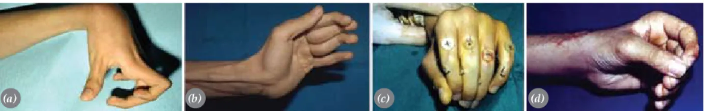
“Swan neck” deformity
The presence of hyperextension of the proximal in-terphalangeal (PIP) joint and flexion deformity of the distal interphalangeal (DIP) joint is known as the
tion and there are progressive extension deformities of the PIP joint that cannot be prevented with splint-ing.[26]
In CP, swan neck deformity is associated with the relatively shorter central slip. This shortness is rela-tive and does not impede the flexion of the PIP joint. Both the spasticity of the intrinsic muscles and the hyperactivity of the extrinsic finger extensors play a role in the pathophysiology of the tenseness on the central slip.[12] The high tension generated by the
spasticity of the intrinsic muscles is conveyed to the central slip via medial interosseous bands, leading to hyperextension on the PIP joint. If loosening of the retinacular ligament accompanies the condition,
dor-Active extension analysis of the fingers while the wrist is in neutral position
Active range of motion analysis of the wrist
Passive joint range of motion analysis of the fingers
Active fist motion The wrist remains in extension No need for transfer Transfer to wrist extensors or PT→ECRB (if FCU is used
for EDC)
If FCU is not used for EDC FCU→ECRB (FCR, PL lengthening) The wrist drops into flexion ECU→ECRB transfer + FCU lengthening or FCU transfer
Passive range of motion assessment of the wrist The wrist dorsiflexion is
above neutral while the fingers are in extension
The wrist dorsiflexion is below neutral while the fingers are in extension
Finger extension while the wrist is in flexion Full Full Full Full FCU→EDC FCU→EDC Limited Limited Limited Deficient FDS, FDP, FPL lengthening Move on to Fig. b Move on to Fig. b No need for transfer
Fig. 9. Assessment and treatment algorithm of wrist and finger extension.
sal subluxation of the extensor mechanism occurs. With simultaneous loosening of the volar plate, the hyperextension of the PIP joint increases and recur-vatum deformity ensues. Meanwhile, the dorsal sub-luxation of the extensor mechanism leads to relative insufficiency of the lateral bands, causing flexion of the DIP joint.
Furthermore, patients with insufficient wrist ex-tensors try to compensate the wrist extension function with the extrinsic finger extensors. Within the frame of the system explained above, the increased load on the central slip augments the severity of the swan neck deformity. In addition, after lengthening opera-tions of the superficialis flexor tendons, the swan neck deformity may become more prominent.
The MP joint flexion observed in swan neck de-formity is explained as follows: The wrist and finger joints make up a kinetic chain. The weakest ring of this chain is the PIP joint, followed by the MP joint. The extension of the PIP joint leads to flexion of the prox-imal and distal joints. Moreover, these forces cause a subluxation of the MP joint in the volar direction. Maintaining flexion of the PIP joint will prevent the deforming effects of these forces, thereby preserving the normal axis of the finger. Based on this rationale, the following treatment methods are recommended for the treatment of swan neck deformity:[10]
1) Decreasing the load on the central slip by cut-ting the ligaments conneccut-ting the intrinsic muscles to the central slip;
2) Keeping the lateral bands on a more volar posi-tion by shortening the retinacular ligament; 3) Preventing load exertion on the central slip by
attaching the extensor tendon to the proximal phalanx, not to the central slip;
4) Limiting joint hyperextension by placing a ten-don graft on the volar side of the PIP joint;
5) Arthrodesis of the PIP joint in flexion;
6) Preventing extension of the joint by using the flexor digitorum sublimis tendon or by per-forming volar capsulorrhaphy: sublimis teno-desis (Fig. 10);
7) Advancing the interosseous muscles;[11]
8) Ulnar motor neurectomy; 9) Sling procedure;[4]
10) Flexion osteotomy of the metacarpal neck.
Surgical treatment of thumb-in-palm deformity
Thumb-in palm deformity is one of the most common and complicated deformities of CP. It is caused by spasticity of the thumb adductor and flexor muscles. Locked inside the palm, the thumb cannot function and hinders the grabbing function of the other fin-gers.
Parallel to the evolution of CP surgery, thumb operations have turned to dynamic soft tissue proce-dures rather than static bone operations. Goldner, In-glis, and Keats, and alike succeeded in forming mo-bile thumbs by maintaining the muscular balance via tendon transfers and selective joint arthrodeses.[27]
The objectives of the surgical treatment of thumb-in-palm deformity are to generate a strong lateral pinch in the mid-phalanx of the second middle finger during fist formation, and to maintain sufficient radial abduction during grabbing.[28] The major challenge of
the treatment is the multifactorial nature of the defor-mity. For this reason, so as to make a systemic defini-tion of the deformity and propose a treatment plan, House[27] and Sakellarides[29] formulated two different
classification systems (Table 2).
The etiology of the thumb deformity is multifacto-rial. There are four main reasons for this deformity:
(i) Spasticity of the adductor and flexor muscles, (ii)
loose paralysis in the extensors and abductors, (iii)
Fig. 10. Correction of swan neck deformity with the sublimis tenodesis technique. (a) The preoperative view of the 13-year-old child with severe spasticity in the left hand. (b) Swan neck deformity occurring as a result of the overcorrection after loosening and tendon transfer. (c) Short-term and (d) long-term results after sublimis teno-desis surgery.
hypermobile MP joint, and (iv) contracture of the first web skin.
Assessing each of these conditions in the preopera-tive term would facilitate the formulation of a surgical plan. The assessment involves physical examination, electromyography, and diagnostic nerve blocks. In gen-eral, increased tonus of the muscles and the thumb’s position at rest point to the muscles mainly responsible for the deformity. (a) In the deformity related to the spasticity of the tenar muscles, there is flexion of the MP joint and extension of the IP joint. (b) In the defor-mity related to the spasticity of the FPL muscle, there is predominating flexion of the IP joint with varying degrees of flexion of the MP joint. (c) Involvement of the adductor pollicis muscle leads to flexion of the MP joint and adduction of the first metacarp towards the center of the palm. (d) Involvement of the opponens pollicis muscle or the first dorsal interosseous muscles results in adduction of the metacarp in line with the palm, trapping the thumb at the web span.
I. Treatment of adductor and flexor muscle spas-ticities. Release of the adductor pollicis, first dorsal
interosseous, and flexor pollicis brevis muscles can be performed as muscle myotomy at the origin, insertion or between the two.[10] Release at the insertion is not
very often preferred since it removes the entire func-tionality of the muscle. Release at origin, on the other hand, preserves the functionality to a certain extent because of the adhesion of the muscle to the adjacent tissues. During muscle myotomy, the muscle is kept at a tense position and the tendinous part is cut while the muscle fibers are protected.
a) Adductor/flexor release. An incision parallel to the
tenar line is made. The dissection is advanced up to the tenar muscles. The flexor pollicis brevis and ab-ductor pollicis brevis are isolated at their beginning point on the transverse carpal ligament and are re-leased from the ligament. Meanwhile, the finger prox-imal phalanx is brought to extension to facilitate the radial movement of the muscles. With proper splinting in the postoperative term, these muscles are adhered to their new place. If spastic, the adductor muscle is also released during the same session. If the muscle is released from its origin, the flexor tendons of the second and third fingers and neurovascular structures are retracted and stripped off over the third metacarp. If the muscle is released from the insertion point, it is followed along the radial side and cut at the tendinous insertion point. In cases where the adductor muscle is to be released without any intervention to the flexor muscles, access to the adductor muscle is via an inci-sion in the first web.
b) Releasing the dorsal interosseous muscle. An
inci-sion is made through the palpable ulnar border of the first metacarp. The radial nerve branches and the exten-sor pollicis longus (EPL) tendon are retracted. The first dorsal interosseous muscle is a bipinnate muscle, with its origin adhering to the first and second metacarps. Its origin in the first metacarp is released, while the segment in the second metacarp is left untouched. This preserved segment helps the flexion and abduction of the second finger. If the incision is advanced distally to the MP joint, the abductor can also be released through this incision.
hyperextension deformity/instability hyperextensible MP joint
Type IV MP and IP joint flexion deformity+ Spastic adductor and first dorsal interosseous+ simple metacarpal adduction contracture flexor pollicis longus muscles
(b) Cause of deformity Treatment
Type I Weak EPL Transferring PL or FCR to EPL
Type II Spastic or contracted intrinsic muscles Releasing tenar muscles - the 1st dorsal interosseous muscle
of the thumb and the carpal tunnel+releasing the 1st web skin, if contracted
Type III Weak abductor pollicis longus Swathing the abductor around the FCR
Fig. 11. Reinforcement of the thumb abductor and extensor tendons: (a) Preoperative and (b) late postoperative appearance of the patient.
(b)
c) FPL tendon lengthening. The FPL spasticity which
is manifest in type IV deformity is treated with frac-tional lengthening or Z-lengthening of the FPL tendon in the proximal wrist, just like in the wrist flexors.
II. Supporting the abductor and extensor tendons.
Tendon reinforcement procedures include abductor pollicis longus (APL) for the abduction of the first metacarp, extensor pollicis brevis (EPB) for the tension of the proximal phalanx, and EPL for the ex-tension of the distal phalanx. This reinforcement can be performed as tendon plication, tendon transfer, or fixing the tendon on a periosteal bone or another ten-don with a proper tension (tenodesis).
1) Tendon transfers. Transfer of the BR, PL, ECLR,
ECRB, FCR, FCU, and FDS tendons have been rec-ommended to restore the thumb extension and remove the thumb from inside the palm. The APL tendon is the most important tendon acting on the carpometacarpal joint. By taking the APL tendon out of the first com-partment and giving it a volar placement, a mechani-cal advantage is provided for the abduction of the first metacarp. In addition, sewing the PL tendon (end-side) on the volarly placed APL tendon may increase the ab-duction.[27] If the extension vector is to be preserved,
the same transfer can be performed by protecting the pulley of the first compartment. As an alternative, the APL is cut and its distal end is reinforced with an ap-propriate motor, while the proximal end is transferred to the EPB tendon (end-side) tensely. This technique provides both metacarp abduction and proximal pha-lanx extension.
In cases where an appropriate motor muscle cannot be found for transfer, the APL is cut and its distal end is re-routed volarly. Then it is fixed on the FCR tendon (end-side). This may create a tenodesis effect and re-store the metacarpal abduction.
Figure 11 shows an a case in which the above-mentioned techniques were combined to reinforce the thumb abductor and extensor tendons.
2) Shortening of the abductor pollicis longus and ex-tensor pollicis brevis tendons via plication
3) Extensor pollicis longus re-routing. The EPL tendon
which surrounds the Lister’s tubercle has an adductor effect due to its trajectory. Therefore, procedures ra-dializing this route are expected to have a positive ef-fect on abduction. Goldner[24] removed the EPL tendon
from its fibro-osseous channel, created a new pulley over the radial styloid, and radialized the route of the tendon. Later, Manske[30] took the EPL tendon through
the first dorsal retinacular compartment, eliminating the need for a new pulley. With further modification of the technique, Rayan and Saccone[31] cut the EPL
tendon at the proximal of the retinaculum and anas-tomosed the proximal end of the tendon to the distal end brought through the first dorsal compartment. The authors reported satisfactory abduction.
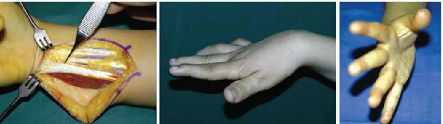
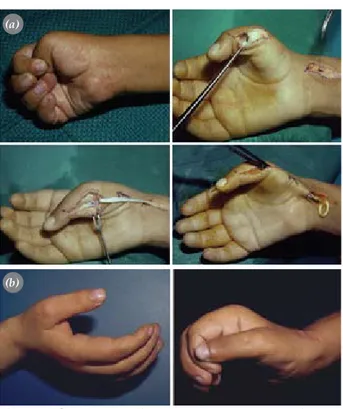
4) Flexor pollicis longus abductorplasty (Fig. 12).
The spastic FPL tendon to be loosened is cut on the proximal phalanx. The distal segment is used for the stabilization of the IP joint in children. The proximal segment is taken through a second incision on the wrist and then radialized through a second subcutaneous tunnel opened radially.
III. Hypermobile MP joint. The stability of the MP
joint is crucial for reinforcements made on the EPL or EPB tendons. If the MP joint already has a hyper-extension exceeding 20º, these reinforcements would result in excessive hyperextension of the MP joint. In order to avoid this, capsulodesis or arthrodesis of the MP joint should be performed in deformities where the MP joint is hyperextensible. Capulodesis is an at-tempt to refrain from arthrodesis, especially in chil-dren younger than 13 years.[7] Filler et al.[32] obtained
satisfactory long-term results by performing capulod-esis in 13 patients with CP. According to their tech-nique, the volar plate is cut and fixed proximally on the metacarp while the MP joint is at 30º flexion. In cases in which capulodesis is insufficient, arthrodesis
of the MP joint may be considered. During arthrod-esis, the growing plate on the epiphysis can be pre-served by performing only joint cartilage resection, enabling this operation to be performed in young children (4-5 years old) without growth arrest.[8]
IV. Release of the first web skin. Any contracture
of the skin can be released via two- or four-flap Z-plasties.
Conclusion
This paper summarizes surgical restorative procedures, in particular active tendon transfers, involving the hand and upper extremity in CP and briefly explains the functional and cosmetic benefits of these procedures. Obviously, these surgical procedures make up only a minimal part of the treatment, compared to lifelong conservative methods and rehabilitation practices.
The prerequisite of the surgical success is to as-sess and follow the patients with a well-equipped team that can offer the best treatment options for CP.
References
1. Özkan T. El cerrahisi. In: Değerli Ü, Bozfakioğlu Y, editör-ler. Genel cerrahi. 7 baskı. İstanbul: Nobel Tıp Kitabevleri;
hemiplegic spastic hand in cerebral palsy. Surg Clin North Am 1981;61:395-406.
5. Keats S. Surgical treatment of the hand in cerebral pal-sy: correction of thumb-in-palm and other deformities. Report of nineteen cases. J Bone Joint Surg [Am] 1965; 47:274-84.
6. Mital MA, Sakellarides HT. Surgery of the upper extrem-ity in the retarded individual with spastic cerebral palsy. Orthop Clin North Am 1981;12:127-41.
7. Skoff H, Woodbury DF. Management of the upper extremity in cerebral palsy. J Bone Joint Surg [Am] 1985;67:500-3. 8. Goldner JL. Upper extremity tendon transfers in cerebral
palsy. Orthop Clin North Am 1974;5:389-414.
9. Beach WR, Strecker WB, Coe J, Manske PR, Schoenecker PL, Dailey L. Use of the Green transfer in treatment of patients with spastic cerebral palsy: 17-year experience. J Pediatr Orthop 1991;11:731-6.
10. Manske PR. Cerebral palsy of the upper extremity. Hand Clin 1990;6:697-709.
11. Carlson MG. Cerebral palsy. In: Green DP, Hotchkiss RN, Pederson WC, Wolfe SW editors. Green’s Operative Hand Surgery. 5th ed. Pennsylvania: Churchill Livingstone; 2005. p. 1197-234.
12. Waters PM, Van Heest A. Spastic hemiplegia of the upper extremity in children. Hand Clin 1998;14:119-34.
13. Strecker WB, Emanuel JP, Dailey L, Manske PR. Compar-ison of pronator tenotomy and pronator rerouting in chil-dren with spastic cerebral palsy. J Hand Surg [Am] 1988; 13:540-3.
14. Miller F. Cerebral palsy. New York: Springer; 2005. 15. Pletcher DF, Hoffer MM, Koffman DM. Non-traumatic
dislocation of the radial head in cerebral palsy. J Bone Joint Surg [Am] 1976;58:104-5.
16. Gschwind C, Tonkin M. Surgery for cerebral palsy: Part 1. Classification and operative procedures for pronation de-formity. J Hand Surg [Br] 1992;17:391-5.
17. Cheema TA, Firoozbakhsh K, De Carvalho AF, Mercer D. Biomechanic comparison of 3 tendon transfers for supina-tion of the forearm. J Hand Surg [Am] 2006;31:1640-4. 18. Sakellarides HT, Mital MA, Lenzi WD. Treatment of
pronation contractures of the forearm in cerebral palsy by changing the insertion of the pronator radii teres. J Bone Joint Surg [Am] 1981;63:645-52.
19. Van Heest AE, Sathy M, Schutte L. Cadaveric modeling of the pronator teres rerouting tendon transfer. J Hand Surg [Am] 1999;24:614-8.
Fig. 12. Correction of the thumb-in-palm deformity with the FPL abductorplasty technique: (a) Preoperative appearance and surgical tech-nique. (b) Postoperative appearance.
20. Özkan T, Tunçer S, Aydın A, Hoşbay Z, Gülgönen A. Bra-chioradialis re-routing for the restoration of active supi-nation and correction of forearm prosupi-nation deformity in cerebral palsy. J Hand Surg [Br] 2004;29:263-8.
21. Colton CL, Ransford AO, Lloyd-Roberts GC. Transpo-sition of the tendon of pronator teres in cerebral palsy. J Bone Joint Surg [Br] 1976;58:220-3.
22. Rayan GM, Young BT. Arthrodesis of the spastic wrist. J Hand Surg [Am] 1999;24:944-52.
23. Inglis AE, Cooper W. Release of the flexor-pronator origin for flexion deformities of the hand and wrist in spastic pa-ralysis. A study of eighteen cases. J Bone Joint Surg [Am] 1966;48:847-57.
24. Goldner JL. Surgical reconstruction of the upper extremity in cerebral palsy. Instr Course Lect 1987;36:207-35. 25. Hoffer MM, Lehman M, Mitani M. Long-term
follow-up on tendon transfers to the extensors of the wrist and fingers in patients with cerebral palsy. J Hand Surg [Am] 1986;11:836-40.
26. Swanson AB. Surgery of the hand in cerebral palsy and the swan-neck deformity. J Bone Joint Surg [Am] 1960;
42:951-64.
27. House JH, Gwathmey FW, Fidler MO. A dynamic ap-proach to the thumb-in palm deformity in cerebral palsy. J Bone Joint Surg [Am] 1981;63:216-25.
28. Tonkin MA, Hatrick NC, Eckersley JR, Couzens G. Sur-gery for cerebral palsy part 3: classification and opera-tive procedures for thumb deformity. J Hand Surg [Br] 2001;26:465-70.
29. Sakellarides HT, Mital MA, Matza RA, Dimakopoulos P. Classification and surgical treatment of the thumb-in-palm deformity in cerebral palsy and spastic paralysis. J Hand Surg [Am] 1995;20:428-31.
30. Manske PR. Redirection of extensor pollicis longus in the treatment of spastic thumb-in-palm deformity. J Hand Surg [Am] 1985;10:553-60.
31. Rayan GM, Saccone PG. Treatment of spastic thumb-in-palm deformity: a modified extensor pollicis longus ten-don rerouting. J Hand Surg [Am] 1996;21:834-9.
32. Filler BC, Stark HH, Boyes JH. Capsulodesis of the meta-carpophalangeal joint of the thumb in children with cere-bral palsy. J Bone Joint Surg [Am] 1976;58:667-70.