https://doi.org/10.1007/s11686-020-00285-0
ORIGINAL PAPER
Overcoming the Challenge; In Vivo Efficacy of Miltefosine for Chronic
Cutaneous Leishmaniasis
Varol Tunalı1,2 · Mehmet Harman3 · İbrahim Çavuş4 · Cumhur Gündüz5 · Ahmet Özbilgin4 · Nevin Turgay2
Received: 22 July 2020 / Accepted: 15 September 2020
© Witold Stefański Institute of Parasitology, Polish Academy of Sciences 2020
Abstract
Background Cutaneous Leishmaniasis (CL) is the most common form of leishmaniasis. CL can be divided into two major groups: acute CL (ACL) and chronic CL (CCL). The aim of this study is to compare the efficacy of miltefosin and pentavalent antimony compounds in vivo with the CCL patient samples.
Materials Three study groups were formed, each consisting of five male Mus musculus (Balb/C) mice. In this model, pro-mastigotes from the culture of a CCL patient were utilized. 100 μL L. tropica promastigote suspension with a density of 108
promastigotes/ml were injected into the hint-right footpad of each experimental animal intradermally. Footpads of the mice were measured every two weeks until 24th week. From the 13th week, miltefosin 50 mg/kg/day was administered orally using gavage for 21 days, Meglumin antimoniate (MA) was administered by intramuscular (IM) injection daily for 21 days at 50 mg/kg/day and saline was administered IM for 21 days for the miltefosine, MA and control group, respectively. Results The footpad measurements of the miltefosine group were lower than the control group statistically. Between the MA group and the miltefosine group and MA group and the control group, there was no statistically significant difference. Giemsa stained slides revealed amastigotes in one, two and all of the slides for the miltefosine, MA and control group, respectively. Molecular tests were performed with the Rotor-Gene device and L. tropica consistent peaks were obtained in one of the miltefosine group, four in the MA group and all mice in the control group.
Conclusions Demonstration of both clinical and laboratory improvement in four of the five experimental animals provides strong evidence that miltefosine is an effective drug in the treatment of CCL. In the literature, no clinical or laboratory stud-ies using miltefosine have been performed with CCL patients only.
Keywords Cutaneous leishmaniasis · Drug resistance · Animal model · Turkey
Introduction
According to the World Health Organisation’s (WHO) list of neglected diseases, Leishmaniasis is the second parasitic dis-ease that causes the highest number of deaths in the World after malaria. Cutaneous Leishmaniasis (CL) is the most common form of leishmaniasis and is one of the few infec-tious diseases of increasing incidence due to conflict and environmental factors [1]. Recently, the disease has reached hyperendemic levels in conflict zones in the Syrian Arab Republic, Iraq, and Afghanistan, also affecting refugees from these regions [2]. Although many cutaneous syndromes associated with CL have been identified, the most common form is localized CL. However, there are other forms of CL; leishmaniasis recidivans, lupoid leishmaniasis, diffuse cuta-neous leishmaniasis, and mucosal leishmaniasis [3, 4].
* Varol Tunalı
1 Department of Emergency Medicine, Faculty of Medicine, Muğla Sıtkı Koçman University, Muğla, Turkey
2 Department of Parasitology, Ege University, Faculty of Medicine, İzmir, Turkey
3 Department of Dermatology, Dicle University, Faculty of Medicine, Diyarbakır, Turkey
4 Department of Parasitology, Celal Bayar University, Faculty of Medicine, Manisa, Turkey
5 Department of Medical Biology, Ege University, Faculty of Medicine, İzmir, Turkey
CL, which does not cause fever or general symptoms, usually with one or more long-term skin lesions, can be divided into two major groups: acute CL (ACL) and chronic CL (CCL). CL lesions are called CCL if they do not heal by treatment or spontaneously within 2 years. Lupoid leishma-niasis and leishmaleishma-niasis recidivans are chronic forms of CL, usually with broader involvement in the face and aestheti-cally wounding forms of CL [5].
Antimony compounds, which have been used as the gold standard treatment option for many years in the treatment of cutaneous leishmaniasis, can be administered by systemic or intra-lesion injection by parenteral route. Although an increasing number of resistant cases have paved the way for alternative treatment methods such as amphotericin-B, the treatment of amphotericin-B has led to the search for treat-ment alternatives, because of the risk of developing serious hepatotoxicity, the need for hospitalization and very high costs. At this point, miltefosine, which has been used safely in VL patients for a long time until some reports of drug resistance from India and Brazil, stands out as an important and valuable treatment alternative in the CCL patient group where treatment difficulties are most evident [6–8].
The purpose of an in-vivo model of CL is to investigate the parasitic basis of human disease and use this information in pharmaceutical studies to prevent the increased risk of infection in the human population. This model can then be used to develop and evaluate new potential anti-leishmanial compounds, options of immunotherapy, therapeutic vaccines [9, 10].
This study aims to compare the efficacy of miltefosine and pentavalent antimony compounds for the CCL patient group, in vivo with a BALB/C mouse model.
Materials and Methods
Patient Information and Sampling
The CCL patient was a 22 year old female with a lesion stretching from the tip of her nose to her right cheek includ-ing the naso-labial sulcus. The lesion was a hemorrhagic crusted erosion that lasted for more than a year. To collect the sample, the healthy skin around the scar was wiped with 70% ethanol and 0.2–0.5 ml of saline solution was injected to the margin of the lesion and then re-aspirated.
Parasite cultivation:
The parasite previously isolated from a CCL patient liv-ing in Turkey was stored in liquid nitrogen after cryopreser-vation and was included in this study. The strain stored in a liquid nitrogen tank was placed in a 37 °C water bath for 5 min after it was removed from the tank. After the viability controls and cell counting with the Thoma slide, the thawed amastigotes were cultivated in media.
10% FCS, 200 U penicillin/ml, and 0.2 mg/ml streptomy-cin were added to the commercially supplied RPMI-1640 medium prior to use and 5 ml of this mix were dispensed in 25 ml flasks. Cultured flasks were incubated in a 25 °C incubator. The media were monitored for the proliferation of promastigotes and a 10 ml promastigote-containing medium was obtained by adding fresh medium every 2–3 days.
A bi-phasic modified Novy-McNeal-Nicolle (NNN) medium called Enriched NNN medium was utilized for cultivation purposes because of the difficulty of cultivat-ing CCL causcultivat-ing Leishmania strain. The Enriched NNN medium was procured by adding cow milk and cow liver extract to the NNN medium as described previously [11].
Molecular Methods
DNA isolation from the isolates was performed with the High Pure PCR Template Preparation Kit (Roche Diagnos-tics GmbH, Mannheim, Germany).
Old World species-specific primers and probes were used for the real-time ribosomal internal transcribed spacer 1 (ITS-1)-PCR method [12]. ITS-1 region of Leishmania parasites separating genes encoding ssu rRNA and 5.8S rRNA, forward primer; 5′-CTG GAT CAT TTT CCG ATG -3ʹ, reverse primer; 5′-GAA GCC AAG TCA TCC ATC GC-3 ′ primers were amplified using the LightCycler-FastStart DNA Master mix using the following specific probes;
Probe 1: CCG TTT ATA CAA AAA ATA TAC GGC GTT TCG
GTTT —FL.
Probe 2: LC640-GCG GGG TGG GTG CGT GTG TG—PH [12].
A reaction mixture of 25 μL was prepared for the real-time PCR test; 1.5 μL H20 (PCR grade water), 1 μL Forward Primer, 1 μL Reverse Primer, 0.5 μL Probe 1, 0.5 μL Probe 2, 12.5 μL QuantiTect Probe PCR Kit Master mix (Qiagen GmbH, Hilden, Germany) and 5 μL of genomic DNA sam-ples were used.
The thermal profile determined for the detection of Leish-mania species separation (L. tropica, L. infantum and L.
major); denaturation, amplification, melt curve analysis and
cooling steps are pre-registered as working protocols in the program of Rotor-Gene (Qiagen GmbH, Hilden, Germany).
Animal Models
For this purpose, three study groups were formed, each consisting of 5 male Mus musculus (Balb/C) mice. In this model, previously cultivated promastigotes were used. Following the introduction of the promastigotes into the logarithmic phase during the amplification step, 100 μL L.
tropica promastigote suspension with a density of 108
pro-mastigotes/ml was injected into the rear-right footpad of each experimental animal intradermally. Lesion development was
monitored for 24 weeks after inoculation regularly every two weeks because of the slow progression of the lesions (Fig. 1). Meglumine antimoniate (Chem-Impex™), (MA) and
miltefosine (BOC Sciences™) stock solutions (10 mM) were
prepared in PBS, and subsequent dilutions were performed in culture media. Stock solutions were kept at − 20 °C and both drugs were prepared daily before the treatment. From the 13th week following the date of inoculation, the follow-ing treatment scheme was applied;
1. Miltefosine group: Miltefosine 50 mg/kg/day, prepared in 1 × PBS (100 µl suspension) daily for 21 days, admin-istered using oral gavage
2. Meglumine antimoniate group: MA a pentavalent anti-mony compound, is administered by intramuscular (IM) injection daily with a dose of 100 µl for 21 days at 50 mg/kg/day, dissolved in PBS.
3. Control group: Saline was administered IM for 21 days. Lesion size measurements were made every 2 weeks during and after treatment, and animals were sacrificed at the end of the 12th week after treatment onset (24th week following infection). Giemsa stained smears prepared from samples taken from the lesion area of sacrificed animals were examined directly under the microscope. The infected foot was kept in 70% alcohol for a while and after aseptic conditions were obtained, the excised tissue was crushed with glass mortar and tissue suspensions were obtained by taking care to preserve the cellular structures in glass tubes containing PBS. The liver, spleen, and lymph nodes were
also suspended together. Prepared tissue suspensions were cultivated in the Enriched NNN media and real-time ITS-1 PCR method was applied.
Statistical Analysis
SPSS 22.0 (Chicago, USA) package program was used for statistical evaluation of the data. The suitability of the vari-ables in the study to the normal distribution was evaluated by the Kolmogorov–Smirnov test. The significance of the difference between the measurement variables matching normal distribution was determined by one-way variance analysis (One-Way ANOVA). The significance of the differ-ence between the variables not conforming to normal distri-bution was evaluated by the Kruskal–Wallis test. Multiple comparisons with posthoc tests were obtained for variables with significant differences between the groups that did not fit the normal distribution. For statistical analyzes, p < 0.05 was considered significant.
Results
Genotyping of the isolate as described in the methods sec-tion revealed L. tropica strain.
Compared to the initial size, it was observed that the foot-pads of the mice in the first 8 weeks were regularly swelling, but from the 10th week till 14th, swelling of the footpads were more prominent. The measurements of the footpads obtained in the first 12 weeks did not differ significantly
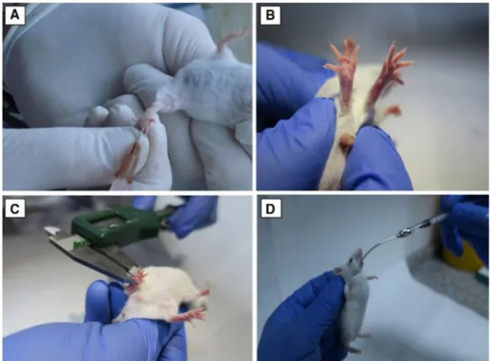
Fig. 1 a Inoculation of
promas-tigotes to mice’s footpads. b Infected footpad of a BALB/C mouse. c Measurement of mice footpads. d Administration of miltefosine using oral gavage technique
between the groups. From the 14th week onwards, the footpad measurements of the miltefosine group were sig-nificantly lower than those in the control group; p = 0.002,
p = 0.001, p = 0.004, p = 0.001, p = 0.001, respectively. No
statistically significant difference was found between the miltefosine group and the MA group and between the MA group and the control group in terms of footpad measure-ments (Fig. 2).
Examination of the Giemsa stained slides prepared from the miltefosine group mice’s footpads revealed amastigotes in one of the slides. In the MA group, amastigotes were observed in two slides, and amastigotes were observed in all samples in the control group (Table 1).
In cultures prepared from tissue swabs from the incision site and tissue suspensions of the foot; promastigotes were present in one, four, and all of the culture media in the milte-fosine group, MA group, and the control group, respectively (Table 1).
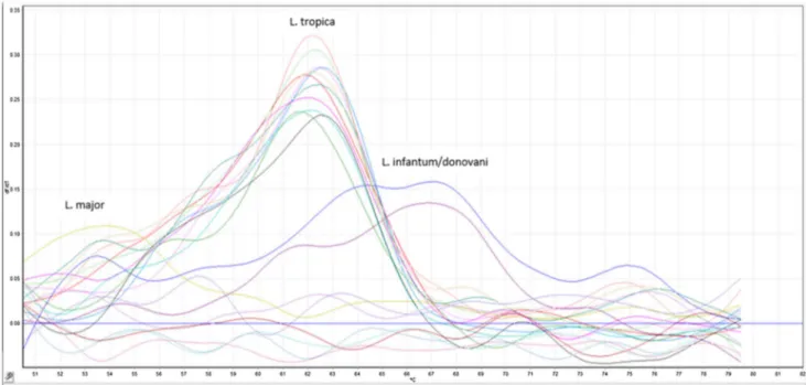
Molecular tests were performed with the Rotor-Gene device using prepared tissue suspensions and L. tropica con-sistent peaks were obtained in one of the miltefosine group, four in the MA group and all mice in the control group (Fig. 3).
Discussion
Drug resistance refers to all processes that is related to leish-mania parasite’s partial or non-responsiveness to a previ-ously effective drug. In human infections, this may be due to the patient’s non-response to treatment or the occurrence of relapses after treatment or in the form of different pres-entations as seen in CCL. Treatment success is influenced by host factors such as immune status, nutrition, age, and sex, as well as the pharmacokinetic properties of the drugs involved [13, 14].
In the literature, there are reports of infection develop-ment in the footpads of the mice, from 7 days to 12 weeks before the initiation of treatment and in different publica-tions treatment initiation is planned based on the measure-ments of the lesions [9, 15]. In our study, the reasons for
Fig. 2 Graphical demonstration of mice footpad measurements before and after treatment. The treatment of the mice starts on the 12th week and continue till the 24th week
Table 1 Performance of each diagnostic method for each group of mice
Diagnostic method Animal group Direct
micros-copy Enriched medium Real time PCR
Miltefosine group 1 1 1
MA group 2 4 4
determining the lesion development period as 12 weeks are; the late onset of lesions in CCL patients and the observation of reproductive dynamics of parasites in culture media as being significantly slower compared to other isolates. At this point, 12 weeks of infection formation process was preferred to eliminate possible infection development problems.
Most of the studies conducted with miltefosine are from South America and with the patients infected with L.
braziliensis [16–18]. In a clinical study from the Nether-lands, treatment outcomes of two CL cases infected with L.
major and L. infantum were evaluated [19]. Another study comparing miltefosine and glucantime efficacy with L.
major-infected CL patients in Iran, both treatment and
post-treatment outcomes were similar, miltefosine tolerance was good except for mild gastrointestinal system (GIS) side effects [20].
Although there are studies investigating the efficacy of miltefosine on L. tropica in vitro [21, 22], in vivo and clini-cal studies with L. tropica are limited. An in vitro study investigating the efficacy of miltefosine for pentavalent anti-monial resistant L. tropica isolates in Turkey found out that these resistant L. tropica strains were susceptible to milte-fosine [23]. To the best of our knowledge, there are just two case reports from Canada and the UK for oral miltefosine treatment in patients who have been shown to have L.
trop-ica infection by genotyping [24, 25].
The development of the leishmania scar was monitored by regular footpad measurements to establish an appropriate model of the CCL clinic. Quantitative evaluation of local infection is provided this way. Regular measurements for
24 weeks from the onset of infection have provided impor-tant data for the clinical follow-up of the infection as well as the treatment and subsequent process. We observed a ces-sation of footpad swelling on the 16th week and decline of the footpad sizes from the 18th week onwards for the drug administered mice. However, we observed a faster decline in the miltefosine-treated group than the MA-treated group. Additionally, in the MA group, the reduction in lesion size almost ceased after 20 weeks. Compared to these, the mice in the control group were observed to have a progressive lesion size. These data provide ampirical evidence of the treatment outcomes.
The positive results obtained from the visceral organs of all mice with positive PCR results were significant in terms of reflecting the character of the CCL isolate or demonstrat-ing that the isolate has the potential of visceralization in mouse models. The possibility of visceralization should be kept in mind in CCL patients and the presence of viscer-alization should be investigated with further studies in this patient group.
The difference between the treated groups can be attrib-uted to several different variables. There is a possibility that the isolate used to establish in-vivo models has developed resistance as a result of repeated treatment with MA. Since MA is administered in accordance with all national and international guidelines, the possibility of incomplete or inadequate treatment is also invalid [8, 26].
In the miltefosine-treated group, only one mouse showed a relatively slower regression of the lesion, and only one mouse was positive in real-time PCR tests. PCR tests were negative
Fig. 3 Positive Real-time PCR melting curve analysis results of all the infected BALB/C mice after 24 weeks. Each piques of the differentially colored curves represent the above labeled strains of leishmania. The curves at the bottom represent the negative samples and negative control
in the remaining four mice, and after a positive result in only one mouse, the test was repeated to exclude the possibility of contamination, but the control was also positive. This condi-tion may be due to the oral gavage administracondi-tion of miltefos-ine. There may be application-related errors in certain points of treatment, or the experimental animal may have vomitted the drug for various reasons. Also, it should be kept in mind that the drug administered to the experimental animal may have reduced efficacy as a result of pharmacokinetic mecha-nisms originating from the animal. That is to say, there is also the possibility that conditions such as infection, malnutrition, metabolic pathologies that may adversely affect the mouse immune system may have developed at any point during the 24-week experimental period [27].
Conclusions
In this study, in-vivo drug efficacy is evaluated on a leishmania isolate from a CCL patient. For this purpose, we compared MA, the most commonly used anti-leishmanial drug in our country and the world, with miltefosine, a new and remarkable anti-leishmanial agent in terms of its low side effect profile, ease of use and cost.
Demonstration of both clinical and laboratory improve-ment in four of the five mice provides strong evidence that miltefosine is an effective drug in the treatment of CCL. In the literature, no clinical or laboratory studies using miltefosine have been performed with CCL patients only. To solve the treatment problems encountered in this special patient group, we conclude that miltefosine may be one of the first drugs that should be considered for the treatment of CCL patients. Considering the results of this study, further clinical studies with miltefosine will reveal valuable data.
Acknowledgements The authors would like to thank Ege University Research Funds for funding this research, and ‘Parasite Bank’ in Celal Bayar University Faculty of Medicine for keeping and supplying the isolates.
Funding This project was supported by Research Funds of Ege Uni-versity with the Project no: 17-TIP-017.
Compliance with Ethical Standards
Ethical Approval This study was approved by the Celal Bayar Uni-versity Local Ethical Committee for Laboratory Animals (No: 77.637.435‐55‐27.09.2016).
References
1. Karimkhani C, Wanga V, Coffeng LE, Naghavi P, Dellavalle RP, Naghavi M (2016) Global burden of cutaneous leishmaniasis: a cross-sectional analysis from the Global Burden of Disease Study
2013. Lancet Infect Dis 16:584–591. https ://doi.org/10.1016/ S1473 -3099(16)00003 -7
2. Du R, Hotez PJ, Al-Salem WS, Acosta-Serrano A (2016) Old world cutaneous leishmaniasis and refugee crises in the Middle East and North Africa. PLoSNegl Trop Dis 10:e0004545. https :// doi.org/10.1371/journ al.pntd.00045 45
3. Abuzaid AA, Abdoon AM, Aldahan MA, Alzahrani AG, Alha-keem RF, Asiri AM et al (2017) Cutaneous leishmaniasis in Saudi Arabia: a comprehensive overview. Vector Borne Zoonotic Dis 17:673–684. https ://doi.org/10.1089/vbz.2017.2119
4. Reithinger R, Dujardin J-C, Louzir H, Pirmez C, Alexander B, Brooker S (2007) Cutaneous leishmaniasis. Lancet Infect Dis 7:581–596. https ://doi.org/10.1016/S1473 -3099(07)70209 -8 5. Hotez PJ, Velasquez RM, Wolf JE (2014) Neglected tropical skin
diseases: their global elimination through integrated mass drug administration? JAMA Dermatology 150:481–482. https ://doi. org/10.1001/jamad ermat ol.2013.8759
6. Ponte-Sucre A, Gamarro F, Dujardin J-C, Barrett MP, López-Vélez R, García-Hernández R et al (2017) Drug resistance and treatment failure in leishmaniasis: a 21st century challenge. PLoSNegl Trop Dis 11:e0006052. https ://doi.org/10.1371/journ al.pntd.00060 52
7. Mathers CD, Ezzati M, Lopez AD (2007) Measuring the burden of neglected tropical diseases: the global burden of disease frame-work. PLoSNegl Trop Dis 1:e114. https ://doi.org/10.1371/journ al.pntd.00001 14
8. World Health Organization. Leishmaniasisn.d.https ://www.who. int/leish mania si/en/.
9. García Bustos MF, Barrio A, Prieto GG, de Raspi EM, Cimino RO, Cardozo RM et al (2014) In vivo antileishmanial efficacy of miltefosine against Leishmania (Leishmania) amazonensis. J Parasitol 100:840–847. https ://doi.org/10.1645/13-376.1 10. Coelho AC, Trinconi CT, Costa CHN, Uliana SRB (2014) In vitro
and in vivo miltefosine susceptibility of a Leishmaniaamazon-ensis isolate from a patient with diffuse cutaneous leishmania-sis. PLoSNegl Trop Dis 8:e2999. https ://doi.org/10.1371/journ al.pntd.00029 99
11. Özbilgin A, Çulha G, Uzun S, Harman M, Topal SG, Okudan F et al (2016) Leishmaniasis in Turkey: first clinical isolation of
Leishmania major from 18 autochthonous cases of cutaneous
leishmaniasis in four geographical regions. Trop Med Int Heal. https ://doi.org/10.1111/tmi.12698
12. Toz SO, Culha G, Zeyrek FY, Ertabaklar H, Alkan MZ, Vardarlı AT et al (2013) A real-time ITS1-PCR based method in the diag-nosis and species identification of leishmania parasite from human and dog clinical samples in Turkey. PLoSNegl Trop Dis 7:e2205. https ://doi.org/10.1371/journ al.pntd.00022 05
13. Wakelin D (1989) Nature and nurture: overcoming constraints on immunity. Parasitology 99(Suppl):S21–35
14. Morrone A, Pitidis A, Pajno MC, Dassoni F, Latini O, Barna-bas GA et al (2011) Epidemiological and geographical aspects of leishmaniasis in Tigray, northern Ethiopia: a retrospective analy-sis of medical records, 2005–2008. Trans R Soc Trop Med Hyg 105:273–280. https ://doi.org/10.1016/j.trstm h.2011.02.003 15. Coelho AC, Trinconi CT, Costa CHN, Uliana SRB (2014) In vitro
and in vivo miltefosine susceptibility of a leishmaniaamazonensis isolate from a patient with diffuse cutaneous leishmaniasis. PLoS-Negl Trop Dis 8:1–11. https ://doi.org/10.1371/journ al.pntd.00029 99
16. Soto J, Arana BA, Tolado J, Rizzo N, Vega JC, Diaz A et al (2004) Miltefosine for new world cutaneous leishmaniasis. Clin Infect Dis 38:1266–1272. https ://doi.org/10.1086/38332 1
17. Monge-Maillo B, López-Vélez R, Saravolatz LD (2015) Miltefos-ine for visceral and cutaneous leishmaniasis: drug characteristics and evidence-based treatment recommendations. Clin Infect Dis 60:1398–1404. https ://doi.org/10.1093/cid/civ00 4
18. Soto J, Rea J, Balderrama M, Toledo J, Soto P, Valda L et al (2008) Efficacy of miltefosine for Bolivian cutaneous leishma-niasis. Am J Trop Med Hyg 78:210–211
19. Dorlo TPC, van Thiel PPAM, Schoone GJ, Stienstra Y, van Vugt M, Beijnen JH et al (2011) Dynamics of parasite clearance in cuta-neous leishmaniasis patients treated with miltefosine. PLoSNegl Trop Dis 5:e1436. https ://doi.org/10.1371/journ al.pntd.00014 36 20. Mohebali M, Fotouhi A, Hooshmand B, Zarei Z, Akhoundi B,
Rahnema A et al (2007) Comparison of miltefosine and meglu-mineantimoniate for the treatment of zoonotic cutaneous leish-maniasis (ZCL) by a randomized clinical trial in Iran. Acta Trop 103:33–40. https ://doi.org/10.1016/j.actat ropic a.2007.05.005 21. Escobar P, Matu S, Marques C, Croft SL (2002) Sensitivities of
Leishmania species to hexadecylphosphocholine (miltefosine), ET-18-OCH(3) (edelfosine) and amphotericin B. Acta Trop 81:151–157
22. RezaeiRiabi T, Sharifi I, MiraminMohammadi A, Khamesipour A, Hakimi PM (2013) Evaluation of a possible synergistic effect of meglumineantimoniate with paromomycin, miltefosine or allopu-rinol on in vitro susceptibility of leishmaniatropica resistant iso-late. Iran J Parasitol 8:396–401
23. Özbilgin A, Çavuş İ, Kaya T, Yıldırım A, Harman M (2020) Com-parison of in vitro resistance of wild leishmania isolates, which are resistant to pentavalent antimonial compounds, against drugs
used in the treatment of leishmaniasis. TurkiyeParazitolojiiDerg 44:12–16. https ://doi.org/10.4274/tpd.galen os.2019.6661 24. Keynan Y, Larios OE, Wiseman MC, Plourde M, Ouellette M,
Rubinstein E (2008) Use of oral miltefosine for cutaneous leish-maniasis in Canadian soldiers returning from Afghanistan. Can J Infect Dis Med Microbiol 19:394–396
25. Killingley B, Lamb LEM, Davidson RN (2009) Miltefosine to treat cutaneous leishmaniasis caused by Leishmania tropica. Ann Trop Med Parasitol 103:171–175. https ://doi.org/10.1179/13648 5909X 39817 7
26. Prevention C-C for DC and CDC—Leishmaniasis—Resources for health Professionals 2017
27. Tuntland T, Ethell B, Kosaka T, Blasco F, Zang R, Jain M et al (2014) Implementation of pharmacokinetic and pharmacodynamic strategies in early research phases of drug discovery and develop-ment at novartis institute of biomedical research. Front Pharma-col. https ://doi.org/10.3389/fphar .2014.00174
Publisher’s Note Springer Nature remains neutral with regard to