238
Makale Kodu/Article code: 1819 Makale Gönderilme tarihi: 11.08.2014 Kabul Tarihi: 14.12.2014
ABSTRACT
This clinical case describes rehabilitation of the a young patient’s anterior teeth via modified laminate veneer restorations with all-ceramic one-piece coronal post technique, crown, and 3-unit fixed dental prosthesis. The modified laminate veneer procedure mentioned in this case report represents an alternative to traditional complete crown restorations via adhesive systems and can yield satisfactory esthetic and functional results.
Keywords: post technique, dental veneers, esthetic.
INTRODUCTION
The maxillary central incisors are the teeth that are most commonly affected by trauma, and enamel and dentine fractures are the most common types of traumatic dental injuries to permanent anterior teeth in young patients.1
Minimally invasive dentistry entails removal of minimal amount of healthy tissues and adopts a philosophy that integrates minimal intervention for the placement and replacement of restorations.2,3
Laminate veneer restorations are compatible with this approach.
Esthetic and functional problems related to the restoration of endodontically treated teeth are particularly relevant to prosthetic dentistry. It has been reported that endodontically treated teeth are vulnerable to fractures, and therefore they frequently require coronal restorations.4-6 Conventional metal-
ÖZET
Bu vaka çalışmasında, genç bir hastanın travmaya bağlı hasar görmüş ön grup dişlerinin farklı bir laminate veneer restorasyon ile beraber tam kuron ve üç üyeli sabit köprü restorayonuyla tedavisi anlatılmaktadır. Bahsedilen modifiye edilmiş laminate veneer tekniği, estetik ve fonksiyonel sonuçlar sunmasıyla, tedavi için uygun olan dişlerde klasik sabit tam kuron protezlerine alternatif bir tedavi seçeneği oluşturmaktadır.
Anahtar Kelimeler: post tekniği, laminate
veneer, estetik diş hekimliği
ceramic complete crown restorations have some disadvantages with regard to esthetics, as they entail a strong risk of restricting translucency, and also of causing discoloration with reference to the associated gingival tissues. They do, however, provide maximum strength.7,8 All-ceramic restorations are metal free and
esthetically more pleasing.9 Furthermore, all-ceramic
restorations result in adequate retention in conjunction with appropriate adhesive techniques, preserving maximum remaining tooth structure.5,7
Sorensen et al10 have reported that if more
than 50% of the intact enamel surface area has been lost due to excessive decay or the application of access preparations for endodontic treatment, laminate veneer restorations would have some limitations with respect to retention and resistance. Therefore, post-like features termed “coronal posts,” consisting of a single-unit porcelain laminate veneer and a short ceramic post extended into the root canal
THE MODIFIED LAMINATE VENEER RESTORATIONS WITH COMBINED PROSTHODONTIC RESTORATIONS ON TRAUMATIC TEETH (CASE REPORT)
HASAR GÖRMÜŞ DİŞLERİN FARKLI BİR LAMINATE VENEER RESTORASYONU İLE KOMBİNE PROTETİK TEDAVİSİ (OLGU SUNUMU)
Yrd. Doç. Dr. Pınar CEVIK* Prof. Dr. Filiz AYKENT**
*Department of Prosthodontics, Gazi University Faculty of Dentistry. **Department of Prosthodontics, Selcuk University Faculty of Dentistry.
≠This case report was presented in the 5th biennial meeting of the European Fedaration of Conservative Dentistry (EFCD) (ConsEuro), 12-15 October 2011, Istanbul, Turkey.
239 veneers.
CASE REPORT
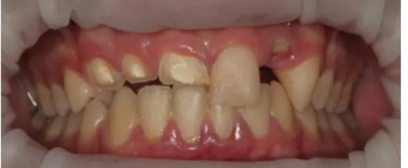
A 14-year-old boy was referred to our clinic with a recent history of a sport-related injury to the maxillary right central and lateral incisors and canines (Fig. 1). After clinical and radiographic examination, endodontic treatment was planned for the fractured teeth. For the missing left lateral incisor, a 3-unit all- ceramic fixed dental prosthesis was considered. Any implant supported prosthesis was not selected because of the economical restriction. A week after endodontic treatment of the fractured teeth, a fiber-reinforced composite post (FRC Postec Plus, Ivoclar Vivadent, Liechtenstein) and composite core (Multicore Flow, Ivoclar Vivadent, Liechtenstein) were fabricated to restore the right canine prior to complete crown preparation. The reason why a complete crown restoration was selected for the right canine was that the patient had the canine guided occlusion. So, a complete crown restoration was selected because of the canine’s position in the occlusal plane. Chamfer finishing lines were generated, as was done for the gingival margin of the relevant canine and the other teeth involved (Fig. 2).
Figure 1. The first appointment of the patient before treatment
Figure 2. Coronal post and laminate preparations
The gutta-percha and endodontic cement of the right central and lateral incisors were removed using a round bur to a depth of 3 mm into the root canal. Glass ionomer cement was placed into the canal orifice to prevent discoloration. To achieve an esthetically favorable border with the gingiva, bipolar electrocauterization was adopted. Retraction cords (Ultrapak 0; Ultradent Products Inc., South Jordan, UT, USA) were used to expose the preparation area around the gingival margin. Laminate veneer preparations were then performed via a conservative approach terminating approximately after 1-mm incisal and labial reduction. Two wooden supports derived from sterile pieces of toothpicks were cut and fitted into the coronal post canal to obtain the required impression. A definitive impression of the prepared teeth was made using polyvinylsiloxane impression material (Virtual Putty and Lightbody, Ivoclar Vivadent Liechtenstein). The interim prostheses were fabricated and inserted into the relevant teeth. The Lava ceramic porcelain system (3M ESPE, St. Paul, MN, USA) was used in the fabrication of the final restorations (Fig. 3). Initially CAD/CAM supported frameworks which include post and core were designed for the treated teeth with CEREC inLab system. In this system, cast model was fabricated from the impression taken from the patient. Then, model was scanned by CEREC camera in the laboratory (CEREC Bluecam, Sirona). Also, the frameworks for the complete crown and 3-unit all- ceramic fixed dental prosthesis were produced by the same method. Prior to veneering, preparation of the frameworks was completed by using CEREC system (CEREC Bluecam, Sirona) with Lava blocks and Lava frameworks were tried on the patient. Then, veneering ceramic was fired on to best fitting frameworks in the right color. Finally, canine guided occlusion was adjusted accordingly.
240 Figure 3. Modified laminate veneer restorations
After final training and finishing, restorations were glazed and cemented using a dual-curing composite system. Initially, all tooth surfaces were etched using 37% phosphoric acid (Total Etch; Ivoclar-Vivadent, Liechtenstein) for 15 s, rinsed for 10 s, and then gently dried for 5 s. Internal surfaces of the Lava restorations were treated with 30-µm silica-modified alumina particles which were silica coated (Cojet Sand, 3M ESPE, St. Paul, MN, USA) at 0.2 Mpa pressure, then rinsed for 60 s and air dried for 20 s. A layer of silane (Monobond S, Ivoclar-Vivadent, Liechtenstein) was applied to the inner surfaces of restorations for 60 s, then air dried. The primer (Syntac Primer; Ivoclar-Vivadent, Liechtenstein) and the adhesive (Syntac Adhesive; Ivoclar-Vivadent, Liechtenstein) were applied to the tooth surfaces consecutively according to the manufacturer’s instruct- tions. An unfilled resin-bonding agent (Heliobond; Ivoclar-Vivadent, Liechtenstein) was applied with a brush and thinned by air drying for 3 s. Dual-polymerizing composite resin cement (Vaiolink II, Ivoclar-Vivadent, Liechtenstein) of the appropriate shade was applied to the restoration. The restoration was positioned appropriately to the restoration area and excess cement was removed. The luting agent was polymerized with an LED device (Bluephase, Ivoclar-Vivadent, Liechtenstein) at 470 mW/cm2 power
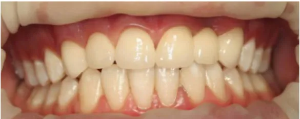
for 40 s. The patient was informed of the importance of oral hygiene and regular follow-up (Fig. 4). He was satisfied with the results after 2 years and did not complain of any functional or coloration-related problems.
Figure 4: Post-operative treatment of the patient
DISCUSSION
This case report describes an alternative method to traditional metal-ceramic restorations, also gives a different solution for the traumatic anterior teeth. This method preserves the remaining tooth structure in the proper traumatic area with the tooth’s retention and resistance properties.
Anterior dental trauma is relatively common among young patients.8 For uncomplicated crown
fractures, treatment strategies may entail reattachment of the fractured crown or prosthetic reconstruction, depending on whether the fracture line is large or small. After deciding the treatment of choice, it is important to maintain the remaining tooth structure, restore the occlusion immediately, and prevent over eruption of opposing incisors.11
Coronal prosthetic restorations are frequently used to prevent fractures in endodontically treated teeth. As a result of access preparations for endodontic treatment, earlier reports have described post-like features termed “coronal posts” in such clinical cases, to enhance retention and resistance of laminate veneers.4,6,7 The restorative materials and
techniques used for modified laminate veneer restoration can be important in endodontically treated teeth, which cannot be treated with conventional metal ceramic restoration owing to compromised esthetics related to limited esthetic of metal-ceramics and to their susceptibility to discoloration.5,7
All-ceramic restorations and esthetic adhesive luting materials offer new solutions in prosthetic dentistry with improved esthetics.8 Moreover, minimally invasive
methods fulfill the need for retention and resistance of all-ceramic restorations.8,9
The application of an MDP-containing silane agent to the inner surface of zirconium oxide ceramic restorations, increased the shear bond strength between restoration and resin luting agents.12-14
241 greater stress resistance with regard to restored teeth.15 Wang et al 16 compared failure modes and
fracture strength of two ceramic structures. They stated that the zirconia cores with high strength and high elastic modulus had better resistance to fracture than the IPS e-max cores. The Lava ceramic framework consists of zirconia supplemented with a specially designed for veneer ceramic. The frameworks are fabricated from pre-sintered zirconia blocks by using CAD/CAM procedures (computer-aided scanning, framework design, and milling) also with chairside solutions and labside solutions.
Sevuk et al5 reported that one-piece all-ceramic
coronal post and laminate veneer restoration with Celay copy-milling system. In celay technique, the pre-models of the restorations are preformed using autopolymerizing resins or composite resins. Celay is less suitable than sintered ceramics for making veneers. Sintered and laminated veneers have best esthetic outcome.17 In the present case report, CEREC
system was selected for the fabrication of frameworks. Use of this technique reduced the time spent on chairside system. Cerec system allows using different kinds of ceramic and zirconia blocks. Also, the additional trying in process and extra materials are not used such as different resin materials so the system eradicates such a polymerization shrinkage in fabrication of pre-models. Frameworks are fabricated directly from the digital scans which provide best results for the patients and the dentists.
Esthetic posts especially fiber-reinforced composite resin posts and zirconia-based ceramic posts are required for the esthetic and biocompatible results.18 Dikbas et al18 also reported that the
reinforcing capacity of zirconium posts in weakened teeth decreasing the risk of fracture. Carvalho et al19
reported that composite resin posts and zirconia posts could increase the structural strength of weakened teeth. In the present case, zirconia based intracanal posts were used in combination of core structure to enhance the esthetic and functional results.
Porcelain laminate veneers can be considered as an alternative choice when the esthetic outcomes
There are some limitations of this case report. Lava zirconia blocks may have adverse effects on translucency of anterior teeth. In this case, the patient is under follow up controls for 2 years. Even so, long term controls more than two years should be done to make a comment about the position of modified laminate veneers on such cases.
In conclusion, the one-piece all-ceramic coronal post with laminate veneer restoration technique investigated for restoration of fractured teeth proved to be minimally invasive, and the esthetic outcome was favorable. We recommend that patients be informed of the importance of regular follow-up in similar clinical cases.
REFERENCES
1. Anchieta RB, Rocha EP, Watanabe MU, et al: Recovering the function and esthetics of fractured teeth using several restorative cosmetic approaches. Three clinical cases. Dent Traumatol 2012;28:166-72.
2. Dawson AS, Makinson OF: Dental treatment and dental health. Part 2. An alternative philosophy and some new treatment modalities in operative dentistry. Aust Dent J 1992;37:205-10.
3. White JM, Eakle WS: Rationale and treatment approach in minimally invasive dentistry. J Am Dent Assoc 2000;131:13S-19S.
4. Caputo AE, Standlee PJ. Biomechanics in clinical dentistry Chicago; Quintessence: 1987. p. 185-203.
5. Sevuk C, Gur H, Akkayan B: Fabrication of one-piece all-ceramic coronal post and laminate veneer restoration: A clinical report. J Prosthet Dent 2002;88:565-8.
6. Walton RE, Torabinejad M. Principles and practice of endodontics Philadelphia; WB Saunders: 1989. p. 249-62.
7. Giachetti L, Bertini F, Bambi C: An 8-year follow-up of a fractured endodontically treated incisor restored with a modified laminate veneer. Dent Traumatol 2008;24:104-7.
242 8. Tayab T, Vizhi K, Srinivasan I: Space maintainer
using fiber-reinforced composite and natural tooth--a non-invasive technique. Dent Traumatol 2011;27:159-62.
9. Bagis B, Satiroglu I, Korkmaz FM, et al: Rehabilitation of an extracted anterior tooth space using fiber-reinforced composite and the natural tooth. Dent Traumatol 2010;26:191-4.
10. Sorensen JA, Strutz JM, Avera SP, et al: Marginal fidelity and microleakage of porcelain veneers made by two techniques. J Prosthet Dent 1992;67:16-22.
11. Turkistani J, Hanno A: Recent trends in the management of dentoalveolar traumatic injuries to primary and young permanent teeth. Dent Traumatol 2011;27:46-54.
12. Wegner SM, Kern M. Long-term resin bond strength to zirconia ceramic. J Adhes Dent 2000;2:139-47.
13. Blatz MB, Sadan A, Kern M. Resin-ceramic bonding: a review of the literature. J Prosthet Dent 2003;89:268-274.
14. Oba Y1, Koizumi H, Nakayama D, Ishii T, Akazawa N, Matsumura H. Effect of silane and phosphate primers on the adhesive performance of a tri-n-butylborane initiated luting agent bonded to zirconia. Dent Mater J 2014;33:226-32.
15. Ozkurt Z, Kazazoglu E: Clinical success of zirconia in dental applications. J Prosthodont 2010;19:64-8. 16. Wang R, Lu C, Arola D, Zhang D. Plastic Damage Induced Fracture Behaviors of Dental Ceramic Layer Structures Subjected to Monotonic Load. J Prosthodont 2013;22:456-64.
17. J. Schmidseder. Color Atlas of Dental Medicine. Thieme Publishing Group;Germany: 2000. p. 10-270.
18. Dikbas I, Tanalp J, Koksal T, Yalnız A, Güngör T. Investigation of the effect of different prefabricated intracanal posts on fracture resistance of simulated immature teeth. Dent Traumatol 2014;30:49-54.
19. Carvalho CA, Valera MC, Olivera LD, Camargo CH. Struc- tural resistance in immature teeth using root reinforcements in vitro. Dent Traumatol 2005;21:155-9.
20. Saridağ S, Akman S, Pekkan G. Esthetic Restoration Of Eroded Teeth Using Porcelain Laminate Veneers With Different Preparation Techniques: Case Report. J Dent Fac Atatürk Uni 2011;21:241-44.
Yazışma Adresi
Dr. Pinar Cevik
Gazi University, Faculty of Dentistry, Department of Prosthodontics, 06510, Ankara, TURKEY Tel: +90 543 7733793 Fax: +90 312 2034192