Bilateral Nasal Polyps Originating From Olfactory
Cleft
ABSTRACT
Nasal polyps generally have similar symptom, they generally originate from ethmoid infundibulum, mucosal contact areas of the uncinate process and middle turbinate. However nasal polyps rarely arise from different and unexpected locations in the nasal cavity such as septum, cribriform plate and olfactory cleft. In this article, we report 63 years old men, had a history of nasal obstruction and loss of smell for six years, diagnosed with bilaterally olfactory cleft originated nasal polyps, which is a very rare site of origin.
Key words: Nasal polyp, cribriform plate, olfactory mucosa, computed tomography, endoscopic surgery
Olfaktör Yarık Kaynaklı Bilateral Nasal Polip ÖZET
Nasal polipler genellikle etmoid infundibulum, uncinat çıkıntı ve orta konkanın mukozal temas yüzeylerinden kaynaklanır ve benzer semptomlara neden olurlar. Nazal polipler ender olarak septum, kribriform bölge, olfaktör yarık gibi bölgelerde de görül-ebilirler. Bu olgu sunumunda 6 yıldır koku alma sorunlarına neden olan bilateral nasal polip tanısı almış 63 yaşındaki erkek hasta sunulmaktadır.
Anahtar kelimeler: Nazal polip, kribriform bölge, olfaktör mukoza, bilgisayarlı tomografi, endoskopik cerrahi
1Istanbul Medipol University Faculty of Medicine, Department of Neurosurgery, Istanbul, 2Canakkale 18 March University Faculty of Medicine, Department of Neurosurgery, Canakkale, 3Istanbul Medipol Hospital, Department of Radiology, Istanbul, Turkey
Received: 15.09.2014, Accepted: 30.09.2014
Correspondence: Adem Bozkurt Aras
Canakkale 18 March University, Faculty of Medicine,Department of Neurosurgery, Canakkale-Turkey
Fax: +90 286 2635957 Phone: +90 505 4785043 E-mail: [email protected]
Gurkan Kayabasoglu1, Kadir Cagdas Kazıkdas2
European Journal of General Medicine
Case Report
INTRODUCTION
Polyps can arise from any mucosal surface in the nasal cavity. Most of the nasal polyps originate from the later-al naslater-al wlater-alls, the anterior ethmoidlater-al sinus, the contact areas of the uncinate process and the middle turbinate. (1,2) Unique locations of origin are septum, cribriform plates or olfactory cleft for nasal polyps. (3,4,5) This is the second case report of a nasal polyp arise directly from the olfactory cleft in the literature.
CASE
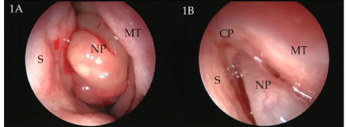
A 63-year-old man had a history of bilateral nasal ob-struction and loss of smell for 6 years. The patient was otherwise healthy and had no history of allergy. Endoscopic nasal examination with 30 degrees tele-scope showed that the bilateral masses were large
pol-ypoid lesions with pedicles arising from the olfactory cleft and were located only at the anterior part of the nose. (Figure 1) Computed tomography scan of the pa-ranasal sinuses confirmed the polypoid masses hanging from cribriform plates at the anterior part of the nasal cavities bilaterally. (Figure 2) Psychophysical testing of olfactory function was performed preoperatively and 6 months postoperatively using the validated “Sniffin’ Sticks” test, where odorants were presented in commer-cially available felt-tip pens (“Sniffin’ Sticks”, Burghart, Wedel, Germany). The test consists of one threshold and two suprathreshold subtests, namely a test for thresh-olds of n-butanol, a test for odor discrimination and one for odor identification. The patient was operated under general anesthesia and polypoid masses were removed totally endoscopically. Histopatologically the excised masses were diagnosed as “allergic nasal polyps” com-posed of loose myxoid stroma and mucous glands cov-Eur J Gen Med 2015; 12(3): 271-272 DOI : 10.15197/ejgm.01436
Eur J Gen Med 2015;12(3):271-272 Nasal polyps originating from olfactory cleft
272
ered with respiratory epitelium. Patient’s smell scores were 14 preoperatively and 35 postoperatively. During one-year follow-up no recurrence was observed.
DISCUSSION
Nasal polyps are benign lesions that are associated with mucosal inflammation of the nasal cavity and paranasal sinuses. Although many theories including allergy, in-flammation, and genetic predisposition have been sug-gested, their etiopathogenesis is not known properly Polyps generally cause nasal obstruction. However, hy-posmia, nasal discharge, snoring, mouth breathing, ob-structive sleep apnea syndrome can be the associated symptoms. (1) Diagnosis of a nasal polyp is made by endoscopic and radiological examinations. Computed tomography is a very helpful tool to diagnose and to de-tect the origin and the extent of the polyp (7). Magnetic resonance imaging can be an alternative in the radio-logic examination. The polyps typically have low to in-termediate T1-weighted and high T2-weighted signal intensities owing to their high water and low protein contents. In cases of an unusual origin or unilaterality,
an inverted papilloma or other malignancies must be considered in the differential diagnosis. (1) Treatment of the nasal polyps is total endoscopic resection of the polyps with the pedicles. The prognosis after the sur-gery is good, and recurrence rate is low. (8)
Sniffin’ Sticks test consists of one threshold and two su-prathreshold subtests, namely a test for thresholds of n-butanol, a test for odor discrimination and one for odor identification. The maximum score of each subtest was 16, resulting in a maximum composite score of 48 (TDI [threshold, discrimination, and identification] score). The normal values for the TDI composite score are > 30.3, with a cut-off between anosmia and hyposmia at 16.5. (8,9) Our patient’s smell scores were 14 (hyposmia level) preoperatively and 35 (normal level) postopera-tively. In conclusion, it should be remembered that na-sal polyps can originate from different and unexpected sites in the nasal cavity. The preoperative detection of the origin is crucial for successful surgical outcomes.
REFERENCES
1. Stammberger H, Hawke M. Essentials of functional endo-scopic surgery. St Louis: Mosby-Year Book; 1993
2. Yanagisawa E, Christmas DA, Yanagisawa R. Endoscopic view of the sites of origin of nasal polyps. Ear Nose Throat J 2000; 79: 490-2.
3. Ozgirgin ON, Kutluay L, Akkuzu G. Choanal polyp origi-nating from the nasal septum: a case report. Am J Otolaryngol 2003; 24: 261-4
4. Arikan OK, Muluk NB, Cirpar O. A case report of a nasal polyp originating in the cribriform plate. Ear Nose Throat J; 2013: 92: 19-22
5. Ozcan C, Duce NM, Gorur K. Choanal Polyp Originating From the Cribriform Plate. J Craniofac Surg 2010; 21: 806-8
6. Aydin O, Keskin G, Ustundag E. Choanal polyps: An evalu-ation of 53 cases. Am J Rhinol 2007; 21:164-8
7. Ozcan C, Zeren H, Talas DU. Antrochoanal polyp: a transmission and light microscopic study. Eur Arch Otorhinolaryngol 2005; 262:55-60
8. Kobal G, Klimek L, Wolfensberger M, et al. Multicenter in-vestigation of 1,036 subjects using a standardized method for the assessment of olfactory function combining tests of odor identification, odor discrimination, and olfactory thresholds. Eur Arch Otorhinolaryngol 2000; 257:205-11. 9. Hummel T, Sekinger B, Wolf S, Pauli E, Kobal G. “Sniffin'
Sticks”: olfactory performance assessed by the combined testing of odor identification, odor discrimination and ol-factory threshold. Chem Senses 1997; 22:39-52.
Figure 1. An endoscopic nasal examination with 30
degree telescope shows large polypoid lesion with pedicle arising from the cribriform plate. (S: septum, NP: nasal polyp, MT: middle turbinate, CP: cribriform plate)
Figure 2. Polypoid masses hanging from cribriform
plates can be seen only at the anterior part of the na-sal cavities bilaterally in computed tomography scans.