CONTROLLED SINGLET OXYGEN GENERATION VIA
PLASMONIC HEATING OF GOLD NANORODS
A THESIS SUBMITTED TO
THE GRADUATE SCHOOL OF ENGINEERING AND SCIENCE OF BILKENT UNIVERSITY
IN PARTIAL FULFILLMENT OF THE REQUIREMENTS FOR THE DEGREE OF MASTER OF SCIENCE IN CHEMISTRY By Tuğçe Karataş August, 2015
ii
CONTROLLED SINGLET OXYGEN GENERATION VIA PLASMONIC HEATING OF GOLD NANORODS
By Tuğçe Karataş August, 2015
We certify that we have read this thesis and that in our opinion it is fully adequate, in scope and in quality, as a thesis for the degree of Master of Science.
_______________________
(Advisor) Prof. Dr. Engin Umut Akkaya
_______________________
Asst. Prof. Dr. Bilge Baytekin
_______________________
Asst. Prof. Dr. Fazlı Sözmen
Approved for the Graduate School of Engineering and Science: _______________________
Prof. Dr. Levent Onural Director of the Graduate School
iii
ABSTRACT
CONTROLLED SINGLET OXYGEN GENERATION VIA
PLASMONIC HEATING OF GOLD NANORODS
TUĞÇE KARATAŞ
M.S. in Chemistry
Supervisor: Prof. Dr. Engin Umut Akkaya August 2015
Photodynamic therapy (PDT) is the up-and-coming and developing methodology in order to treat various cancer tissues. The success of therapeutic action is directly related to the presence of cytotoxic singlet oxygen (SO) in tumor tissues. However, the feasibility of PDT is bounded by two major factors, hypoxia and the requirement of incident light penetration through cancer tissue. With these considerations, we have combined aromatic endoperoxides and gold nanorods so as to accomplish the possible restrictions. In this project, we synthesized and characterized both PEGylated anthracenic endoperoxides and gold nanorods separately and then further characterization was achieved for the combination of gold nanorods and aromatic endoperoxides. We have successfully proved that the thermal decomposition of endoperoxide molecule was carried out by irradiation of gold nanorods that resulted in the generation of both singlet and molecular oxygen.
Keywords: Photodynamic therapy, gold nanorods, aromatic endoperoxides, singlet
iv
ÖZET
ALTIN NANOÇUBUKLARIN PLASMONİK ISITILMASI İLE
SİNGLET OKSİJEN ÜRETİMİ
TUĞÇE KARATAŞ
Kimya Bölümü Yüksek Lisans Tezi Tez Yöneticisi: Prof. Dr. Engin Umut Akkaya
Ağustos 2015
Fotodinamik terapi (FDT) son zamanlarda pek çok araştırmacı tarafından derinlemesine incelenen, gelecek vaat eden ve gelişmekte olan kanser tedavisi için uygulanan yeni bir tedavi şeklidir. Bu tedavi yöntemi ile kanserli hücrelerin yok edilebilmesi ortamda bulunan zararlı singlet oksijenin (SO) varlığıyla doğrudan ilişkilidir. Yapılan araştırmalar sonunda FDT’nin etkisini artırabilmenin iki önemli faktöre bağlı olduğu görülmüştür. Bunlardan biri hipoksiya, bir başka deyişle kanser dokularındaki moleküler oksijen yetersizliğidir. Oldukça önemli olan diğer problem ise kanserli hücreye uygulanan ışığın dokulardan ancak oldukça az bir kısmının geçebilmesidir. Bu iki sorunu göz önünde bulundurarak ve bu problemleri en aza indirebilmek için aromatik endoperoksitler ile fonksiyonlandırılmış altın nanoçubuklar kullanmayı hedefledik. Bu proje kapsamında suda çözünebilen endoperoksit molekülleri ve altın nanoparçacıklar sentezlendi ve her bir aşamada tüm ürünler karakterize edildi. Ve bu proje ile ışıkla uyarılan altın nanoparçacıkların ısınması ve yüzeylerinde bulunan endoperoksitlerin bozunarak zararlı singlet oxygen açığa çıkarması ile amaçlandığı üzere kanser hücrelerinin ölümü gerçekleşmiştir.
Anahtar Kelimeler: Fotodinamik terapi, altın nanoçubuklar, aromatik
v
vi
ACKNOWLEDGEMENTS
I would like to present my sincere gratitude to my supervisor Prof. Engin U. Akkaya for his endless support, guidance, and patience. He is the most intelligent, foreseeing, and the funniest person in my life. Being a member of Prof. Akkaya’s research group is the most right decision in my life. I will never forget his support throughout my life. I would like to thank Dr. Safacan Kölemen for being a brother for me and for his endless support, patience, and guidance to everything present in this thesis. He presented all his experience to improve my skills and knowledge in that laboratory process. I feel myself very lucky to meet him and to work with him.
I owe a special thanks to Dr. Tuğba Özdemir Kütük, Cansu Kaya, Ceren Çamur, Darika Okeev, Hale Atılgan, Jose Luis Bila, Melek Baydar and Tuğçe Durgut for their close friendships, endless support, help and understanding. We share everything and help to each other whenever we need. I will miss being and working with them. I am sincerely grateful to our past and present members Dr. Fazlı Sözmen, Dr. Murat Işık, Dr. Ruslan Guliyev, Dr. Sündüs Erbaş Çakmak, Dr. Esra Tanrıverdi, Dr. Onur Büyükçakır, Dr. Yusuf Çakmak, Nisa Yeşilgül, Işın Sakallıoğlu, Taha Bilal Uyar, Ahmet Atılgan, Yiğit Altay, Dr. Dilek Taşgın, Dr. Özlem Seven, Özge Yılmaz, Deniz Yıldız, Abdurrahman Türksoy, Dr. İlke Şimşek Turan, Dr. Tuna Subaşı and rest of the SCL (Supramolecular Chemistry Laboratory) members.
I would like to express special thanks to my mother Fatma Arzu Karataş and father Muammer Karataş for their endless love, support and understanding. I owe them a lot. Finally, I would like to thank my love Sinan Altuğ Ataş for his great love, support, patience and understanding.
vii LIST OF ABBREVIATIONS
PDT : Photodynamic therapy
ROS : Reactive oxygen species
HPD : Haematoporphyrin derivative
PS : Photosensitizer
LEDs : Light-emitting diodes
ISC : Intersystem crossing
BPD : Benzoporphyrin derivative
LSPR : Localized surface plasmon resonance
GNR : Gold nanorod
CTAB : Cetyltrimethylammonium bromide
PEG : Poly (ethylene glycol)
DMSO : Dimethyl sulfoxide
DMF : Dimethylformamide
EPO : Endoperoxide
TMS : Tetramethylsilane
CHCl3 : Chloroform
MS : Mass Spectroscopy
TLC : Thin layer chromotography
DPBF : Diphenylisobenzofuran
viii
Contents
1. INTRODUCTION ... 1
1.1. Photodynamic Therapy ... 1
1.1.1. General Information ... 1
1.1.2. History of Photodynamic Therapy ... 2
1.1.3. Major components of Photodynamic Therapy ... 6
1.1.4. Mechanism of Photodynamic Therapy ... 7
1.1.5. The effects of Photodynamic therapy on tumor tissues ... 9
1.2. Gold Nanorods ... 10
1.2.1. Plasmonic Properties of Gold Nanorods ... 13
1.2.2. Synthesis of Gold Nanorods ... 17
1.2.3. Functionalization of Gold Nanorods ... 20
1.2.4. Biological and Biomedical Applications of Gold Nanorods ... 22
1.3. Polycyclic Aromatic Endoperoxides ... 24
1.3.1. General Information ... 24
1.3.2. Preparation of Endoperoxides ... 25
1.3.3. Thermal Dissociation of Endoperoxides ... 27
2. EXPERIMENTAL PROCEDURE ... 30
2.1. General ... 30
2.2. Singlet Oxygen Trap Experiments ... 30
2.3. Synthesis of (2) ... 31 2.4. Synthesis of (3) ... 32 2.5. Synthesis of (5) ... 33 2.6. Synthesis of (6) ... 34 2.7. Synthesis of (7) ... 35 2.8. Synthesis of (8) ... 35 2.9. Synthesis of (9) ... 36
2.10. Synthesis of Gold Nanorod ... 37
2.10.1. Preparation of Seed Solution... 37
2.10.2. Growth of Gold Nanorods at longitunal LSPR Band ... 37
2.10.3. Decorating of Gold Nanorods with EPOs ... 38
3. RESULTS & DISCUSSION ... 39
4. CONCLUSION ... 54
REFERENCES ... 55
ix
A.1 Controlled Singlet Oxygen Generation via Plasmonic Heating of Gold Nanorods ... 63
A.1.1. 1H and 1CH
3 NMR Spectra ... 63
x
List of Figures
Figure 1. The molecular structure of Photofrin ... 3
Figure 2. Modified Jablonski diagram for illustrating photosensitization processes ... 8
Figure 3. Gold nanoparticles with various size and shape. Copyright © 2011, Royal Society of Chemistry. Reprinted with permission from ref (62)62 ... 12
Figure 4. Plasmonic properties of gold nanorods with their different aspect ratios. Copyright © 2009 WILEY-VCH Verlag GmbH & Co. KGaA, Weinheim. Reprinted with permission from ref (56)56... 15
Figure 5. Mechanism of seed-mediated growth method for preparation of gold nanorods. ... 18
Figure 6. The photothermal therapy of malignant cells with anti-EGFR/Au by applying Ti: sapphire laser. Copyright © 2006, American Chemical Society. Reprinted with permission from ref (93)93 ... 22
Figure 7. Photothermal therapy of A549 malignant cells with anti-EGFR/ Au by using NIR CW lasers. Copyright © 2010 WILEY-VCH Verlag GmbH & Co. KGaA, Weinheim. Reprinted with permission from ref (94).94 ... 23
Figure 8. Mechanism of cycloaddition reaction of singlet oxygen. ... 27
Figure 9. Thermal dissociation of aromatic endoperoxides. ... 28
Figure 10. Thermolysis of cycloreversion pathway of aromatic endoperoxides. ... 28
Figure 11.TEM images of gold nanorods (top) and electronic absorption spectrum of gold nanorods in HEPES buffer solution (bottom). ... 40
Figure 12. Representation of reaction pathways of our target structure. ... 41
Figure 13. Formation and thermolysis of endoperoxide molecule (8). ... 43
Figure 14.Left: Thermolysis of 5x10⁻⁵ M PEGylated endoperoxide molecule (9) in DMSO at various temperature. Right: Thermolysis of (9) represented as absorbance vs. temperature graph. For all selected temperatures the reaction was heated for 30 min. ... 44
Figure 15.Thermolysis of 5x10⁻⁵ M PEGylated endoperoxide molecule (9) in DMSO at 95oC for 6 hours. Inset: Absorbance of 5x10⁻⁵ M (9) in DMSO at 37oC and again the molecule was heated for 6 hours at body temperature. ... 45
Figure 16.Thermolysis of 1x10⁻⁵ M PEGylated endoperoxide molecule (9) in HEPES buffer (pH 7.2, 20 mM) at 100oC for 1 hr. ... 46
Figure 17. The results related to the decomposition of trap molecule (DPBF) owing to existence of singlet oxygen. ... 46
Figure 18. Left: TEM image after the conjugation of PEGylated endoperoxide (9) with gold NRs. Right: Normalized electronic absorption spectra of CTAB-GNR, (9)-GNR (in HEPES (pH 7.2, 20 mM) and (9)-(9)-GNR in DMSO... 48
Figure 19. Heating 1x10⁻⁵ M GNR-(9) conjugate at 70 ◦C in DMSO resulted in the absorbance decrease of DPBF. ... 48
Figure 20. The irradiation of gold NR-(9) with the laser at 830 nm resulted in the decrease of absorbance maximum of DPBF at 414 nm. ... 49
Figure 21. The effect of unconjugated GNRs on the absorbance of DPBF after excition with 830 nm laser light. ... 50
Figure 22. The differences in the singlet oxygen yield of GNR and GNR-(9) conjugate in DMSO. ... 50
xi
Figure 23. The confocal microscopy of HeLa cells represented ROS generation (top: ROS sensor, middle: DAPI, bottom: merged with DIC ). ... 52 Figure 24. The confocal microscopy of HeLa cells represented apoptotic cell death by Annexin V (top: Annexin V, middle: DAPI, bottom: merged with DIC ). ... 52 Figure 25. Hela cell's viability was assayed by MTT test after irradiation with 808 nm laser (2 W/cm2 , 10 min). ... 53
xii
List of Tables
Table 1. Clinically applied photosensitizers for malignant diseases1, 36 ... 5
Table 2. Activation parameters for thermal decomposition of several endoperoxide molecules... 29
1
1. INTRODUCTION
1.1. Photodynamic Therapy
1.1.1. General Information
Photodynamic therapy (PDT) has been accepted as a new therapeutic action for a number of disorders, especially for several forms of cancer such as malignant (head and neck, brain, urological, gynecological, dermatological, gastrointestinal cancers) and non-malignant tumors (age related macular degeneration-AMD, psoriasis).1, 2, 3 The therapeutic action is affiliated with the photochemical reaction, which culminates the generation of singlet oxygen. Cytotoxic singlet oxygen is produced by irradiation of loaded photosensitizer in a targeted cell that leads to an energy transfer to molecular oxygen on cancer cells.4 Thus, the basis of PDT requires the existence of three non-toxic components; molecular oxygen, photosensitizer, and light which initiate the production of highly reactive cytotoxic singlet oxygen. Singlet oxygen, and other reactive oxygen species (ROS) lead to the cell death because of their significant amount of toxicity.5 So that, the cell death in PDT can be occurred in two different
ways, apoptosis and necrosis. Furthermore, PDT can be applied any cancer patient either before or after the use of radiotherapy, chemotherapy, ionizing radiation or surgery which have serious side-effects resulted in damaging the functions of healthy cells.4, 6 PDT is a non-invasive treatment method and patients can be treated many times with this therapeutic action. PDT will be investigated deeply in the following sections.
2
1.1.2. History of Photodynamic Therapy
Light-dependent therapeutic models, which have been applied by many researchers, date back more than three thousand years.7, 8 The first attempts of using light in PDT
were carried out by ancient Egyptian, Chinese and Indian in order to cure some diseases, such as psoriasis, rickets, vitiligo and even psychosis.2 In the 2nd century BC, light-dependent therapy was defined as “heliotherapy” by Herodotes and it was used for restoration of human health.9 At the nineteenth century lots of studies related to modern light therapy has been accomplished by different researchers. In 1900, the relation between the light at certain wavelength and chemicals was investigated by Oscar Raab.10, 11 One of his experiment was related to the toxic effects of acridine on paramecia and in that study he investigated light-dependent treatment method accidentally thanks to strong thunderstorm.12, 13 Four years later Herman Von Tappeiner who was the director of the Pharmacological Institute of the Ludwig-Maximilians University and A. Jesionek represented a new term “photodynamic
action” after they treated skin tumors by collaborating white light and eosin.14, 15 In
the same year, Niels Finsen was rewarded with a Nobel Prize thanks to his studies related to treatment of cutaneous tuberculosis with the ultraviolet light. Thus, Niels Finsen evolved “phototherapy” which can be expressed as treatment of diseases by using light.16
3
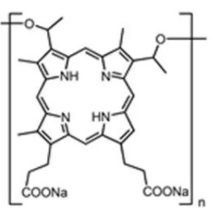
Figure 1. The molecular structure of Photofrin
The most important photosensitizer to the enhancement of PDT is the haematoporphyrin that was firstly fabricated in an impure form by Scherer in 1841.17
In the first 15 years of nineteenth century haematoporphyrin was used for many photobiological investigations, basically in order to demonstrate how it sensitized paramecia, mice18, and humans19 to light. Porphyrins can be described as a cyclic porphin based structure with a four pyrrole rings. These compounds was studied by W. Hausmann and his studies included haematoporphyrin and light in order to cure red blood cells and paramecium in 1911.18 W. Hausmann’s studies highlighted the first attempt to treat humans by using porphyrin derivatives. The German doctor Friedrich Meyer-Betz had very interesting experience after he injected haematoporphyrin to his own body. After the light interaction with this photosensitizer he reported swelling and pain.19 PDT was accelerated by Richard Lipson and his collaborators at the Mayo
Clinic in the 1960s.20-23 They synthesized more improved haematoporphyrin derivative (HPD) which is called photofrin 1 (figure 1).20 Besides Friedrich Meyer-Betz’s experiences with haematoporphyrin, these former version of HPD could be injected in small amounts, because it can easily accumulate in tumor tissues. I.
4
Diamond and coworkers reported the first in vivo studies which demonstrated the retardation of brain-tumor growth in rats for almost 20 days.24 In 1975 Thomas Dougherty and his colleagues represented so important improvement in PDT. Their studies involved injection of HPD and using red light which resulted in the extinction of tumor in mice.25 After the remarkable improvements of animal experiments in PDT, first attempt with HPD for cancer patients was effectuated by Kelly and colleagues in 1976. They used HPD for both diagnosis and treatment of cancer tissue. According to their report the bladder cancer tissue could be diagnosed in five patients. Furthermore, this sensitizer was applied to cure one patient who experienced failures of radiotherapy and chemotherapy for recurrent bladder tumor. HPD succeeded to decelerate the growth of bladder cancer and it leaded to necrosis of the tissue.26 Further studies
performed by Dougherty et al.27, O.J. Balchum and co-workers28 and J.S. McCaughan
et al.29 demonstrated that PDT had promising results in different type of cancers such
as breast30, head and neck31, pancreatic32, gynaecological33, brain tumors34 if they were treated at their early-stages.
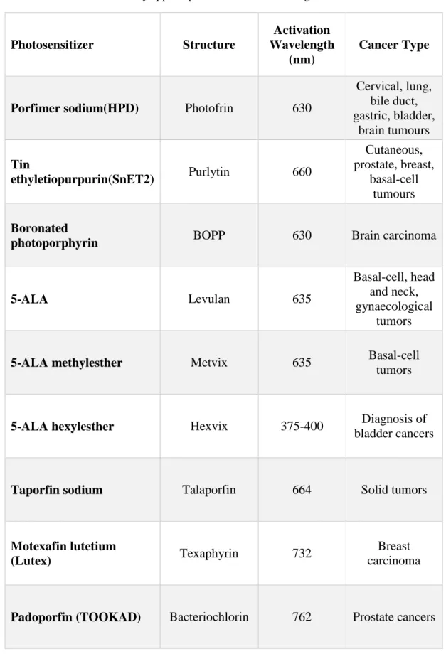
The most widely used photosensitizer, Photofrin (figure 1), was approved especially for the bladder cancer therapy first time in 1993.1 As of the synthesizing partially purified derivative of HPD (Photofrin), it was applied for early-stage lung, gastric, cervical dysplasia, oesophageal tumors and also for advanced-stage lung and oesophageal cancers in many countries. Furthermore, there are many photosensitizers which got FDA approvals (Table 1) for photodynamic therapy can be denominated as m-THPC (Foscan) for head and neck tumors, 5-ALA benzylesther (Benzvix) for gastrointestinal tumor, 5-ALA hexylesther (Hexvix) for diagnosis of bladder tumors.35
5
Table 1. Clinically applied photosensitizers for malignant diseases1, 36
Photosensitizer Structure
Activation Wavelength
(nm)
Cancer Type
Porfimer sodium(HPD) Photofrin 630
Cervical, lung, bile duct, gastric, bladder, brain tumours Tin ethyletiopurpurin(SnET2) Purlytin 660 Cutaneous, prostate, breast, basal-cell tumours Boronated
photoporphyrin BOPP 630 Brain carcinoma
5-ALA Levulan 635
Basal-cell, head and neck, gynaecological
tumors
5-ALA methylesther Metvix 635 Basal-cell
tumors
5-ALA hexylesther Hexvix 375-400 Diagnosis of
bladder cancers
Taporfin sodium Talaporfin 664 Solid tumors
Motexafin lutetium
(Lutex) Texaphyrin 732
Breast carcinoma
6
1.1.3. Major components of Photodynamic Therapy
In order to understand the PDT action extensively, it is important to note that the principle of PDT involves three fundamental components: a photosensitizer, visible light and molecular oxygen (3O2). The efficiency of therapeutic action is directly
related to the feature of photosensitizer (PS). Easy production process with low cost and good stability after manufacturing make PS an ideal agent for PDT. High absorption peak at longer wavelengths between 600-900 nm which is called therapeutic window is a critical interval for efficiency of PS. For example, 400 nm blue light can be used to illuminate superficial skin lesion, however it can only penetrate 1mm through lesion.37, 38 Furthermore, the wavelengths of incident light longer than 900 nm do not have enough energy to generate cytotoxic singlet oxygen through energy transfer. Thus, it has been considered that penetration of incoming light through tissue is quite good at this therapeutic window.39 PS should be individually
non-toxic under dark condition that implies dark toxicity. Also, it should have minimum phototoxic side effect for healthy tissues. Another toxicity related property is having low systemic toxicity which defines longer retention time in tumor tissues and rapidly excretion from the body. The exquisite photochemical reactivity which directly affects the yield of singlet oxygen is another required property of a PS. Briefly, increasing the efficiency of PS during therapeutic action and decreasing its toxicity can be managed by functionalizing sensitizer.
The second requirement for PDT is light source. In this therapeutic action light is significant component both for inducing photochemical reaction and for the efficiency of PDT. Therapeutic window (600-900 nm) is the most effective interval for the
7
penetration of light through tissue as discussed before. Thus, as blue light could not penetrate easily through tissue, red light can penetrate more deeply.39 Furthermore, longer wavelengths are not suitable to initiate the production of singlet oxygen because of their insufficient energy even if they can penetrate more deeply. There are lots of light sources for PDT depending on absorption spectrum of PS and the type of disease. The argon-pumped dye lasers and light-emitting diodes (LEDs) are most favored light sources for PDT. During the irradiation period in PDT its efficiency also depends on the total light dose, light delivery, and light exposure time.36 The last significant component of PDT is molecular oxygen (3O2). It is necessary to generate cytotoxic
singlet oxygen after irradiation of PS.
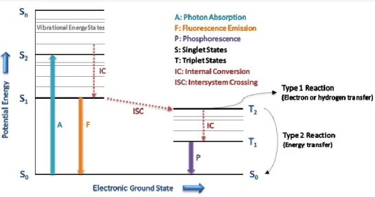
1.1.4. Mechanism of Photodynamic Therapy
In PDT action, the absorption of light by PS eventuates with the excitation of one electron from ground state of PS to a higher energy orbitals of singlet-excited state. At that point, this excited PS is very unstable and it will relax in two possible pathways. It can relax as emitting fluorescence so, it can turn back to ground state which is more stable. Alternatively, inter-system crossing (ISC) from singlet excited state to more stable triplet excited state is another most possible pathway if the photosensitizer involves transition metal complexes (Pt, Ru, Ir, Os)40 or heavy atoms (iodine, bromine) or intramolecular spin convertors (C60) without heavy atoms.41 After ISC takes place
and the excited PS transferred to the triplet state, there are also two possible reaction pathways for this excited triplet state sensitizer.Firstly, the activated PS can react with organic molecule in cell membrane so, PS transfer electron to molecule in order to
8
produce a radical. Then, unstable radicals will react with molecular oxygen (3O 2) to
form reactive singlet oxygen (1O2). In the second reaction, the excited PS can directly
transfer its energy to molecular oxygen (3O2) that results in the formation of reactive 1O
2 (figure 2).40, 42 One should note that each of reactions is molecular oxygen
dependent so that hypoxia (the deficiency of oxygen) can prevent the efficiency of singlet oxygen in tumor tissues.
Figure 2. Modified Jablonski diagram for illustrating photosensitization processes
The highly reactive singlet oxygen has very short lifetime almost 10-320 ns and its diffusion in the aqueous medium is limited around 10 nm to 55 nm.43 Photodamage with regard to the presence of reactive singlet oxygen can be carried out only if the cell and generated 1O
2 are close enough to each other. The photodynamic damage and
cytotoxicity of PDT action are multifactorial and they are dependent on the type of sensitizer and its location, the injected dose of PS, the total dose of incoming light, and the presence of molecular oxygen in tumor tissue.36 Thus, in order to increase the efficiency of PDT action all factor should be optimum coherency.
9
1.1.5.
The effects of Photodynamic therapy on tumor tissues
In PDT action the product of photochemical reaction can activate tumor destruction in three possible ways. First one is the direct tumor death due to the presence of cytotoxic singlet oxygen. PDT action can also result in tumor destruction because of the damage in vascular system. The last case involves stimulation of immune system to damage tumor cell.1 These all three results of PDT action serve for the tumor destruction and they provide long-term tumor control. The eradication of tumor cells can be occurred by direct photodamage after PDT action, but it is not the sufficient mechanism for the destruction of total tumor cells due to many reasons. One of the reason is that injected PS cannot distribute uniformly in cancer tissue.44 Additionally, another possible reason, was reported by Mladen Korbelik and co-workers in 1995, is related to the insufficient tumor-cell killing because of the increase in the distance between vascular supply and cancer cells.45, 46 The availability of molecular oxygen which can be
directly used for the photochemical reaction during PDT action is the most possible limiting parameter for cancer cell eradication.47 Thus, hypoxia and vascular damage after therapeutic action influence long-term tumor control negatively.
Further promising result of PDT for cancer treatment is damaging tumor vascular system. The existence and reproduction of tumor cells are directly dependent on blood vessels which carrying nutrients and oxygen. In 1989, vascular shutdown mechanism was performed with Photofrin-based PDT by Barbara Henderson and co-workers. They used a mouse with fibrosarcoma and therapeutic action triggered the damage in vascular system which decrease the oxygen supply to tumor cells.48 One should note
10
growth. PDT based vascular damage and inhibition of tumor growth were experienced with other photosensitizers, Photofrin, HPD, and benzoporphyrin derivative (BPD). However, these studies reported that cycloxygenase and vascular endothelial growth factor were upregulated during PDT because of the generation of singlet oxygen and hypoxia resulted in PDT.49
The last parameter is the activation of immune system to strive against tumor cells. At the end of nineteenth century, the studies about the relation between PDT action and immune system showed that the penetration of lymphocytes, macrophages, and leukocytes through tissue was capable of activating the immune response for the destruction of tumor cells.50, 51 Thus, immune system activated inflammatory process
between healthy and cancerous cells will contribute damage of PDT-activated tumor tissue. For instance, in 1996 the retardation of tumor growth due to neutrophil accumulation, which can be determined as an immune response in PDT-treated tissue, was reported by Wil de Vree and co-workers.52 The correlation between the immune system and PDT strengthen the effect of therapeutic action on tumor tissues so, PDT can be considered as a potential immune therapy.1
1.2. Gold Nanorods
The prime mover of nanotechnology, Richard P. Feynman, introduced his imaginary thoughts in his well-known talk, called as “There’s Plenty of Room at the Bottom” in 1959. It is the first time to realize the possibilities of working with a nano-level by using atoms as a building blocks. In this manner, Feynman gave the opportunity to
11
many researchers to think and to develop the new, interesting, and fascinating area “nanotechnology”. In the light of Feynman’s thoughts, E. Dexter enhanced the idea of nanotechnology in his book which was entitled as “Vehicles of creations: the arrival
of the nanotechnology era” in 1986.53 Nanoscience, considering its birthdate which is
around half century ago, is a highly promising field, and it is being used and developing in different business sectors like cosmetics, electronics, textile, medicine and many more. For the nanotechnological developments, nanoparticles have great attention due to their promising applications in optical devices, plasmon-enhanced spectroscopies, and biomedical technologies.54 The chemical and physical properties of nano-scale matters are different than bulk system and these properties are mostly defined by the free electron motions.55 In other words, the reduction of dimensions of bulk materials
in 1-100nm scale affects its physical and chemical properties owing to the restriction of electron motions. Thus, it is obvious that the free electron motions of a matter are contingent upon its size and shape. For instance, the materials with 2-10 nm scale which has smaller dimension than their Bohr radius exhibit new properties due to quantum confinement and large surface area-to-volume ratio.56 In the case of metal
nanoparticle the surface effect become considerable when its size is smaller than the electron mean free path. In accordance with these explanations, noble metal nanoparticles including gold nanoparticles have these properties. For instance, Au nanoparticles, which have the large surface area-to-volume ratio resulted in higher number of surface sites with more dangling bonds, exhibit higher degree of chemical reactivity and bonding properties.55 Furthermore, Au nanoparticles have several properties, such as nonlocal dielectric responses, confinement-induced shifts of energy levels, and enhanced optical transition probabilities owing to the electron confinement
12
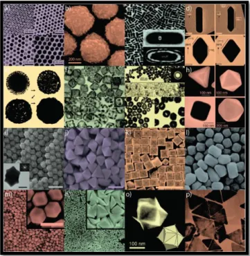
in the material.57 With regard to all of these remarkable features, gold nanoparticles
have been investigated in many research groups deeply (figure 3).
Figure 3. Gold nanoparticles with various size and shape. Copyright © 2011, Royal Society of Chemistry. Reprinted with permission from ref (62)62
Among all plasmonic nanoparticles, gold nanocrystals have a very important and remarkable property, localized surface plasmon resonance. In accordance with this property, Au nanoparticles can initiate the localized surface plasmon oscillations of the free electrons in a conduction band after the confinement of photons in their nanoscale size. Owing to the confinement of photons, Au nanoparticles have the ability to increase the amplitude of the light wave that provides the enhancement of electrical field.58 Furthermore, localized surface plasmon resonance (LSPR) properties of Au
nanoparticles can be accommodated in a desired range by tuning the shape and size of the particles in the course of chemical synthesis. Besides all other type of Au nanoparticles, gold nanorods (NRs) have an opportunity in the case of LSPR. Since, these rod-like shaped nanoparticles have two plasmon modes which are longitudinal
13
LSPR and transverse LSPR. Longitudinal one is comprised of the electron oscillations along the length axis, and transverse mode is induced by the light polarization along the transverse directions of Au nanorods. It is essential to note that; during chemical synthesis it is possible to tune the longitudinal LSPR wavelength of nanorods by changing the aspect ratios in order to cover the visible and near-IR regions.56
1.2.1. Plasmonic Properties of Gold Nanorods
The most exceptional and intriguing property of plasmonic nanoparticles is the localized surface plasmon resonances (LSPRs) that is the result of the confinement of conduction band electrons in a nano-scale as defined in a previous section. Owing to the resonant excitation, the ability to concentrate optical field in a region which is very close to the surfaces of nanoparticles enables the enhancement of electrical field, the scattering of light in higher degree, and the ability of plasmonic photothermal conversion.54 Furthermore, the geometry of nanoparticle directly affects the plasmonic properties of the nanostructure. For instance, the plasmonic properties of symmetrical and dissymmetrical metal nanoparticles are quite different. Compared to their symmetrical counterparts, rod-like shape provides nanoparticles to strengthen their plasmonic behaviours.54, 58 Regarding to the cylindrically symmetric gold nanorods, the conduction band electrons can move in two different directions that results in two distinct modes of gold NRs. In the transverse LSPRs of gold nanorods, the excitation of conduction electron oscillation along short axis exhibits a blue-shifted LSPR peaks around 500nm. However, in the longitudinal LSPRs of nanorods, the electrons move at the longer direction of gold NRs that result in the presence of longitudinal LSPR
14
peaks at red side of electromagnetic spectrum.56 Furthermore, the energy of the
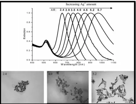
longitudinal LSPRs of gold NRs is extremely affiliated with the aspect ratios.59 So that by tailoring the ratio between the diameter and length of nanorods during chemical synthesis, it is possible to shift the maximum absorbance of longitunal LSPRs peaks from the middle of the visible region (~600 nm) to the near-IR region of electromagnetic spectrum (~1100 nm) (figure 4).56 In addition to aspect
ratio-dependence of longitudinal LSPR peaks the optical properties of gold NRs are strongly dependent on the dimensions of the particles.59 In 2006, the dimension dependency of the scattering efficiency of gold NRs was investigated via calculations by Link and co-workers.60 In pursuit of theoretical studies, the gold NRs with the same aspect ratio but different magnitude of dimensions were synthesized by Ali and collaborators in 2012. Their investigation showed that the larger gold NRs exhibited a greater extinction coefficient rather than gold NRs with small directions because of the scattering efficiency.61, 62 It should be noted that the light scattering efficiency of larger gold NRs is quite better than smaller gold NRs.As a result of this feature, gold NRs with larger dimensions can perform better in optical imaging applications owing to its better scattering efficiency and smaller gold NRs can be applied in photothermal therapies owing to its better absorption efficiency.59
15
Figure 4. Plasmonic properties of gold nanorods with their different aspect ratios. Copyright © 2009 WILEY-VCH Verlag GmbH & Co. KGaA, Weinheim. Reprinted with permission
from ref (56)56
The relaxation of absorbed energy through thermalization in gold nanoparticles will yield with the generation of heat which might lead to temperature increase on the surface of nanoparticles.54 So that, this ability of generation of heat due to the plasmon resonance decay make gold nanoparticles to be excellent contributor for photothermal therapy, drug delivery systems and controllable drug/ gene release.54, 56 By
synthetically tailoring LSPR peaks of gold nanorods into the red region of electromagnetic spectrum, it can have strong light absorption and better tissue penetration in comparison with other light absorption species, such as spherical gold nanoparticles and organic dyes.63 In this manner, gold nanorods have been investigated for photothermal conversion-based biomedical applications in recent years. For that applications the most important factor is the photothermal conversion efficiency of nanorods that involves the competition between radiative and non-radiative decays and the conversion efficiency is contingent upon the geometries and the plasmonic properties of gold nanorods.54 The study of Chen and co-workers showed that gold
16
NRs will have highest photothermal conversion in case of better proximity between the wavelength of incident light and longitudinal LSPR wavelength of nanorods.64 This condition provides the efficient excitation of gold NRs by incident laser. In addition to the dependency of plasmonic properties, geometry is another important factor for efficient photothermal conversion. According to theoretical and experimental investigations the volume of gold nanoparticles inversely affects the conversion efficiency. So that, effective photothermal conversion takes place in a small nanorods due to the reduced radiative decay that gives the ability of converting absorbed energy into heat. Furthermore, the concentration of applied gold NRs has additional influence on the photothermal conversion behaviors.54 This concentration effects was investigated by Choi and collaborators and they showed that higher concentration of nanorods resulted in better photothermal conversion.65, 66
The determination of temperature distribution and heat formation on the surface of gold nanorods for photothermal conversion-based application, such as catalysis, hyperthermia, and microfluidics is very important. Until the study of Quidant and co-workers, only theoretical studies have been made and many efforts focused on spherical nanostructures.54 In 2010 Quidant et al. presented a new technique to determine the local temperature on a single gold nanorod, thermal microscopy technique.67, 68 Their method involves the determination of heat distribution on gold nanorod by using the molecular fluorescence polarization anisotropy and they showed that the local temperature of gold nanorod distributes uniformly in all directions.
17
1.2.2. Synthesis of Gold Nanorods
The first attempt for the synthesis of colloidal gold nanoparticles by reducing gold chloride in the existence of phosphorus was reported by Michael Faraday in 1857.69 In
the light of Faraday’s study there have been many efforts in order to generate gold nanoparticles, involving nanoshells, nanorods, nanospheres, nanocubes, and other various type of nanoparticles. Thus, many methods that can be exemplified as photochemical reduction, electrochemical reduction, and chemical reduction have been achieved.56, 70 Regarding to fabricating gold nanorods with high uniformity,
quality, and yield two well-known procedures have been developed, top-down and bottom-up methods. The production of gold nanorods is obtained by combining lithography process with the deposition of gold crystals in top-down methods. However, in bottom-up method gold nanorod formation begins from the nucleation and growing of nanorods is achieved by the reduction of additional gold salts.54
The fabrication of gold nanorods by bottom-up methods involves some typical techniques, categorized as wet-chemical,71 photochemical reduction,72 electrochemical,73 solvothermal,74 and sonochemical techniques.75 In these bottom-up methods, the growth of gold nanorods is carried out by reducing gold salts within some reducing agents, consisting of ascorbic acid and sodium borohydride. Furthermore, the elongation of gold nanorods along one direction during the reduction process is achieved by using templates. The first study related to templates was reported by Masuda and co-workers. In their study, anodic aluminium oxide membranes were used as a template in several bottom-up techniques.76 Even if gold nanorods with high yield
18
efforts should be made in order to obtain pure nanorods without any residual of template.54 So that, recent studies represented the more developed version of templates. The widely used template for the growth of gold NRs is cetyltrimethylammonium bromide (CTAB) which acts as a stabilizing agent and direct the longitudinal growth of gold nanorods.77
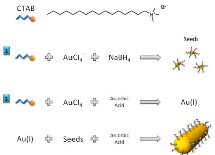
In literature, the most favored method for the fabrication of gold nanorods is the bottom-up, seed-mediated growth method was investigated by El-Sayed et al and Murphy et al. independently.78, 79 Wet chemical seed-mediated growth method enables to obtain almost monodispersed, high yield and quality, and uniform gold nanorods.54
Figure 5. Mechanism of seed-mediated growth method for preparation of gold nanorods.
As it is represented in figure 5, seed solution which involves nearly 1.5 nm small gold nanoparticles are first synthesized by reducing chloroauric acid (HAuCl4) by
additional cold sodium borohydride (NaBH4) in a CTAB solution. Afterwards, a little
19
reducing HAuCl4 supported by ascorbic acid in the existence of AgNO3 in an aqueous
CTAB solution. At that point, ascorbic acid provides the reduction of Au(III) to Au(I) and then, supplementary reduction of Au(I) to Au(0) is actuated by the addition of seed solution in order to fabricate gold nanorods. During the preparation of gold NRs the addition of AgNO3 plays an important role to obtain high yield gold nanorods that
refers to the proportion between the amount of rod-shaped nanoparticles to the total amount of nanostructures, including rods, spheres, shells, triangular plates in a solution.77, 80 So that, the presence of AgNO3 enables to control over the aspect ratio
of nanorods.81 In a detailed perspective, it is supposed that Ag+ ions are attached to the {110} faces of gold nanoparticles; hence additional gold atoms will be accumulated on the {100} faces of nanocrystals that lead to the longitudinal growth of gold nanorods.77 Consequently, seed-mediated growth method can produce nanorods with 99% yield. Furthermore, in this method it is possible to tailor the shape and size of gold nanorods while attentively arranging the growth conditions. The variable parameters for tuning size and shape of nanorods can be defined as the pH of the solution, temperature, the composition and concentration of surfactant, and the structure of the seed in the growth method.82 In addition to the size and shape tuning of gold nanorods, the aspect ratios can be diverging between 2.4 and 8.5. For example, the aspect ratio of gold NRs is directly dependent on the concentration of AgNO3,
because Ag+ ions are responsible for binding selectively to the high energy facets of nanorods. As well as many advantages of bottom-up method such as fabricating uniform and mono-dispersed nanorods with high yield, this method has also some drawbacks.54 First possible problem is the unrestrained placement of gold nanorods during the growth method due to the random behavior of Au ions during the reduction
20
and deposition processes. Secondly, even if the same growth procedure is applied, the shape and size of gold nanorods can differ in each trial.
1.2.3. Functionalization of Gold Nanorods
The stability of gold nanorods under different conditions is achieved by functionalizing CTAB-coated gold nanorods with convenient inorganic and organic additives, also the functionalization provides to obtain intented reaction sites for other modifications.58 Before introducing detailed informations regarding to functionalization of gold NRs through different methods, the influence of CTAB capping on the surface of Au nanorods should be emphasized. It is believed that the surface of nanorod is negatively charged as a result of the strong adsorption between gold surface and bromide ions. So that, the positively charged ammonium end of CTAB is capable of binding to bromide ions on the surface of gold nanorods by means of electrostatic interactions which yield with forming inner layer. The structure of CTAB molecule possesses two ends, ammonium head group and hydrophobic alkyl tails which does not like to be situated in water. Thus, another supplementary CTAB layer is formed, with hydrophobic carbon-chain end placing inside and ammonium head groups locating outside.83 Ultimately, the existence of CTAB bilayer on gold nanorods provides the positive charge on the surface of rods and better stability in aqueous medium.84 The interdigitated bilayer can be distorted as in the case of the lower CTAB concentration than the critical concentration for formation of micelle.
21
In general, for the functionalization of gold nanorods the affinity between gold and thiols is utilized. Small thiol-terminated molecules are not suitable due to the steric effect such that they are not capable to handle with the large attractive forces on rods. Thus, the reaction between small thiol molecules with gold NRs causes the aggregation of nanorods. Gold-thiol bonding chemistry mostly prefers thiol-terminated molecules with high molecular weights, such as poly(ethylene glycol)s (PEG)s to functionalize the gold NRs. Thiol-terminated molecules prefer to react with Au nanorods at the two ends that resulting in small packing density of CTAB at both ends.85 Functionalization of gold NRs with thiol-terminated PEGs yields with PEGylated gold NRs with high stability and better biocompatibility, hence the retention time of these nanorods in aqueous medium is extended. Furthermore, PEGylated nanorods are capable of disperse in aqueous and several polar organic mediums, such as acetone, alcholos, acetonitrile, dimethyl sulfoxide (DMSO), dimethylformamide (DMF), and phosphate buffered saline solutions.86 Surface modification of gold NRs can be obtained by disulfides and dithiocarbamates.87 Besides Gold-thiol bond chemistry, there are some possible interactions, such as antibody-antigen interaction, electrostatic attraction, and DNA sequence recognition to modify the surface of gold nanorods.54 For instance, the electrostatic attraction is observed in the deposition of negatively and positively charged polyelectrolytes on the positively charged surface of nanorods via a layer-by-layer deposition method.88 Moreover, biological polyelectrolytes and proteins can be used to functionalize the surface of CTAB-coated nanorods by virtue of electrostatic adsorption. Also, further modification of gold nanorods can be accomplished with functional groups, like amine groups or carboxylic acid through the layer-by-layer technique.54, 89
22
1.2.4.
Biological and Biomedical Applications of Gold Nanorods
One of the most important non-invasive therapeutic actions of metal nanoparticles is hyperthermia which depends on the photothermal conversion efficiency of nanocrystals. The irradiation of gold nanoparticles can be achieved by several items, such as ultrasound (acoustic waves), microwave irradiation, and near-infrared laser that allow local heating on cancer tissue.90 Among all other metal nanocrystals, gold NRs are used commonly as a photothermal agent owing to high photothermal conversion ability, strong light absorption in near-infrared region, which causes the laser light penetration through deeper tissues, small size, and ease functionalizing for targeted cells.54 Photothermal heating of nanorods results in the temperature increment on targeted site more than the normal body temperature (37C), hence controlled increase in the local temperature has positive effects in cancer cells such that it triggers the tumor tissue death by disrupting the functionalities of DNA and RNA, denaturation of proteins, apoptosis/necrosis.91, 92
Figure 6. The photothermal therapy of malignant cells with anti-EGFR/Au by applying Ti: sapphire laser. Copyright © 2006, American Chemical Society. Reprinted with permission
from ref (93)93
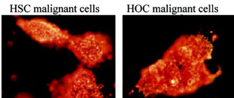
The first study for hyperthermic effect of gold nanorods was reported by El-Sayed and co-workers in 2006. The conjugated gold nanorods with anti-EGFR antibodies on
23
human oral tumor cells was irradiated with near-IR laser light(figure 6).93 So that the
study demonstrated that the theranostic effect of their designed nanorod resulted in both imaging of targeted region and hyperthermia on tumor tissue. The selective tumor region was firstly exposed by light scattering images of conjugated gold nanorods, and then it was irradiated by Ti: sapphire laser (red light at 800 nm).
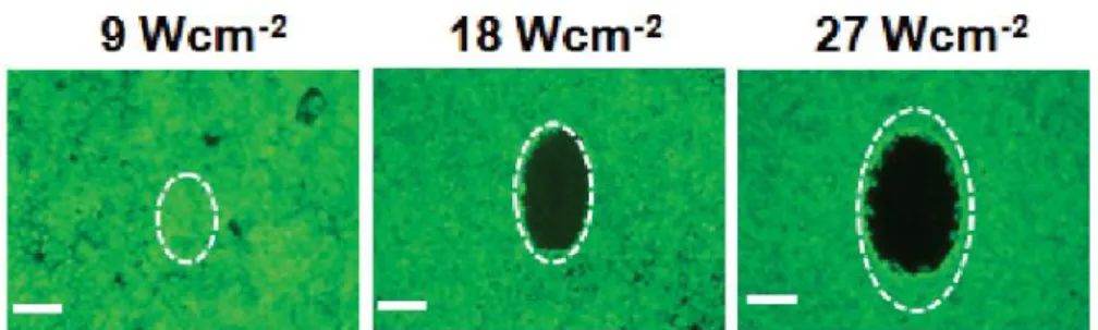
Figure 7. Photothermal therapy of A549 malignant cells with anti-EGFR/ Au by using NIR CW lasers. Copyright © 2010 WILEY-VCH Verlag GmbH & Co. KGaA, Weinheim.
Reprinted with permission from ref (94).94
In 2009 Yeh and co-workers conducted experiment on photothermal effects of PEI/PSS- modified gold nanorods which were targeted on the death of A549 malignant cells. Figure 7 represented that the power of incident light is very important to damage the cancer cells. Since, it is obvious that even if 9 W cm-2 is not sufficient for cell
death, 18 W cm-2 and 27 W cm-2 can damage the malignant cells.
The promising feature of the combination of photothermal therapy (PTT) and photodynamic therapy (PDT) was investigated by Choi and co-workers in 2011.95 In their study PDT drug (AlPcS4) and gold nanorods were combined to observe the dual
therapeutic effect of PDT and PTT at the same cancerous region. So that, the complex of gold nanorod and photosensitizer can be used both to visualize and to treat various type of malignant tissues.
24
1.3. Polycyclic Aromatic Endoperoxides
1.3.1. General Information
The well-known property of polycyclic aromatic hydrocarbons is the ability of trapping reactive singlet oxygen in order to generate endoperoxide (EPO) molecules. In this manner, some of these generated endoperoxide molecules can release either singlet oxygen or triplet oxygen in their excited states in case of heating or irradiation of light.96 In 1926 first studies related to endoperoxide compounds were carried out to investigate the dissociation process of rubrene. In the light of first investigation of endoperoxides, further studies included the dissociation behavior of 9,10-diphenylanthracene.96 Among many other anthracenes, especially the endoperoxide form of 1,4-dimethoxy-9,10-diphenylanthracene can produce singlet oxygen at room temperature.96 In 1964 Foote showed the further investigations regarding to singlet oxygen which was considered as an active species in photooxidation after irradiation of EPOs.97 Foote’s researches was improved by Rigaudy in order to demonstrate the relation between electron donating groups and dissociation temperature of EPOs. So that, Rigaudy showed that the dissociation temperature of dimethyl- and 1,4-dimethoxy- naphthalene derivatives is lower than the EPOs of 9,10-diphenylanthracene.98 Afterwards, many research groups have taken advantage of the reactive singlet oxygen in biological environments by sensitizing water-soluble endoperoxide of naphthalene derivatives.99 In the light of several researches the substitution of polycyclic aromatic hydrocarbons in different position provides the control on dissociation of endoperoxides.
25
1.3.2. Preparation of Endoperoxides
In the literature there are almost 500 endoperoxide molecules which have been derived from polycyclic aromatic hydrocarbons and most of EPOs were generated by photosensitized oxygenation. This photosensitized oxygenation was described as a [4+2] cycloaddition of singlet oxygen to aromatic hydrocarbon.100 Among all endoperoxide derivatives 3 fused benzenic compounds (anthracene) and higher members of acene compounds can easily undergo [4+2] cycloaddition reaction with
1O
2 whereas the aromatic molecules which has less than 3 fused benzenic core such as
benzene and naphthalene cannot react with singlet oxygen.96 Regarding to many studies it is obvious that the efficiency of reaction between singlet oxygen and the carbons of polycyclic aromatic compound depends on structural, steric factors and solvent effects.
The electron density of hydrocarbons directly enhances the efficiency of reaction between aromatic hydrocarbons and 1O2 owing to the electrophilic nature of 1O2. The
number of fused ring of the aromatic hydrocarbons is one of the structural effects. In literature it is well known that EPOs can be synthesized from hydrocarbons which has fused benzenic cores range from 1 to 9. The studies with different ring number of acene series demonstrated that the reactivity of the substrate towards 1O2 increases with the
number of fused ring directly. For instance, Stevens and coworkers worked with different acene molecules such as anthracene, tetracene, and pentacene. Their studies showed that the number of fused ring increased the reactivity by about 2-fold.101 Following factor which affects the reactivity of aromatic compound toward 1O
2 is
26
demonstrated the influence of electron donating groups on the reaction rate of 1O 2
addition. Regarding to their studies binding electron-releasing groups to the aromatic hydrocarbons, which will give the cycloaddition reaction with 1O2, increases the
reactivity of that hydrocarbons in the order H < C6H5 < CH3 ≤ OCH3.96
Steric factor is another significant parameter which is able to affect both the reaction side of 1O
2 and the reactivity of aromatic hydrocarbon toward 1O2. According to Erden
and collaborators investigations [2,2] paracyclophane diene can be given as an example of steric effects on endoperoxide formation. Thus, this molecule, which has 2 benzene rings, is distorted in order to undergo the cycloaddition reaction of 1O2.102
Furthermore, peri interaction in 1,8-dimethylnaphthalene also plays an important role in the reactivity of aromatic compounds.103 The presence of two methyl groups in that
molecule relaxes the steric strain in the transition state in this manner peri interaction increases the reaction rate for endoperoxide formation. The last factor that affects the reaction rate of cycloaddition of 1O2 is solvent effects. Regarding to many
investigation with different solvent molecules, it is obvious that highly polar solvents increases the reaction rate more than other solvents. In 1995 Aubry and coworkers investigated the solvent dependency of rate constant of endoperoxide formation. They showed that the rate constant of 1O
2 cycloaddition to aromatic substrate increases from
cyclohexane to water by more than 100-fold in case of water-soluble derivatives of aromatic compounds.104
In 2000 Bobrowski and collaborators performed the study of cycloaddition of singlet oxygen to benzene and this investigation revealed some differences between Diels-Alder reaction and [4+2] cycloaddtion of 1O2 to aromatic hydrocarbon.105 According
27
to their studies the suggested cycloaddition mechanism involves the single-step reaction between aromatic ring and singlet oxygen that is accomplished with the charge transfer from the organic donor to 1O2.96
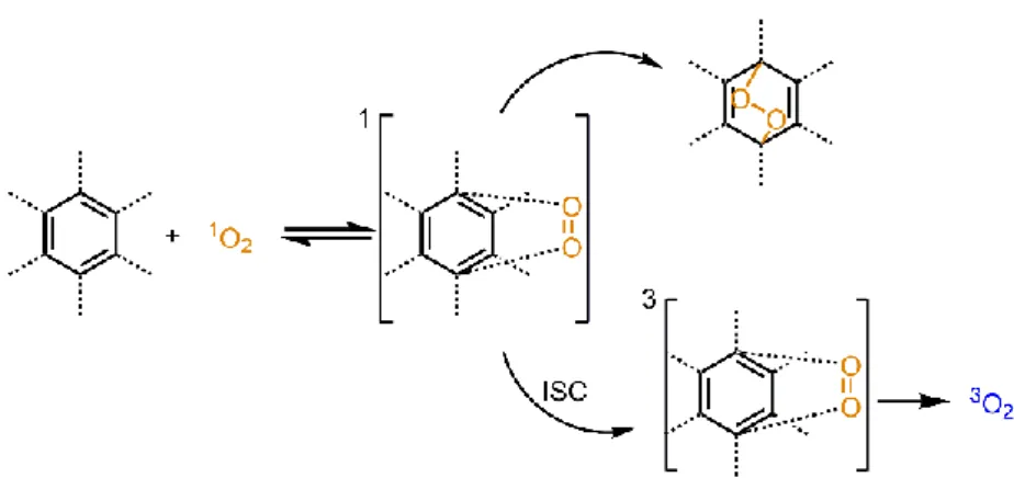
Figure 8. Mechanism of cycloaddition reaction of singlet oxygen.
The cycloaddition reaction involves mainly two steps (figure 8). In the first step reaction between aromatic ring and singlet oxygen will resulted in the reversible intermediate which is in the singlet state. Then, this intermediate can resulted in either the endoperoxide formation or dissociation into starting molecule and 3O2. Molecular
oxygen formation takes place because of spin-forbidden intersystem crossing (ISC).106
1.3.3.
Thermal Dissociation of Endoperoxides
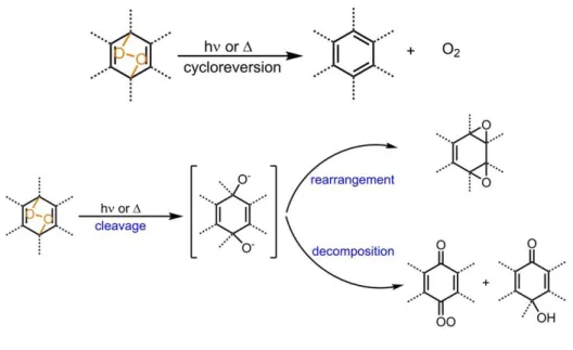
The thermal decomposition of endoperoxides results in two possible pathways; cycloreversion and homolytic cleavage. In a detailed explanation of two primary pathways: cycloreversion leads to starting compound and oxygen, in a triplet (3O2) or
a singlet state (1O
2), and homolytic cleavage of peroxide bond in EPO that leads to
28
Figure 9. Thermal dissociation of aromatic endoperoxides.
Figure 10. Thermolysis of cycloreversion pathway of aromatic endoperoxides.
After the thermal decomposition of EPOs, the competition between resulted pathways, cycloreversion and cleavage, is affected by the relative activation energies. The activation parameters for thermal decomposition of some endoperoxide molecules are listed in table 2.96 So that regarding to that table, for cycloreversion pathways the magnitude of activation enthalpy of endoperoxide increases from unsubstituted benzene to naphthalene derivatives and to substituted anthracenic derivatives of endoperoxides. Furthermore, the higher values of activation enthalpies of anthracenic
29
derivatives leads to competition between cleavage and cycloreversion. However, the aromatic substitiution at bridgehead meso positions of endoperoxide provides the more favored cycloreversion process upon heating.96
In point of thermal cycloreversion process of endoperoxides, it also divides into two possible pathways. Cycloreversion can result in either homolytic cleavage, leading to oxygen molecules in singlet and triplet state due to ISC, or concerted cycloreversion mechanism which only produces singlet oxygen (figure 10). The singlet oxygen yield was investigated by Turro and coworkers and they found that 1,4- substituted naphthalenic and anthracenic EPO derivatives generated singlet oxygen in a high yield, however 9,10- type anthracenic EPOs decompose both singlet (1O
2) and molecular
oxygen (3O2) (table 2).107
Table 2. Activation parameters for thermal decomposition of several endoperoxide molecules.
30
2. EXPERIMENTAL PROCEDURE
2.1. General
In that project, all 1H NMR and 13C NMR spectra were recorded by using Bruker DPX-400, which is operating at 100 MHz for 13C NMR and 400 MHz for 1H NMR, in CDCl3
or DMSO-d6 with tetramethylsilane (TMS) as internal reference. All characterizations
with NMR spectroscopy were performed at 25 C. Coupling constants are presented as Hz and chemical shifts were recorded in parts per million (ppm). Absorption spectrometry was acted using a Varian Cary-100. Mass spectrometry measurements were performed with Agilent Technologies 6224 TOF LC/MS and 6530 Accurate Mass Q-TOF LC/MS. TEM images were obtained by using FEI Technai G2 F30 high-resolution transmission electron microscope and carbon grid. All reactions with regarding to aromatic endoperoxide derivative were monitored by thin layer chromatography using Merck TLC Silica gel 60 F254. Silica gel column
chromatography of all products was performed over Merck Silica gel 60 with particle size: 0.040-0.063 mm, 230-400 mesh ASTM.
2.2. Singlet Oxygen Trap Experiments
Singlet oxygen experiments were performed in organic solvent by using 1,3-Diphenylisobenzofuran (DPBF) as a singlet oxygen trap molecule. First of all,
(9)-GNR conjugate and a trap molecule (DPBF) were mixed in DMSO, then several dark
31
Afterwards, (9)-GNR conjugate was irradiated at absorption maximum of GNR using laser light. Singlet oxygen generation was observed while monitoring absorbance decrease of trap molecule after the irradiation of the conjugate by incoming laser light. The instrument for irradiation of (9)-GNR conjugate was Tsunami, SpectraPhysics, USA laser system in combination with Spectrum Analyzer, SpectraSuite, Ocean Optics, USA. Excitation wavelength and power of light were adjusted to 830 nm and 1 W/ cm2 respectively. Laser irradiation was studied in Dr. Bulend Ortac research
group at UNAM, Bilkent University, Ankara, Turkey.
2.3. Synthesis of (2)
A commercial 4-formylboronic acid (1.0 g, 6.67 mmol) was dissolved in aqueous sodium hydroxide solution (5.4 mL of a 2.5 M solution). Then, the solution of 4-formylboronic acid was diluted with 40 mL distilled water. A freshly prepared solution of KMnO4 (246 mg, 1.56 mmol, 0.23 eq.) in distilled water (7.5 mL) was added two
times into the diluted 4-formylboronic acid solution at 1-hour intervals. The mixture was stirred overnight, after that ethanol (3 mL) was added and further stirring was maintained for 10 min at 50 C. After cooling the reaction to room temperature, it was
32
filtrated over celite. The filtrate was acidified to pH 2.5 by the agency of 0.5 M HCl. After all procedure, the pure white solid product was obtained with 90% yield. 1H NMR (400 MHz, DMSO-d6): H 8.30 (2H, s), 7.80 (4H, s); 13C NMR (100 MHz,
DMSO-d6): C 194.0, 167.9, 134.9, 134.5, 132.4, 128.7, 128.5, ppm. MS HRMS
(TOF-ESI): m/z calcd: 164.040; found: 164.037 [M-H]-, = 17.0 ppm.
2.4. Synthesis of (3)
25 mL MeOH was used to dissolve (2) (1.0 gr, 6.02 mmol), then 5 drops of concentrated H2SO4 was added to the solution of (2). After stirring at reflux for 1 day,
the solvent was evaporated under vacuum. Then aqueous Na2CO3 was added. The
extraction of mixture was carried out by using ethyl acetate and organic phase was washed with water. After the solvent was removed, the pure product (60%) was obtained by recrystallization with toluene. 1H NMR (400 MHz, DMSO-d6): H 8.20
(2H, s), 7.80 (4H, s), 3.75 (3H, s); 13C NMR (100 MHz, DMSO-d6): C 164.8, 132.6,
129.1, 126.3, 50.4 ppm. MS HRMS (TOF-ESI): m/z calcd: 178.056; found: 178.053 [M-H]-, = 13.2 ppm.
33
2.5. Synthesis of (5)
The Suzuki coupling reaction was performed with phenylboronic acid (0.61 g, 5mmol), 9,10-Dibromoanthracene (1.68 g, 5 mmol), Pd(PPh3)4 (0.58 mg, 0.5mmol)
and 2M Na2CO3. They were mixed and dissolved in degassed toluene (30 mL), ethanol
(10 mL) and distilled water (12 mL). The mixture was stirred for 3 hours at 100 C. The reaction mixture was extracted with ethyl acetate and the organic phase dried over anhydrous Na2SO4. The solvent of organic phase was evaporated under vacuum and
compound was purified by silica gel FCC by using hexane as an eluent. The pure white solid was acquired (35%). 1H NMR (400 MHz, CDCl3): H 8.61 (2H, d, J = 8.8 Hz),
7.71-7.62 (2H, m), 7.60-7.53 (5H, m), 7.45-7.30 (4H, m); 13C NMR (100 MHz, CDCl3): C 138.4, 137.8, 131.1, 131.0, 130.0, 128.4, 127.8, 127.7, 127.4, 126.9, 125.5
34
2.6. Synthesis of (6)
The future Suzuki coupling reaction involved (3) (80 mg, 0.44 mmol), (5) (150 mg, 0.44 mmol), Pd(PPh3)4 (61 mg, 0.044 mmol) and 2M Na2CO3. They were mixed and
dissolved in degassed toluene (6 mL), ethanol (2 mL) and distilled water (3 mL). Reaction was heated for 3 hours at 100 C. The reaction mixture was extracted with ethyl acetate and the organic phase dried over anhydrous Na2SO4. The solvent of
organic phase was evaporated under vacuum and compound was purified by silica gel FCC by using 3:1 (DCM: hexane) as an eluent. The pure white solid was acquired (53%). 1H NMR (400 MHz, CDCl3): H 8.32 (2H, d, J = 1.8 Hz), 7.78-7.73 (2H, m),
7.69-7.60 (7H, m), 7.55 (2H, d, J = 1.3 Hz), 7.39-7.32 (4H, m), 4.05 (3H, s); 13C NMR (100 MHz, CDCl3): C 167.1, 144.4, 138.9, 137.8, 135.8, 131.6, 131.5, 131.3, 129.9,
129.7, 129.5, 128.5, 127.6, 127.1, 126.5, 125.4, 125.1, 52.3 ppm. MS HRMS (TOF-ESI): m/z calcd: 388.146; found: 388.149 [M], = 6.1 ppm.
35
2.7. Synthesis of (7)
The solution of (6) (80 mg, 0.21 mmol) in 5 mL MeOH was mixed with 2M LiOH in 1 mL water. After stirring the mixture for 3 hours at 60 C, the mixture was acidified to pH 2.0. The precipitate was filtrated and dried (48%). 1H NMR (400 MHz, CDCl3):
H 8.22 (2H, d, J = 8.0 Hz), 7.71-7.62 (2H, m), 7.60-7.55 (5H, m), 7.53-7.48 (2H, m), 7.47-7.37 (6H, m); 13C NMR (100 MHz, CDCl3): C 167.8, 143.5, 138.5, 137.8, 136.1,
131.8, 131.3, 130.7, 130.1, 129.6, 129.4, 129.2, 128.3, 126.9, 126.5, 126.3, 126.0 ppm. MS HRMS (TOF-ESI): m/z calcd: 373.123; found: 373.121 [M-H]-, = 5.6 ppm.
36
(7) (50 mg, 0.13 mmol) was dissolved in co-solvent DCM-THF (10 mL-5mL) and the
solution was cooled to -78 C. A little amount of methylene blue (0.013 mmol) was added to the solution, then mixture was stirred for 3 hours under O2 atmosphere. Water
cooled 400W Hg arc lamp (white light) irradiation was carried our throughout the reaction. Solvent was evaporated in vacuum and compound was purified by performing silica gel FCC in 95:5 (DCM: MeOH) as an eluent (55%). 1H NMR (400 MHz, CDCl3): H 8.25 (2H, d, J = 1.5 Hz), 7.78 (2H, d, J = 1.7 Hz), 7.71 (2H, d, J =
1.5 Hz), 7.68-7.59 (3H, m), 7.35-7.29 (4H, m), 7.11-7.02 (4H, m); 13C NMR (100 MHz, CDCl3): C 140.1, 139.8, 139.5, 132.6, 130.1, 129.1, 128.5, 127.9, 127.5, 127.2,
123.6, 123.4 ppm. MS HRMS (TOF-ESI): m/z calcd: 407.129; found: 407.118 [M-H] -, = 25.7 ppm.
2.9. Synthesis of (9)
(8) (50 mg, 0.12 mmol), H2N-PEG-SH (292 mg, 0.086 mmol, MW: 3400 g/mol) were
mixed and dissolved in 5 mL dry THF. DCC-DMAP coupling procedure was achieved by the addition of DMAP (11mg, 0.15 mmol) and DCC (26mg, 0.12 mmol) into the solution. The mixture was stirred for 2 hours at room temperature and precipitate
37
formation was observed. Precipitate was filtered in order to remove side products and cold diethyl ether was added to the solution in order to precipitate the product. After the filtration of the final mixture, the pure white solid product was obtained (42%). 1H NMR (400 MHz, CDCl3): H 8.12 (2H, d, J = 8.2 Hz), 7.78 (2H, d, J = 8.2 Hz), 7.71-7.60 (5H, m), 7.21-7.12 (6H, m), 7.11-7.07 (2H, m), 3.78-3.49 (m, PEG), 2.90 (2H, t, J = 8.2), 1.22 (2H, s), 0.90 (1H,s). 13C NMR (100 MHz, CDCl 3): C 167.0, 157.2, 140.9, 140.4, 140.1, 139.9, 136.0, 134.5, 132.9, 128.3, 127.7, 127.3, 127.2, 123.5, 123.2, 70.4, 70.3, 70.2, 70.0, 69.9, 69.5, 66.9, 38.5, 33.8, 25.6, 24.9 ppm.
2.10. Synthesis of Gold Nanorod
2.10.1. Preparation of Seed Solution
CTAB solution (5.0 mL, 0.20 M) was mixed with HAuCl4 solution (5.0 mL, 0.00050
M) and the mixture was gently stirred. Then, ice-cold NaBH4 (0.60 mL, 0.010 M) was
added that yielded with brownish yellow solution. The mixture was vigorously stirred for 2 min and it was stored at room temperature.
2.10.2. Growth of Gold Nanorods at longitunal LSPR Band
After the preparation of seed solution, CTAB solution (5.0 mL, 0.20 M) was added to AgNO3 solution (0.20 mL, 0.0040 M) at 25 C. Then, further addition involved
38
(70.0 µL, 0.0788 M) was added and this addition changed the color of solution from dark yellow to colorless. Final addition contained 12.0 mL of the seed solution to the growth solution at 27-30 C. During the reaction proceeded, the color change of the solution was observed within 10-20 min. The color of the solution was purple as expected. The temperature of the growth medium was kept at 27-30 C for 12 hours. The solution was centrifuged at 6000 rpm for 10 min so as to remove excess CTAB solution and the solution dispersed in distilled water. With this seed-mediated growth method, 1.8E+10 nps/mL, 0.060 mg/mL GNRs were obtained.
2.10.3. Decorating of Gold Nanorods with EPOs
In order to acquire effective conjugation between GNRs and EPOs, GNR solution was centrifuged at 14000 rpm for further removal of CTAB. After all excess CTAB solution was removed, equi-volume solutions of (9) and GNR (O.D1.0, 0.25 nM) were mixed. The mixture was stirred vigorously and sonicated for 1 min. Then, the reaction was left to react for 2 hours. The unreacted (9) was removed by centrifugation at 7000 rpm for 10 min and (9)-GNR was dispersed in buffer or DMSO.