INSTITUTE OF SCIENCE
THE EFFECT OF CURCUMIN ON P53 /TIGAR AND AUTOPHAGY
PATHWAY IN HUMAN OSTEOSARCOMA U2OS CELL LINE
MASTER THESIS OTHMAN RASW ABDALLA
BIOLOGY
THESIS SUPERVISOR Assist. Prof. Dr. CAN ALI AĞCA
ii
I want t offer this endeavor to ALLAH Almighty to give me ability, knowledge, strength and opportunity to undertake this research study. For their support and helping during my study I sincerely thank the following in particular.
I should express my deep thanks and gratitude to my supervisor Assist. Prof. Dr. Can Ali AĞCA for their help, continuous support and excellent guidance during entire my study, I want to say Allah razı olsun.
I express my sincere thanks to my Prof. Dr. Victor NEDVZVETSKI, who contributed his time within the lab and helping me to understand and appreciate the techniques used.
I owe my special thanks to my principal Assoc. Prof. Dr. Artem TIKHOMIROV for motivating and support me to pursue.
I express my warm thanks to dear Aryan Mahmood FARAJ for his help and support during my study. I wish to thanks Yusuf TOY for his help in the laboratory.
I also would like to refer the BUBAP project office of Bingol University for supporting my thesis (project no: BAP-FEF.2017.00.005).
Last but not least, I sincerely thank to my partner Sanaa for his personal support and infinite patience at all times. This thesis is a small tribute to my mother, brother, and sister, I should also express my sincere thanks to my family-in-law has given me their unequivocal support.
Othman Rasw ABDALLA Bingol 2018
iii
PREFACE ... ii
CONTENTS ... iii
LIST OF ABBREVIATIONS ... v
LIST OF FIGURES ... vi
LIST OF TABLES... vii
ÖZET ... viii
ABSTRACT ... ix
1.INTRODUCTION ... 1
2.LITERATURE REVIEW ... 5
2.1.Osteosarcoma U2OS cell ... 5
2.2.The P53 roles in cancer... 5
2.3.TP53-induced glycolysis regulator (TIGAR) ... 7
2.3.1.The role of TIGAR in program cell death ... 7
2.4.P53/TIGAR axis ... 8
2.5.Curcumin ... 9
2.5.1.Curcumin and cancer ... 10
2.5.2.The role of curcumin on autophagy in cancer ... 11
3.1.Cell culture ... 13
3.1.1.Cell Thawing ... 13
3.1.2.Cell splitting ... 14
3.2.Cell proliferation assay (MTT assay) ... 15
3.3.Immunocytochemistry ... 16
3.4.Western blotting assay ... 17
iv 3.4.4.Protein transfer ... 19 3.4.5.Membrane blocking ... 20 3.4.6.Antibody ... 20 3.4.7.Observation ... 21 3.4.8.Statistical analysis ... 21 4.RESULT ... 22
4.1.Curcumin inhibited Osteosarcoma U2OS cell proliferation ... 22
4.2.Curcumin modulated both p53 marker and TIGAR protein expression in human osteosarcoma U2OS line. ... 25
4.3. Curcumin modulated both Beclin-1 and LC3 protein expression in human osteosarcoma U2OS cell line………. 28
5.DISCUSSION ... 34
6. CONCLUSION……….. 39
REFERENCES ... .. 40
v
LIST OF ABBREVIATIONS
ROS : Reactive oxygen species
kDa : Kilodalton
PFK2 : Phosphofructokinase 2
SCO : Synthesis Cytochrome C
TEM : Transmission electron microscopy
FBS : Fetal bovine serum
PBS : Phosphate buffered saline
EDTA : Ethylenediaminetetracetic acid
PFA : Paraformaldehyde SDS : Sodium dodecyl sulfate
PAG : Polyacrylamide gel
DPBS : Dulbecco's Phosphate-Buffered Saline
TEMED : Tetramethylethylenediamine
PVDF : polyvinylidene difluoride
TBS : Tris-buffered saline
BSA : Bis(trimethylsilyl)acetamide
ECL : Electrogenerated chemiluminescence
PCD : Program cell death
DAPI : Diamidino-2-phenylindole
DEMEM : Dulbecco's Modified Eagle's medium
vi
LIST OF FIGURES
Figure 2.5. Structure of curcumin... 10 Figure 4.1. Curcumin inhibited cell proliferation osteosarcoma U2OS cell line……. 23 Figure 4.2. Curcumin decreased cell viability of osteosarcoma U2OS cell line…….. 24 Figure 4.3. Confocal laser scanning microscope pictures of control and treated
osteosarcoma U2OS cell line with curcumin. ... 25 Figure 4.4. Curcumin inhibits both P53 and TIGAR expression in U2OS cell line…. 28 Figure 4.5. Curcumin modulated both Beclin 1 and LC3 expression in U2OS cell…. 29 Figure 4.6. Curcumin induced LC3 protein expression in U2OS cells line. ... 33
vii
LIST OF TABLES
Table 3.1. Primary antibodies used in immunoblotting and immuno-cytochemistry assay…..……….………... 21 Table 3.2. Secondary antibodies used in immunoblotting and
viii
İNSAN OSTEOSARCOM U2OS HÜCRESINDE CURCUMIN'in P53 /
TİGAR VE OTOPHAJI UZURINE ETKISI
ÖZET
Osteosarkom, kemiklerde en sık rastlanan primer tümörlerdir ve genellikle tibia, femur, humerus gibi uzun boru şekilli kemiklerin metafizi çevresinde ortaya çıkar. Curcumin,
Curcuma longa L'nin kurutulmuş toprak rizomlarındaki en önemli etkiye sahip
bileşendir. Yara iyileşmesini destekler, karaciğer hasarına, katarakt oluşumuna ve pulmoner toksisiteye karşı koruma sağlar. Curcumin (zerdeçal) geleneksel Çin tıbbında binlerce yıldır kullanılmaktadır. Anti-bakteriyel, inflamatuvar, oksidan ve anti-kanser özellikleri de dahil olmak üzere birçok farmakolojik etkiue sahip olduğu bildirilmiştir. Curcumin’in, hücre invazyonunu engellemek, migrasyonu ve inflamatuar yanıtları bastırmak, apoptoz veya otofajiyi tetiklemek birçok etkisinin olduğu kanıtlanmıştır. Kansere karşı curcumin etkilerinin kapsamlı bir şekilde araştırılmış olmasına rağmen curcumin'in p53 / TIGAR ve otofajik yolak üzerindeki etkisi U2OS hücre hattında henüz ele alınmamıştır. Bu nedenle insan osteosarkom U2OS hücre hattında curcuminin p53 / TIGAR ve otofaji yolu üzerindeki etkisininin araştırılması amaçlanmıştır. Osteosarkom U2OS hücreleri 24 saat süreyle farklı konsantrasyonlarda (10, 15, 20 ve 25 μM) curcumin ile tedavi edildi. Hücre çoğalması hücre canlılık testi olan MTT ile saptanmıştır. Bu proteinlerin (p53, TIGAR, Beclin-1 ve LC3) ekspresyon seviyeleri, western blotting ile analiz edilmiştir. MTT analizinin sonuçları, curcumin'in osteosarkom hücre proliferasyonunun ve hücre canlılığının inhibisyonuna neden olduğunu göstermiştir. Curcumin aralığı 15-25 μM doz ve p53, TIGAR, Beclin1 ve LC3 modülasyonu için doza / bağımlı doğrusal bir etkisinin olduğu belirlenmiştir. Osteosarkom U2OS hücre hattı 15-25 uM curcumin ile tedavi edildiğinde p53 ve TIGAR protein seviyelerinde bir düşüş olduğu ortaya konmuştur. Beclin-1'in curcumin tarafından inhibe edilmesi otofajiye spesifik belirteç olan mikrotübüle bağlı protein LC3’nin artmasına yol açmaktadır. Immünositokimya sonucunda curcumin dozunun artmasıyla total LC3 proteininin ekspresyonunun belirgin şekilde arttığı gösterilmiştir. Bu bulgular, curcumin'in antikanser özelliklerinin altında yatan mekanizmanın anlaşılmasını geliştirmiştir.
ix
THE EFFECT OF CURCUMIN ON P53 /TIGAR AND AUTOPHAGY
PATHWAY IN HUMAN OSTEOSARCOMA U2OS CELL LINE
ABSTRACT
Osteosarcoma is a cancer disease from primitive bone tumor initiation and evolution of tubular long bones include femur, humerus and tibia. Osteosarcoma disease is defined and diagnosed throughout the inspection of tumor osteoblast cells that produce osteoid. Turmeric (Curcuma longa) is the most effective component in dried ground rhizomes of a member Zingiberaceae family. Curcumin have excellent free radical scavenging properties, however curcumin is almost the strongest antioxidant present in turmeric. Turmeric seasonal herbaceous plant with long lateral ramifications originally comes from Southeast Asia. Curcumin properties include anticancer, bacterial, anti-inflammatory and antioxidant. Curcumin as chemical therapeutic agent demonstrated to suppress or inhibit various signaling pathways includes arrest tumor cell proliferation, inducing autophagy or apoptosis, inhibit malignant cell invasion and suppressing inflammatory responses. Despite the fact that effects of curcumin in cancer has been broadly reported, but the weather of curcumin on p53/TIGAR and autophagy has not been addressed in osteosarcoma U2OS cell line. In this present study, we screened the weather of curcumin after treated U2OS cells with curcumin, cultured cell were treated by various concentrations (10, 15, 20 and 25) µMolars curcumin intended for 24h. After cultured and treatment cells proliferation and viability were detected by MTT assay. The expression levels of these proteins (p53, TIGAR, Beclin-1 and LC3) were analyzed by immunoblotting assay and the expression of LC3 protein autophagy marker were analyzed by immunocytochemistry assay. The MTT assay results demonstrated that curcumin lead to distinct suppression of osteosarcoma cell proliferation and cell viability. There were observed a linear dose/depends effect for curcumin range 15-25 µM dose cause to modulate of p53, TIGAR, Beclin1 and LC3. Analysed protein reveals a relation reduction in the levels of p53 and TIGAR proteins above treatment with 10–25 µM curcumin. The result of immunocytochemistry showed that by increasing curcumin dose significantly increased the expression of the total amount LC3 protein in cytoplasm osteosarcoma U2OS cells. Our studied has improved the perceptive of the fundamental technique anticancer properties of curcumin.
1. INTRODUCTION
Cancer is a band of disease that is controlled generally by way of a chemotherapeutic chemical which are harmful to normal cells and tumor cells (Ravindran et al. 2009). Cancer cells vary from normal cells by six different characters includes evading of apoptosis, metastasis, unlimited replicative potential, able to invade to the neighbor tissues, sustained angiogenesis and insensitivity to anti-growth signals (Weinberg and Hanhan 2000). The cells grow and divide under controlled condition, but sometimes the control is lost and the cells grow and divide uncontrolled and form a tumor may be benign (non-cancerous) or malignant (cancerous) (Schneider 2001). The growth factor of a signaling pathway in cancer cell based on three most important cellular strategies includes changes in trans-cellular signals, changes extracellular growth signals and signaling of messengers. In the other hand, many cancer cells depend on autocrine stimulation, over expression of growth factor receptors induced more intracellular receptors than extracellular growth receptors (Hejmadi 2010). Bone cancer is a kind of malignant that is able to affect each bone in the body. Ther are two types, primary and secondary bone cancer that are arises from the cells that formulate up the skeleton of the body. Bone cancers can also occur when tumors start in other organs, such as lung, prostate, and breasts metastasize (spread) to the bone (Buijs and Plujim 2009).
Osteosarcoma also termed (osteogenic sarcoma) is the most wide-ranging primary malignancy of skeletal sarcoma and infrequently in the soft tissues, identified by spindle cells of mesenchymal source depositing young osteoid matrix and expansion of osteoid or immature bone tissue through the cancer cells (Durfee et al. 2016; Lamoureux et al. 2007). The infection age mostly presented among 10-25 years age, and it is the mainly common factors of malignant-associated deaths in childhood and babyhood (Swan et al. 2004). Although, osteosarcoma is the general primary tumor bone in children, proportion is bimodally distributed across age (Durfee et al. 2016). The occurrence of (OS)
obviously elevated in males compare with females and slightly higher occurrence in Blacks and Hispanics than Caucasians (Savage et al. 2004).
Program cell death (PCD) occurs under physiological condition as a result cell death concerned in the selective removal of unwanted cell (Elis et al. 1991). Apoptosis is a process of defend system for the discriminating elimination of harmful, injured, aging, dysfunctional and unwanted cells (Abe et al. 2000). It is commonly recognized by distinct morphological characteristics and fundamental component of a lot of ordinary physiological action such as, immune response, embryogenesis and the growth of normal tissue (Korsmeyer and Vau 1999). Therefore, control of apoptosis is important for tissue homeostasis which cause to a deregulation selection of pathological situation as well as chemoresistance and carcinogenesis (El-Deiry and Burns 2003).
Apoptosis is derivative from the Greek word for regulated and concerned in regulation of numerous pathological and physiological stimulation processes. Throughout development process, apoptosis removed structures that are no longer desired. During life, cells that are potentially harmful or useless such as, infected, aged, mutated or injured cells eliminates by apoptosis mechanisms. Apoptosis deregulation lead to cancer (Jin and El-Deiry 2005). Algeciras-Schimnich et al (2002) demonstrated that apoptosis induced generally during the stimulation of definite (enzymes) proteases known caspases. Caspases are cleaving multiple substrates and effectors of cell suicide, most important to morphological and biochemical alters which are properties of apoptotic cell mechanism (Abe et al. 2000). Kumar et al (2010) are confirmed three kinds of biochemical changes which occur in apoptosis, activation of caspases-2, a breakdown of proteins DNA and membrane alter and identification by phagosytic cells. These morphological changes end up of the construction of apoptotic bodies (ApoBDs) which are usually degradated by phagocytes cell after fusion with lysosome known phagocytosis process (Geske and Gerschenson 2001).
A second line of programmed cell death is autophagy (Gozuacik and Kimchi 2004). Vojo in (2014) was considered that the autophagy induction through definite developmental states in reply to different hormones by deprivation of nutrient or by the extra type of stress. The induction process start with sequential steps bulk cytoplasm termed
(autophagosome) within a cytosolic double-membrane vesicles, fuses with lysosomal degradation, then delivered into internal single membrane vesicle into the lysosome lumen known an autophagic body. Autophagy, dissimilar of apoptosis, has a homeostatic function as a degradation method through which cellular components are turnover. During stress or starvation, such as malnourishment, autophagy starts to engulfed, digested and turnover of cellular proteins, cytoplasm and organelles in to metabolism and homeostasis (Bialik and Kimchi 2008). Normal cells have the autophagic capability to remove organelles and chemical toxin, the degradation of mitochondria due to oxidative stress and radiation, subsequently defensive cellular against DNA damage from reducing the incidence of reactive oxygen species, cell malignant transformation and hereditary stability, therefore autophagy is a key to maintaining genome stability (Mathew et al. 2007). Ouyang et al (2012) reported that autophagy can participate a pro-tumour function by regulating a number of pathway such as Beclin-1, Bcl2, PI3K class III and I, mTORC1/2 and p53.
Generally, autophagy has two roles for cancer cells, the first role is anti-metastatic roles, during the early stages of metastasis, and autophagy inhibits the metastasis by restricting tumour necrosis and oncogene-induced senescence. The second role is pro-metastatic roles by make easy the self-alteration of pro-metastatic cells (Su et al 2015). Aita et al (1999) demonstrated that autophagy-associated genes LC3 and Beclin-l can abolish tumor growth by induction of autophagy process. Since, LC3 and Beclin-l concerned a prospective beneficial marker in malignancy managing. It was described that monoallelically deleted Beclin-1 gene were reported over 40-75% human cancer of the breast, ovary and prostate. Microtubule-associated protein (MAP1A and MAP1B), light chain 3 (LC3) is a mammalian homolog of yeast Atg8 and a main ingredient of autophagosome. Immunoblotting of LC3 typically reveals two bands (LC3-I, 18 kDa) located in the cytosol and (LC3-II, 16 kDa), located in autophagosomal membranes. In addition, Beclin-1 autophagy related gene is the mammalian orthologue of yeast Atg6, (Jackson et al. 1999). Kihara et al (2001) refer to Beclin-1 (autophagy related gene 6) well-established center autophagy and other membrane-trafficking actions, localizes to the trans-Golgi apparatus, including the class 3 phosphatidylinositol 3-kinase complex (PI3K-III), plays a key role in membrane trafficking and complex reformation of endocytosis, cytokinesis and autophagosome.
The P53 gene is a fundamental of tumor suppressor (Vogelstein 2000; Vousden and Prives 2009). The p53 protein acts in reply to different cellular stress to adjust target genes that influence cell cycle arrest, cell differentiation, cell senescence, cell death, DNA repair and neovascularization. The p53 protein activates after DNA damage and induces gene transcription such as p21Waf1/Cip1 that function as controllers of cell cycle progression at G1 phase (Kita et al. 2011). Palikaras et al (2013) reported the role of p53 as apoptosis promoter; p53 activated under a broad range of stress conditions like DNA damage, hypoxia, cellular senescence, encourage cell cycle checkpoint by abnormal oncogene expression, apoptosis and DNA repair. The TIGAR protein display a resemblance to the bisphosphatase domain of (PFK-2/FBPase-2) an enzyme that play an important role in promotes respiration and inhibit of glycolysis (Bensaad et al. 2006; Li et al. 2009). TIGAR is a different protein which applies a negative contact on autophagy by decreasing intracellular (ROS) levels (Cheung et al. 2009). Selak et al (2006) considered that TIGAR is a p53-induced protein that drives a metabolic alters from glycolysis in to the pentose phosphate pathway (PPP) which causes the regeneration of NADPH. Curcumin is a golden-yellow color, hydrophobic (insoluble in water) polyphenol compound, originated from turmeric root of the plant Curcuma longa L. The anti-cancer beneficial of turmeric has been well demonstrated in a many of human cancers, includes melanoma, breast and prostate cancer (Burgos-Moron et al. 2010).
The purpose of this research is the effect of curcumin on p53/TIGAR pathway and autophagy on human osteosarcoma U2OS cells. However, the role of Curcumin in apoptosis has been described, but the potential regulation of p53/TIGAR pathway and autophagy has not been addressed.
2. LITERATURE REVIEW
2.1. Osteosarcoma U2OS Cell
The human cancer U2OS cell was originated in 1964 as a slightly differentiated mesenchymal connective tissue sarcoma of the tibia of girl fifteen years old. It was regarded one of the first developed cell lines and is utilized more commonly. Spectral cytogenetic analysis and karyotyping analysis has explained structural rearrangements, alterations and high incidence of aneuploidy and chromosomal instability (Bayani et al. 2003). Spectral analysis indicated the near-triploidy state of U2OS cells which appeared more frequently to be arrangement of chromosomal damage and tetraploidization. Other rearrangements mainly involved chromosomes 20 and 8, from simple translocations to highly complex rearrangements, but also to a less extent chromosome 17. Most of the centromeric breakpoints occurred on chromosomes 6, 13 and 17, but at least four on chromosomes 2, 4, 5, 8, 19 and 21. Two tumor suppressive genes pRb and p53 are play important function in U2OS cells, while in other, more combative, osteosarcoma cell lines such as SAOS2 these two genes are mutated (Wesierska-Gadek 2005; Schmid 2005; Isfort et al. 1995). Allied with other mesenchymal osteosarcoma cell lines, the U2OS cells have the minimum point of chromosomal alteration and no more than 2% of cells posses multipolar mitoses, similar to normal control fibroblasts, probably due to functional pRb and p53 (Isfort et al. 1995). The osteosarcoma U2OS cell line is widely utilized to day includes biochemistry, biomedical studies, molecular biology, bone formation and arthritis (Bartkova et al. 2006).
2.2. The P53 Roles in Cancer
The p53 genes cellular tumor antigen p53 encoded by a p53 gene at chromosome band 17q13.1. The p53 identified a cellular gatekeeper and guardian of the cellular genome
That composed 11 exons the first one is non-coding gene. The tumor protein p53 is the mainly mutated tumor inhibitor gene in human cancer. In respond to different stresses include oxidative stress, overexcited proliferative signals, DNA damage, oncogenic activation, ionizing radiation, nutrient deprivation, hypoxia, neovascularization and other mode of stress, the p53 gene start to activates and then roles as a transcription factor to modulate various biological action such as cellular senescence, transient cell cycle arrest, apoptosis, differentiation, metabolism of cell, at the same time regulation of autophagy (Bieging et al. 2014). Hollstein et al (1999) investigate that mutation of p53 lead to the progress of most malignant cell since p53 preserve security of cell by protecting genome from mutation. The p53 gene inactivation can progress the capability of cells to stay alive metabolic stress, by an increase level of baseline autophagy (Tasdemir 2007; Strachan and Read 1999.) It is necessary to stimulation of autophagy by p53 gene inactivation (deletion, inhibition or, depletion) can activate autophagy which are measured as (GFP-LC3) puncta is a cell cycle-dependent experience that arise in the S phases and G1 phase excluding the M and G2 phases The transcription factor regarded most important roles in p53 gene. Vogelstein et al (2000) studied the induction stress make activate of p53 which cause to the stimulation of expression of a large amount of p53 target genes. Villunger et al. (2003) and Jeffers et al (2003) demonstrated that the expression of the BH3-domain protein PUMA play cell type-specific roles in p53-activated apoptotic or differentiation in many types of cells. Martindale and Holbrook (2002) demonstrated that one more significant role of p53 gene that identified the live or death of cells in the controlling of intracellular (ROS) levels. The p53 gene can induce several genes that outcome in up regulation of reactive oxygen spices generation, which cause to apoptosis (Polyak et al. 1997; Johnson et al. 1996; Li et al. 1999). Lander and Weinberg showed that during defective p53 tumor suppressor protein and a few of the DNA repair genes cause to inherited predispositions in the growth of malignant. The p53 protein has a vital role in cell cycle arrest after DNA damage by activating p21, start to DNA repair and induce apoptosis by overexpression of Bax (Gomez-manzano et al. 1997). Jin , Levine (2001) and Vogelstein et al (2000) considered that nearly 31 genes has been discovered to be controlled or regulated by p53 gene and to have a p53 RES that binds the p53 tumor protein. The central role of p53 that is starting to come out it is an ability to regulate the stability among oxidative phosphorylation and glycolysis. ATP and ADP can directly alter p53 action; ATP is inhibiting the capability of p53 tumor gene to bind DNA and
with ADP promoting (Okorokov and Milner 1999). The mammalian cells have two types of p53, nucleus and cytoplasmic p53. Nucleus p53 is autophagic inducer by interacting with its objectives damage-regulated autophagy modulator (DRAM) and sestrin1/2. The DRAM is p53 target gene induces macroautophagy and trigger autophagy under p53 control. The sestrin1/2 is another target of DRAM, negative regulator of mTOR1 by activating AMPK and cytoplasmic p53 described as an autophagic inhibitor (Ouyang et al. 2012), in the other hand autophagy promotes by AMPK protein (Shojaei et al 2014).
2.3. TP53-induced Glycolysis Regulator (TIGAR)
The genomic TIGAR location on chromosome 12p13-3 and contains two possible p53 binding sites and six possible coding exons, one inside the first intron and the other upstream of the first exon (Bensad et al. 2006). Bensaad et al (2009) reported that TIGAR protein shows resemblance to the functions as a Fructose-bisphosphatase (Fru-2,6-BPase) an enzyme that has a vital role in glycolysis and apoptosis regulator. TIGAR also functions an overall decrease level of (ROS) by elevating NADPH production. The ROS-adaptive effect can act a crucial role in preserving cells against cytotoxic property of anticancer promoter (Maddalena et al. 2009; Moellering et al. 2011) and elevated intracellular glutathione (GSH) concentrations of have been concerned against too many chemotherapeutic agents (Bensaad et al . 2006). The Warburg effect theory in (1920) describes that malignant cells preferentially uses the metabolize glucose by glycolysis pathway to generate ATP despite being the existence of oxygen, therefore the capability of TIGAR protein to suppress glycolysis suggest expected danger for malignant cell continuity (Courtnay et al 2015).
2.3.1. The Role of TIGAR in Program Cell Death
Bensaad et al (2006) and Jen et al (2005) studied that TIGAR protein is apoptosis regulator and TP53-induced glycolysis was identified during microarray investigation of gene expression subsequent the activation of p53 tumor protein .Colleagues and Bensaad showed the TIGAR gene save cells from reactive oxygen spices (ROS) related apoptosis (Bensaad et al. 2006). It may be the reason why the malignant cells necessitate to high amount of TIGAR for continuity. Furthermore, TIGAR-mediated ROS reduction is able
to restriction autophagy action. Considering autophagy is able to function to decrease the amount of reactive oxygen levels, sustain energy production throughout starvation or metabolic stress situation and inhibit apoptosis (Bensaad et al. 2009). Wanka et al (2012) has recorded that TIGAR expression level was radically goes up in human malignant such as, colorectal cancers, glioblastoma and invasive breast cancers. Li and Jogl (2009) recorded that the TIGAR has been displayed to function to hydrolyze fructose 2,6bisphosphate and fructose 1,6bisphosphate. These two mechanisms actions cause to the similar effects on glycolysis process. Downregulation of glycolytic level, which in various cells be reported to pro-apoptotic and expression of TIGAR, as a result reduce levels of fructose 2,6bisphosphate and fructose 1,6 bisphosphate. One importance role of TIGAR is elevated NADPH production that supply to the scavenging of (ROS) through decreasing of glutathione level (Bensaad et al. 2006).
2.4. P53/TIGAR Axis
Crighton et al and Bensaad et al in (2006) were investigated that in reply to cellular stresses like DNA destruction or oncogene activation, cause to mutate tumor suppressor protein p53. Wild-typep53 stimulates the expression of target genes and becomes stabilized. These target genes make a different of cellular responses to stress counting cell-cycle arrest, apoptosis, DNA repair, and senescence metabolism angiogenesis, differentiation and autophagy. The tumor protein P53 play an essential function in the regulation of cellular metabolism action, especially oxidative phosphorylation and glycolysis through transcriptional control of its downstream genes by synthesis of cytochrome c oxidase release SCO2 and TP53-activated glycolysis regulator (TIGAR) (Jones et al. 2009; Kang et al. 2006). Zhang et al (2010) has been branded that malignant cells use breakdown of glucose by glycolysis process that produce less ATP and is able to happen in hypoxic tissues that cannot gain enough ATP by oxidation and phosphorylation. The roles of TP53 gene by the regulation of energy metabolism through SCO2 and TIGAR implement recent observation into the puzzles for metabolism of malignant cell and approach for cancer treatment. TIGAR was originally recognized as a transcriptional target of tumor protein p53 (Bensaad et al. 2006). TIGAR considered to play an important function in tumour inhibition and the antioxidant function of TIGAR would be dependable with a role in protecting from the addition of injure. The TIGAR
expression has been organized to be prominent in a numeral of tumour types and malignant models (Lee et al.2015). Throughout process independent on the preservation of wild-type p53. Furthermore, in human breast cancer the expression of TIGAR was found vice versa corresponded to the amount of p53 tumor protein.13 Overall, TIGAR can role in a tumour inhibitor as part of a p53 response, however be able to supply to cancer development after TIGAR expression is uncoupled and deregulated from p53 (Lee et al.2015).
2.5. Curcumin
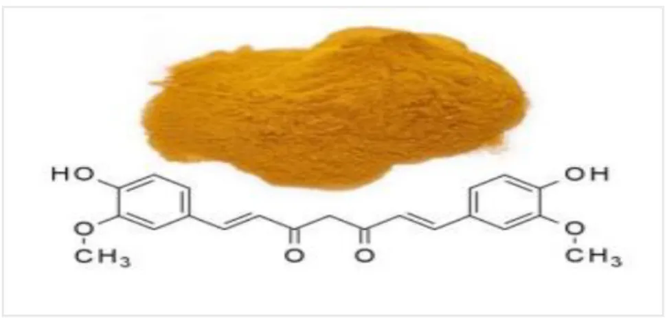
Curcumin is known scientifically as or Curcuma Longa the major effective component originated from the rhizome of the herbaceous plant and has a long time of consumption in Chinese and Ayurvedic medicine as a handling for provocative situation. The common name of curcumin is (turmeric) belong to ginger family of Zingiberaceae curcuma longa its perennial plant and is cultivated in India and centuries in East Asian countries (Jurenka 2009; Ammon et al. 1991). The earlier research reported that curcumin has been associated many pharmacological actions including, cardio-protection, diabetic, anti-inflammation, Neuro-protection and antimicrobial properties (Robert et al. 2014; Aggarwal 2010; May et al. 2014; Nishiyama et al. 2005). Curcumin played a potential function in inhibiting metastasis and malignant tumor cell production (Chen et al. 20016). Odot et al 2004 has reported that curcumin ranked as a third generation of anti-tumor and Chemopreventive agent in National Cancer Institute of United States. Until now there are no researches reported in humans or animals have important toxicity associated to curcumin, except very high doses (Jurenka et al. 2008; Goel et al 2008). Curcumin permanent plan has many components, such as curcuminoids, the main active component for medicinal use. Curcuminoids give the golden yellow-color to curcumin, consist of (curcumin I) diferuloylmethane, (curcumin II) demethoxycurcumin, bisdemethoxycurcumin (curcumin III) (figure 2.1). (Hanai et al 2009; Goel et al. 2008). The predominant compound turmeric is curcumin I (77%),is an important bioactive compound, curcumin II and curcumin III (~17% and ~3%) respectively (Goel et al. 2008; Changtam et al. 2010). Curcumin is the predominant curcuminoid that composed about 2-6% of the turmeric spice (Shishodia et al. 2005).
Figure 2.5. Structure of Curcumin, golden yellow pigment curcuminoid is the predominant compound curcumin I (Curcuma longa L) MW: 368.37, MF: C21H20O6. (Changtam et al. 2010) Wikipedia/Ronhjones and Brovi PL
2.5.1. Curcumin and Cancer
Cancer is a genetic infection, characterized by stop obeying of normal cells and outgrowth of unbalance side, grow continuously without control and divers throughout the body (Pelengaris and Khan 2013). Interestingly, curcumin can modulate targeting critical processes which are mainly concerned in cancer development, such as initiation, promotion and progression (Brennan 1975). The chemotherapeutic and chemo-preventive induces by curcumin in different kinds of cancer cell (Lambrechts et al. 2011). Nagaraju et al (2012) recorded that anti-carcinogenic reaction of curcumin has been attended in brain cancer, lymphomas, leukemia, pancreatic, cervix, gastrointestinal tract, and colorectal epithelial, melanoma, multiple myeloma malignant. The effect of curcumin action in cell growth and induction of signal transduction pathways in both dependent and independent androgen prostate cancer cell lines has been reported (Shankar et al. 2007; Gao et al. 2005). Curcumin suppressed the expression of cyclin D1, CDK-1 and CDC-25 as a result arrested the cell cycle (Kuttan et al 2007). Zhang et al (2010) confirmed that curcumin activate apoptosis in Cutaneous T-Cell Lymphoma (CTCL) cell lines by blocking the activation NF-kB and STAT-3. The effective anti-cancer properties of curcumin are recognized to its antioxidant consequence that limits free lipid peroxidation and radical–mediated DNA damage (Shukla e al. 2003). Interestingly, colleagues and Kang (2005) discovered that anticarcinogenic process apply by curcumin depend on various concentration.
2.5.2. The Role of Curcumin on Autophagy in Cancer
Autophagy is a molecular and cellular mechanism dependent on circumstance can induce or block cell death. But autophagy promotes cell death or survival remains unclear (Fan et al. 2010). Autophagy is an active cellular mechanism occurs as a cellular response to starvation or pathogen infection that degrades proteins or organelles. Autophagy has been involved in both immunity and development, any abnormality in this process associated diseases such as cancer (Jiang et al. 2009). Some studies are co-agreed together about an autophagic role in cancer that autophagy may act as an killer or a guardian; depend on carcinogenesis stage, surrounding cellular environment or therapy (Ouyang et al. 2012). Several proteins have been involved in autophagosome formation, beclin-1, Atg-5, 7, 10 and 12 required to the autophagic vacuole. PI3K-III is required in the early stages of autophagosome generation and Atg6 for the vacuolar formation and transport, but PI3K-I act as autophagic inhibitor-mediated through mTOR (Lefranc et al. 2007). The autophagic cell death may reduce cancer metastasis, and autophagy regulates the release of HMGB1 from cancer cells to trigger the dendritic cells mediated anti-cancer immune responses. Autophagy negatively regulated by PI3K/Akt/mTOR pathway, results in the increase of tumour stage and grade and promotes cancer progression. Su et al (2015) studied autophagy can inhibit in a hepato cellular carcinoma (HCC) lung cancer, by silencing of beclin-1 and Atg5, decreased the pulmonary metastasis. Ultimately, Lefranc et al demonstrated in (2007), the premature stage of tumour growth, autophagy act as a tumour inhibitor, although autophagy suppressed in early stages of tumour development, it seems to be taking a charge in later stages of cancer as a defensive mechanism against stress condition. The autophagic regulators inside the cells described in scientific literature, the autophagic regulators are the target of rapamycin complex-1 (mTORC1), PI3K class I, III, Beclin-1 and p53, which targeted by autophagy-related drug design (Ghavami et al. 2014). Jiang et al (2009) expected some biochemical markers used to autophagic detection such as LC3-II, GFP-LC3, PI3K/AKT/mTOR, p62 and Atg12–Atg5 negative regulator of autophagy. Consequential from the rhizome of plant Curcuma
longa, curcumin have anti-malignant, anti-inflammatory, antioxidative, anti proliferate,
and antimicrobial properties (Devassy et al. 2015). Thongrakard et al (2014) ;Shinojima et al (2007) demonstrated that curcumin have effect on autophagy by activating ERK1/2 pathway and down regulating Akt/mTOR, and this process as well mediated through
AMPK and associated to the general degradation of P53 .Castration-resistant prostate cancer (CRPC) cells induced by curcumin subsequently cultured in 50 μMolars curcumin for 24 h, some of the (autophagosomes) membrane observed in CRPC cells by Transmission electron microscopy, These investigation were validate through the increasing expression of LC3-II in a dose-dependent manner (Yang et al. 2017). Curcumin was previously reported to induce apoptosis; however, the aspect of curcumin on autophagy in U2OS cell was not reported. In this present study, we want to evaluate the role of curcumin on autophagy marker (LC3 and Beclin-1) in U2OS human osteosarcoma cell line. Autophagy has a role in inhibiting apoptosis by damaging mitochondria and preventing the release of pro-apoptotic from mitochondria (Kim et al. 2015).
3. MATERIAL AND METHOD
3.1. Cell Culture
Is a technique of culturing the cells under controlled conditions, the field where cell culture technology is now playing an important action in cell biology, generally, cells or tissues must be grown in days or one week to obtain a number of cells to analyze. Cells need to cultivate a number of special proficiencies in order to protect the structure, behavior, function, and biology of the cells. The basic techniques need to maintaining sterilized technique, preparing media with the appropriate characteristics, recovering frozen stocks, passaging freezing, storage, and counting. Cell culture should be doing in appropriate techniques and using sterile equipment. Cell culture generally includes some techniques such as below.
3.1.1. Cell Thawing
Remove the vials from liquid nitrogen tank (LN) tank immediately hold it into a 37 °C heated water bath (Nuve from Turkey) and upset vial continuously until sides are thawed but center remain frozen, the medium should be thawed as quickly as possible approximately in 1-2 minute, for preventing ice crystals formation that can cause cell lysis. Clean the top of the vial with 70% ethanol (Sigma-Aldrich from Germany) and air dry before opening, transferred thawed cell suspension gently into sterile (15ml) falcon tube (ISO Lab from Germany) was containing (3 or 4) ml pre-warmed complete growth medium (DMEM) (Gibco by life technology from UK) which contained 10% (FBS) fetal bovine serum (Gibco by life technology from south America) and 2% Pen/Strep (10 unit/ml, 10Mg/ml) (Gibco by life technology from USA), centrifuged (Hettich from Germany) for (3) minutes at (1100) round per minute (rpm) in 4°C. Remove supernatant,
by aspirator then re-suspended cell gently by add (3) ml fresh growth medium (DMEM) (Gibco by life technology from UK), 10% (FBS), (Gibco by life technology from south America) and 2%Pen/Strep (10 unit/ml, 10Mg/ml) (Gibco by life technology from USA) and transferred medium from falcon tube which contain suspended cells to (75) cm2 flask (Sigma-Aldrich from Germany) with an appropriate amount of medium. Incubated the cells in a humidified CO2 incubator (Esco from Singapore), with 5% CO2 at (37) °C. To
be ensure checked the cell after 24 hours the cells are attached to the bottom of the plate and changed the medium when the color indicator (phenol red) changed 2%.
3.1.2. Cell Splitting
Look at the cells under the microscope when the confluence of cells reached to approximately eighty-five percent in the flask. After aspirate off the old media by sterile serologic pipette (Sigma-Aldrich from Germany) inside the biosafety cabinet (Esco from Singapore), added roughly (3-4ml) phosphate buffered saline 1x (PBS) (Sigma-Aldrich from Germany), rinse the surface of the flask for removing the detached cells and washing the attached cells from remained old medium which contain (FBS) should stop the activity of trypsin enzyme. After washed the cells once time, the PBS discarded by sterile pipette, after that added (2-3 ml) of 0.05% Trypsin-EDTA (Gibco by life technology from UK) to cover the surface of attached cells, incubated for about five minutes in the humidified incubator (Esco from Singapore) with 5% CO2 at 37°C. The
flask should be checked under an inverted microscope (Nikon from Japan) to be sure that all cells detached completely. Added growth medium (DMEM) which contained 10% (FBS) (Gibco by life technology from south America) and 100 units/ml pen,100 micro-g/ml strep (Gibco by life technology from USA) 1:4 ratio with trypsin inside the biosafety cabinet, the (FBS) (Gibco by life technology from south America) must be inactivate the trypsin action, then mixed the trypsinized cells and media by sterile serologic pipette and collected within sterile (15) ml falcon tube (ISO Lab from Germany), then centrifuged (Hettich from Germany) at (1100) rpm at 4°C for 4 minutes. Inside the bio-safety cabinet the supernatant removed by aspirator and resuspended cells with new fresh growth media (DMEM) pre-heated in 37°C water bath and add into two new culture flasks or Petri dish (Sigma-Aldrich from Germany), place flask back into the
37°C CO2 incubator for (24) hours. After (24) hours, check cells under an inverted
microscope (Nikon from Japan) to be known the cells attached or not, then the medium should be changed one time in two days or when a color of the indicator (phenol red) changed until the confluence of cells reached 80-85%.
3.2. Cell Proliferation Assay (MTT Assay)
Remove culture from the incubator into bio cabinet (laminar flow hood), then collected cells from cultured flask counted under an inverted microscope (Nikon from Japan) by counting chamber (hemocytometer) slide (Microb Hunter from Germany). The cells cultured within 96 well plates (Sigma-Aldrich from Germany), at a density of 5*103 per each well in 100 µl of culture medium (DMEM), divided into five groups and took three replications for each group. Culture the cells in a 5% CO2 humidified incubator at 37 °C (Esco from Singapore) overnight for attaching the cells to the basement of the well plates. In second day, the cells will be treated with curcumin different concentration (10, 15, 20 and 25) μMolars 24 hours. The growth medium removed gently and then added the fresh pre-warmed growth medium with chemicals by different concentrations according to five groups. The first one is control without any Curcumin; the last four groups were treated with Curcumin by different concentration (10, 15, 20 and 25µm). In the third day, after the incubation period, added 10µl the first reagent of MTT labeling (concentration 0.2-0.5 mg/ml) (Roche from Germany) for each well, incubated the cells in an incubator (Esco from Singapore), with 5% CO2 at 37 °C for 3-4 hours, the cells
should away from light. After that, added (100) µl the second MTT reagent kit solubilization reagent (Roche from Germany) for each well, place the plate to stand overnight away from light. Checked for complete solubilization of the formazan crystals and measured the Spectra Max series absorbance microplate readers (Molecular devices LLC from the USA), the wavelength measured for formazan was between 570-600 nm according to microplate ELISA reader filters, sometimes more than 630 nm is used.
3.3. Immunocytochemistry
The U2OS cells (2*105) cultured in 6 well-plates (Sigma-Aldrich from Germany), which cover slip fixed before, the cells are controlled under the inverted microscope to ensure that the cells attached on the cover slip inside 6 well plate, discard the old media and rinse with (PBS), then added 1 ml fresh pre-warmed complete growth medium (DMEM) (Gibco by life technology from UK) which contained 10% (FBS) fetal bovine serum (Gibco by life technology from south America) and 2% Pen/Strep (10 unit/ml, 10Mg/ml) (Gibco by life technology from USA), for each five Eppendorf tube (1.5ml), after that added different concentration of curcumin to the 1.5ml Eppendorf tube(10, 15, 20, and 25) µMolars, mixed very well, then added to each well plate, except one well contain 2 ml growth pre-wormed medium (DMEM) without curcumin service as a control for 24 h. After 24 h the culture medium aspirate gently by aspirator, then at comfortable room temperature (RT) rinse the cells twice (PBS), Fixed cells incubated in 4% (v/v) paraformaldehyde (Formalin) or formaldehyde in phosphate-buffered saline (PBS), intended for 20 minutes at comfortable room temperature or absolute methanol 100% (chilled at -20°C) at comfortable room temperature (RT) for five minute. Discarded the paraformaldehyde (Formalin), after rinse the cells three time with cold PBS, then incubate with 0.1% triton X-100 in PBS for 15 minute at comfortable room temperature (RT), aspirate 0.1% triton X-100 in PBS, wash the cells three time in PBS, then the cells were blocked with 5% BSA for 1h at comfortable room temperature after that diluted primary antibodies to the appropriate concentration (1:500) anti-LC3 antibody obtain from (Santacruz biotechnology) added (1.5 ml) to each cover slip inside the 6 well plate for 2 hour overnight at 4°C or at comfortable room temperature, then collect the primary antibodies solution be able using again by adding (2-3 µl) of sodium azide solution, after that rinse the cells in 1%BSA in PBS attached cover slip inside the well plate for three time for 10 minute , dilute the fluorescent -conjugated secondary antibody (anti-rabbit-FITC IgG) in 5 % BSA in PBS, incubate the cells on the cover slip inside well plate in conjugated secondary antibody for 2 hours against from light, then discard the dilute fluorescent secondary antibody, rinse cells in 1% BSA for three times to 10 minutes, away from light, finally, the cells were visualized under confocal microscopy, most commonly confocal laser scanning microscope (CLSM), (Zeiss, Jena, GERMANY)
prepared with a camera subsequent by counter staining with 5 μg/ml 4′,6-diamidino-2-phenylindole (DAPI) in1% BSA.
3.4. Western Blotting Assay
The SDS-page gel electrophoresis (Bio-Rad, Singapore) is an electrical field used for separating or purifying protein according to size and molecular weight, by using polyacrylamide and bis-acrylamide (Sigma-Life science, China) for preparing the gel. The protein molecule goes from anode to cathode or vice versa according to the electric charge present on the molecules by using an electrical source Power Pack (Bio-Rad, Singapore). This technique used for determining the amount of proteins expressed or suppressed inside the cells treated with chemicals as treatment, this technique comprises some steps and techniques as below:
3.4.1. Cell Treatment
The cells treated with chemicals Curcumin (Sigma-Aldrich from Germany) for 24 hours. The cells (1.25*106) Cells cultured in six-well plates (Sigma-Aldrich from Germany), place it in a humidified incubator (Esco from Singapore) with 5% CO2 at 37°C overnight
for attaching the cells to the bottom of the plate. After an incubation period discarded the medium with a new sterile serological pipette, washed cells one or two times by (DPBS) Dulbecco's phosphate buffered saline exclusive of Ca+2 and Mg+2 (Sigma-Aldrich from Germany), then discarded with a sterile serologic pipette. Added (2) ml of new fresh pre-warmed medium with chemicals as a treatment in different concentration, separated into five groups, the first group control added growth medium without curcumin, the last four groups were treated with curcumin by different concentration (10, 15, 20 and 25 µm), after a period (24) hours for treatment collected the cells within labeled (1.5ml) Eppendorf tube.
3.4.2. Cell Lysate Preparation
After collected the cells within Eppendorf tubes 1.5ml centrifuged Eppendorf tubes (Sigma-Aldrich from Germany), discarded the media then washed the cells one or two
times with cold(DPBS), Dulbecco's phosphate buffered saline exclusive of Ca+2 and Mg+2 (Sigma-Aldrich from Germany), then centrifuged at 6600 rpm for 10 minutes after removal the supernatant quietly. During that time should be prepare cell lysis solution,which composed from lysis buffer 50 mM Tris (Sigma Life Science from USA) PH 7.4, 0.1% sodium dodecyl sulfate (SDS) (Merck from Germany),150 mM NaCl (Sigma-Aldrich from Germany), 1% NP-40 (Sigma-Aldrich from Germany), 0.5% sodium deoxycholate (Sigma-Aldrich from Germany), 0.01% protease and phosphate inhibitor (Cell Signaling from USA)) in (15ml) falcon tube in ice and then added to Eppendorf tubes which contain the cells as a pellet in ice and resuspended the cells within lyses solution, mixed well by pipette two or three times, place the cells with lyses solution in ice +4 °C for one hour, mixed the cells three or four times during the incubation period. Cell debris and un-degraded protein particles separated by centrifugation, (14000) rpm for 10 minutes at +4 °C. Collected the supernatant contained total protein within a new labeled Eppendorf tubes and discarded the pellet. The UV-1800 spectrophotometer (SHIMADZU from Japan) used for detecting the absorbance of protein at (595 nm) by using Bradford solution, which composed from Bradford buffer (Commasie blue) (Merck from Germany), 85% phosphoric acid (H3PO4) (Balmumcu
Kimiya from Turkey), absolute ethanol (Merck from Germany), and double distilled water), to determine the concentration of protein for each cell lysate.
3.4.3. Electrophoresis (SDS-Page)
After preparation of cell lysis solution, protein solution boiled with sample Laemmle buffer solution composed from 60 Mm Tris-HCl (Sigma-Aldrich from Germany) PH 6.8, 10% glycerol(Fisher from USA), 2% sodium dodecyl sulfate (SDS) (Merck from Germany), 5% b-mercaptoethanol (Sigma-Life Science from Germany), 0.01% bromophenol blue(Merck from Germany)) for 5 minutes at 95-100 °C by using (thermo mixer Eppendorf from Germany), for denature protein and giving minus charge to protein molecules by sodium dodecyl sulfate, for every 100 µl of a protein sample, used the 30µl sample buffer. After that, the protein solution samples loaded to the well of polyacrylamide gel ,gel 30% acrylamide and bis-acrylamide (Sigma-Aldrich from Germany), 1 M Tris-HCl (PH-6.8), SDS, 10% ammonium persulfate (Merck from
Germany), N,N,N,N-Tetramethyl-ethylenediamine (TEMED) (Sigma-Aldrich from China) nearly the same concentration of protein, for separating protein according to size. The gel was running inside the tank by giving electricity (25 mA), by using power supply (Power Pack from Singapore) till the sample loaded protein reached the last destination of a polyacrylamide gel.
3.4.4. Protein Transfer
The protein from gel polyacrylamide transferred to membrane polyvinylidene difluoride (PVDF) membrane (Thermo Fisher from the USA), has a high binding affinity for protein or nucleic acid. polyvinylidene difluoride (PVDF) (Thermo Fisher Scientific from the USA) membrane are highly hydrophobic and should be pre-wetted with 100% methanol, the gel placed in transfer buffer, the transfer sandwich constructed in transfer cassette by using two pre-wetted extra-thick filter paper approximately (3 mm).The polyacrylamide gel should be in the cathode (red electrode) side relative to a membrane in the anode (black electrode) side, between the membrane and gel should be avoided the air bubble. The cassette loaded into a wet tank with transfer buffer and used electric supply 195 mA for 110 minutes; the temperature of transfer buffer inside the tank should be saved in +4°C. After transferring protein from polyacrylamide gel to Polyvinylidene fluoride (PVDF) (Thermo Fisher Scientific from USA) membrane, the membrane took and stayed wet with ultrapure water or TBS-T (0.1 Tween-20 (Merck from France), 25 mM Tris (Sigma Life Science from USA), 150mM NaCl (Sigma-Aldrich from Germany), 2mM KCl (Riedel-de Haen) and adjust pH 7.4). The membrane stained with a rapid reversible stain like Ponceau stain (Sigma-Aldrich from Germany) for locating proteins band in the Polyvinylidene fluoride (PVDF) (Thermo Fisher Scientific from USA) membrane and then washed three time by TBS-T (0.1 Tween-20 (Merck from France), 25 mM Tris (Sigma Life Science from USA), 150mM NaCl (Sigma-Aldrich from Germany), 2mM KCl (Riedel-de Haen) and adjust pH 7.4).
3.4.5. Membrane Blocking
After transfer processes, the membrane must be blocked to prevent any non-specific binding antibody. The membrane incubated with 5% bovine serum albumin (BSA) (Sigma-Aldrich from USA) dissolved in TBS-T (25 mM Tris (Sigma-Life Science From USA), 150 mM NaCl (Sigma-Aldrich from Germany), 2mM KCl (Riedel-de Haen) and adjust (pH7.4), or 5% skim milk powder (Sigma-Aldrich from Germany) in5ml TBS-T (25 mM Tris (Sigma-Life Science From USA), 150 mM NaCl (Sigma-Aldrich from Germany), 2mM KCl (Riedel-de Haen) and adjust (pH7.4), then incubated for one hours in comfortable room temperature or overnight in +4°C on shaker or roller.
3.4.6. Antibody
After incubated membrane wash 3 time for 5 minutes on incubator shaker (Sigma-Aldrich from Korea) in comfortable room temperature (RT), then for detecting the effect of treatment on autophagy pathway in U2OS cell, used two antibody against some types of autophagy marker proteins by the different specific dilution factor. Antibody solved within 3% bovine serum albumin (BSA) (Sigma-Aldrich from Germany) in TBS-T solution (25 mM Tris (Sigma-Life Science from USA), 150 mM NaCl (Sigma-Aldrich from Germany), 2mM KCl (Riedel-de Haen) and adjust (pH 7.4) and set (PH 7.4), then incubated polyvinylidene difluoride (PVDF) membrane over night within the primary antibodies solution in +4 on roller or shaker as shown in (Table 3.1) after primary antibody, the membrane washed with TBS-T solution 3 times five minutes except first time 10 minutes, then incubated 1 hour with secondary antibody (anti mouse and anti rabbit according to primary antibody) solved in 5% skim milk(Sigma-Aldrich from Germany ) in TBST or 3% bovine serum albumin (BSA) (Sigma-Aldrich from the USA) dissolved in TBS-T solution as shown (Table 3.2) Washed membrane five times with TBS-T solution, one time ten minutes and the rest of them five minutes.
Table 2.1. The primary antibodies used in immunoblotting and immunocytochemistry. The amount of desired proteins inside the cells was determined by using the primary antibodies in different concentration as mentioned in the table and the origin of issued
Name of antibody Dilution Obtained from
anti P53
1:500 Santacruz biotechnology
anti TIGAR anti LC3-I-II anti BECN1
Table 2.2. Secondary antibodies used in immunoblotting and immunocytochemistry
3.4.7. Observation
The PVDF membrane incubated with ECL solution (Abcam, UK) three minutes, and then stayed with medical film (Konica, USA) in dark place side by side by using cassette, exposure times are 3 seconds to 5 minutes according to different types of antibodies and then washed film with medical X-Ray machine (Carestream, USA).
3.4.8. Statistical Analysis
All data from different two replicate were statistically analysed. The significance assessed by one-way analysis of variance with Tukey nonparametric non-parametric
analysis Graph Pad prism5, a P value less than <0.05 was measured statistically significant.
Name of antibody Dilution Obtained from
Anti-mouse 1:5000 Santacruz biotechnology
4.
RESULT
4.1. Curcumin Inhibited Osteosarcoma U2OS Cell Proliferation
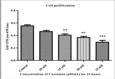
Cell proliferation assay depends on decreasing of yellow tetrazolium salts by metabolic actions of active cells, moderately by the function of dehydrogenase enzymes. The intracellular formazan was measured and solubilized by spectrophotometric means consequently purifications of detergent-solubilized (American Type Culture Collection, 2011). To observe the consequence of curcumin on U2OS cell proliferation, cell treated with dissimilar concentration of curcumin (10, 15, 20 and 25) μMolars for 24 h. Cell proliferation measured by MTT assay. The curcumin has an anti-proliferation cancer effect in malignant osteosarcoma U2OS cells dose-dependent manner (Figure 4). The curcumin low doses 10 μMolars inhibited but not significantly 16.5% ± 0.05 cell proliferation rate (P<0.05) but curcumin( 15 and20) μM significantly inhibited (27.1% ± 34% ± 0.1)U2OS cell proliferation rate (**P<0.05). Curcumin high doses 25 μM perspective inhibited osteosarcoma cell proliferation (47.5% ± 0.1) (***P<0.05), as shown in (Figure 4.1).
C e l l prol i fe rati on Con trol 10 uM 15 uM 20 uM 25 uM 0.0 0.2 0.4 0.6 0.8 *** ** **
C once ntration of C urcumin (µM/mL) for 24 hours
O D ( 57 0 n m -6 70 n m )
Figure 4.1. Curcumin inhibited cell proliferation Osteosarcoma U2OS cell line. The cells treated with curcumin different concentration (10, 15, 20 and 25) μMolars for 24 hours. (1) Untreated cells (control); cells incubated with ideal high glucose growth medium (DMEM) with 10% FBS (2) Curcumin 10 µM, (3) Curcumin 15 µM, (4) Curcumin 20 µM, and (5) Curcumin 25 µM. MTT assay reagent added after 24 hours (Roche, Germany) then measured the absorbance by ELISA reader using two different absorbencies (A570 - A670). The result analyzed by one-way ANOVA Graph Pad prism5
To refined the consequence of curcumin on human osteosarcoma U2OS cell. The cell viability results estimated from MTT assay. The curcumin plays an important role as anti-Cell viability in a malignant osteosarcoma U2OS cell line after treated with curcumin different concentration (10, 15, 20 and 25 ) μMolars 24 hours incubation time, as shown in figure 3. Curcumin high dose 25μMolars, intensely decreased the cell viability of U2OS cell line 47.5% ± 0.1 when measured to free curcumin control group (***P<0.05). Consequently, curcumin (20) μMolars doses considerably decreased cell viability (33.6% ± 0.1) (***P<0.05). Curcumin 15 μM notably decreased cell viability 26.85% ± 0.05 (**P<0.05). Curcumin low dose 10 μM decreased cell viability noticeable, 16.4%± percentage (*P<0.05), as shown in (Figure 4.2).
C e l l vi abi l i ty Con trol 10 uM 15 uM 20 uM 25 uM 0 50 100 150 * ** *** ***
C on ce n trati on of C u rcu m i n (µM/m L) for 24 h ou rs
10
0%
r
at
e
Figure 4.2. Curcumin decreased cell viability of osteosarcoma U2OS cell line. Cells treated with curcumin by different concentration (10, 15, 20 and 25) μMolars for 24 hours. (1) Untreated cells (control); cells incubated with ideal high glucose growth medium (DMEM) with 10% FBS (2) Curcumin 10 µM, (3) Curcumin 15 µM, (4) Curcumin 20 µM, and (5) Curcumin 25 µM. The results analyzed by dividing the group results to the control group and multiplying by 100 and then analyzed by one-way ANOVA Graph Pad prism 5
A flow pictures, explain further whether curcumin effectiveness were captured by confocal microscopy laser scanning microscope (CLSM510) (Zeiss, Jena, GERMANY) of human osteosarcoma U2OS cells from control to different concentration dose (10, 15, 20 and 25) μM after treated with curcumin for 24 hours as shown in (figure 4.3) In particular by increasing the concentration of curcumin significantly inhibit cell growth (proliferation) compare with free curcumin as a control group shown in figure 4.3. The high doses curcumin (25) μM powerfully inhibited the U2OS cell proliferation compare to the control group. Curcumin 20 μM significantly inhibited U2OS cell proliferation. Surprisingly, low doses curcumin (10) μM rarly effect on U2OS cell after 24 hours incubation.
Figure 4.3. Confocal laser scanning microscope (CLSM510) pictures of control and treated osteosarcoma U2OS cell with or without curcumin treatment after 24 h period, (15-25) μM curcumin lead to cell decline and partial detachment
4.2. Curcumin Modulated Both P53 Marker and TIGAR Protein Expression in Human Osteosarcoma U2OS Line
Affect of Curcumin on autophagy pathway and p53/TIGAR axis in human U2OS osteosarcoma cell line. To evaluate further Curcumin affects on autophagy pathway and p53/TIGAR axis, Human Osteosarcoma U2OS cell treated with Curcumin and then results complicated by using western blot and immunocytochemistery for analyzing the proteins amount.To visualize the stimulation of p53 and TIGAR proteins in human osteosarcoma U2OS cell line and elaborate further expression those two proteins, cell treated with curcumin different concentration (10, 15, 20 and 25) μMolars for 24 h period and then analyzed the level of TIGAR and p53 proteins using western blotting.
The p53 tumor protein has a vital function in cell cycle arrest after DNA damage by activating p21, start to DNA repair and induce apoptosis by overexpression of Bax (Espinosa et al. 2003; Liu et al. 2011; Prives and Vousden 2009; Vogelstein et al. 2000). Levine (1997) considers p53 important studied proteins because of it have fundamental task in cell continuity and cell death. In recent studies, p53 identified as a cellular core in responding and regulating to cellular metabolism (Maddocks and Vousden 2011; Jiang et al. 2013). The p53 protein initiates to regulating glucose degradation by glycolysis process regulating The expression of fructose-2,6-bisphosphatase (Matoba et al. 2006; Bensaad et al 2006). The p53 as a product of Tp53 gene has a critical role in p53 induce Bax mediator of mitochondrial apoptosis or cell cycle regulator p21 (Eisele and Weller 2013). The most essential role of p53 tumor protein is starting to emerge to help control the stability among oxidative phosphorylation and glycolysis. ATP and ADP can directly change p53 activity, through ATP block the ability of p53 to attach DNA and ADP build up (Lee et al. 2014).
Western blotting analysis result showed that curcumin inhibited p53 protein expression in malignant osteosarcoma U2OS cell line in dose-dependent method. The high doses curcumin (25) μM extremely inhibited p53 protein expression (*** P < 0.05). Curcumin 20 μM significantly inhibited p53 protein expression after 24 hours incubation (**P < 0.05). Surprisingly, low doses curcumin (10 and 15) μM did not change p53 protein expression inside osteosarcoma U2OS cell line after 24 hours treatment (P < 0.05). As shown in (Figure 4.4.A). The description of TIGAR as a p53 target gene show various roles in antioxidant functions of TIGAR and malignant suppression. It has regulation role in the protecting p53 response to repairable or passing stress (Lee et al. 2014). Lee et al (2014) Depending on type of cell and context the TIGAR protein can regulate the expression of glycolysis and reactive oxygen spices (ROS). The p53 independent of TIGAR protein expression has reported in a lot of human cancer (Cheung et al. 2013; Won et al. 2011). In order to understand mechanistically how curcumin inhibits cell proliferation and cell viability, finally dictate the cancer cells in culture towards autophagy; we observed the expression of TIGAR, in human cancer osteosarcoma U2OS cell line, after treated with curcumin. Decreasing of the TIGAR protein was found in osteosarcoma U2OS cell line when treated with curcumin different doses (10, 15, 20 and 25) µM 24-hour incubation and the number of autophagic marker proteins identified
determined the amount of them by western blot. Curcumin inhibited TIGAR protein expression inside osteosarcoma U2OS cell line in a dose-dependent manner after 24-hour incubation. Curcumin 25 µM dramatically inhibited TIGAR expression (***P < 0.05). Curcumin 20 µM after 24-hour incubation significantly inhibited TIGAR expression (***P < 0.05). However, Curcumin 15 µM significantly inhibited TIGAR expression (**P < 0.05), but Curcumin 10 µM without any effect on TIGAR protein expression. As shown in (Figure 4.4.B).
control M 10 15M 20M 25M 0.0 0.5 1.0 1.5 Cont rol 10 M 15 M 20 M 25 M Kda 53 37 p53 GAPDH ** *** C on ce n trati on of C u rcu m i n p53/GAPDH Pe rc en t o f c on tr ol M 0 10M 15M 20M 25M 0.0 0.5 1.0 1.5 Cont rol 10 M 15 M 20 M 25 M k da 37 30 TIGAR GAPDH ** ** *** C on ce n trati on of C u rcu m i n TIGAR/GAPDH Pe rc en t o f c on tr ol
Figure 4.4. Curcumin inhibits both P53 and TIGAR expression in U2OS cell line. The human malignant osteosarcoma U2OS cell treated with curcumin (10, 15, 20 and 25) μMolars for 24 hours, harvested cells used in western blotting for detecting two protein markers, (A) The p53 protein expression levels after extracting GADPH and compared to free curcumin as a control group (***P < 0.05), (B) The expression of TIGAR protein extracted GADPH protein amount (***P < 0.05), the results data showed by one-way ANOVA Tukey non-parametric analysis Graph Pad prism5
A
4.3. Curcumin Modulated Both Autophagy Marker Beclin-1 and LC3
To studied whether autophagic marker consequential in the activation of autophagy process, we measured amount of autophagic protein markers of beclin1 and LC3, as shown in Figure 4.5(A and B).The existence of autophagy in human osteosarcoma has been a proved by many studies. On the other hand, the pragmatic aspect of autophagy in osteosarcoma is controversial. Beclin1 plays a essential role in the phagophore construction, which is the first step of autophagosome biogenesis (Von Hoven et al. 2012). In osteosarcoma cell, Beclin-related autophagy act a controller key (Barkor) has been revealed to play an important function in autophagy stimulation. Decreasing of Barkor in osteosarcoma U2OS cell lead to decreased autophagy as demonstrated through blocking autophagosome creation. Liang et al (1998) reported that Beclin-1 is autophagic protein marker in humans is encoded by the BECN1 gene. While Beclin-1 able to stimulates autophagy, inhibit oncogenesis by interacting with PI3K-III, and binding to Bcl-2, while the interaction of Beclin-1 with Bcl-2 suppresses autophagy and stimulates oncogenesis. P53 and down-regulated autophagy gene (DRAM) can activate autophagy, which mutated in 50% of cancers (Lefranc et al. 2007). The western blot results illustrated curcumin inhibited beclin-1 protein expression when treated with high-doses (Figure 3.5). High dose (25) μM Curcumin, interestingly inhibited the expression of beclin-1 protein inside osteosarcoma U2OS cells (**P < 0.05), and curcumin 20 μM significantly inhibited the beclin-1 protein expression (*P < 0.05). In another hand, strangely Curcumin 15 μM significantly induced the expression of beclin-1 (**P < 0.05) beside it, 10 μM Curcumin didn’t have any significant affect on beclin-1 protein expression (P < 0.05) as shown in (Figure 4.5.A).
The conversion of the resident cytosolic structure of microtubule-related protein 1 light chain 3 (LC3-I) to membrane-bound lapidated (LC3-II) is recognized as an essential method of autophagosome formation (Mizushima et al. 2010; Sirichanchuen et al. 2012) and generally the expression of LC3-II protein is markedly a maker of autophagy (Von Hoven et al 2012). In mammalian cells, LC3-II protein is a key for starting autophagy. While, the LC3-II protein degrades quickly by the lysosomal enzyme, throughout autophagy followed by autolysosome formation. LC3-II protein uses protease inhibitor and pepstatin-A during starvation-induced autophagy, which causes of lysosomal income
after autophagosome formation (Tanida et al. 2005). Furthermore, a current study verified that Atg5/Atg7 is an independent pathway of autophagy and not related to LC3 formation but appeared to involve autophagosome formation (Kang et al. 2011). The total LC3 protein expression evaluated by western blot and immunocytochemistery after incubated the cells 24 hours with different concentration of Curcumin. The western blot result shows, which Curcumin treatment induced the total amount of LC3 protein expression when treated with 20 and 25 μMolars (*P < 0.05 and **P < 0.05) respectively for 24 hours. There isn’t any significant change in total amount LC3 protein expression however the LC3 protein expression increased in 15 μMolars shown in (Figure 4.5.B).