ISSN 2167-8677 (online) DOI 10.5195/d3000.2019.89
Investigation of surface topography of different root-end filling
materials: An in vitro study
Mine Koruyucu1, Hazal Özcan1, Merve Bayram2, Abdullah Burak Çankaya3, Nurullah Keklikoglu4, Figen Seymen1
1Istanbul University, Faculty of Dentistry, Department of Pedodontics, Istanbul, Turkey 2Istanbul Medipol University, Faculty of Dentistry, Department of Pedodontics, Istanbul, Turkey 3Istanbul University, Faculty of Dentistry, Department of Oral and Maxillofacial Surgery, Istanbul, Turkey 4 University, Faculty of Dentistry, Department of Histology and Embriology, Istanbul, Turkey
Abstract
Aim: Although there are many materials that can be used for retrograde filling in surgical endodontics, none of
them can be regarded as an ideal material yet. The purpose of this study was to compare the surface topography of three different root-end filling materials. Methods: 36 extracted single rooted human incisor teeth were cleaned and decoronated to standardized 10 mm root lengths. The root segments were prepared and 2 mm apical resection were performed. The samples were randomly separeted to three groups (Group A: Ca(OH)2, Group B:
MTA Angelus, Group C: ProRoot MTA), each one of them was comprised of 12 roots. Materials were placed as 2 mm apical barriers and obturated with guttapercha and AH-Plus sealer. Each group was dimidiated into two subgroups (A1,A2,B1,B2,C1,C2). Groups A1,B1,C1 were stored in normal saline (NS), groups A2,B2,C2 were stored in neutral phosphate buffer saline (NPBS) solution and samples were incubated at 370C for 2 weeks.
Stereomicroscope (32X) was used to photograph the root-end filling. Results: All specimens demonstrated white crystals formation and sediment over the root-end filling materials and on the superficial border of the root-end cavities’ wall as a white plaque. A2,B2,C2 samples had more crystal sediment on root-end fillings than samples A1,B1,C1. Dissolution and corrosion were observed in groups A1, A2. Conclusions: The results of this study revealed that calcium hydroxide is more resorbable than MTA Angelus and ProRoot MTA. The crystals formation and precipitation were observed in neutral phosphate buffer saline solution which were more than normal saline solution for all groups as a hydroxiapatite crystals.
Key words: Root canal filling materials, root canal
medicaments, Mineral trioxide aggregate
Citation: Kuruyucu M, et al. (2019) Investigation of surface topography of different root-end filling materials: An in vitro study. Dentistry 3000. 1:a001 doi:10.5195/d3000.2019.89
Received: August 7, 2018 Accepted: September 18, 2018 Published: July 25, 2019
Copyright: ©2019 Kuruyucu M, et al. This is an open access article licensed under a Creative Commons Attribution Work 4.0 United States License.
Email:[email protected]
Introduction
Endodontic surgery is performed in cases when treatment with orthograde root canal is
treatment-resistant to
periradicular inflammation or non-surgical root canal treatment is unsuccessful [1,2]. In periradicular surgery, a root-end cavity preparation is performed then a retrograde filling material is used
for filling. The periradicular surgery aims to block the ways in which transmission can occur between the root canal system and surrounding tissues [3]. Surgical endodontic treatment technique is a feasible treatment option, and the type of root-end filling material can affect the end result [4]. Root-end filling materials get in touch with periapical bone tissue after apex resection surgery.
Various materials are invariably used for instance amalgam, bonding systems, zinc oxide eugenol cements, glass ionomer cements, and calcium-silicate cements, all of which are commonly named as mineral trioxide aggregate based cements [5,6].
An ideal root-end filling material should demonstrate particular characteristics. The material
should adjust to the living tissue with its biocompatibility and therefore be able to start tissue repairing of periodontal ligament complex and most importantly its own cementogenesis. It should also be radiopaque like a natural tooth while being antibacterial, non-corrosive, non-resorbable and moisture indifferent. While being easy to handle and able to adapt to the dentinal walls, they should still be dimensionally stable and cost effective as well as safe to use with its non-toxic characteristics [7]. The problems encountered during pulpal healing have been treated with calcium hydroxide for many decades. Calcium hydroxide does not lead to periapical reactions, it has predictable results. Another advantage of calcium hydroxide is its mixability with certain liquids. However, calcium hydroxide may have got some disadvantages. These are resorpsion, variable treatment time, unpredictable treatment period, apexification situation, increased risk of tooth fracture, and poor patient compliance due to the long treatment time. Disadvantages of calcium hydroxide can affect treatment results [8,9].
Calcium silicate based materials have received great interest due to their high sealing ability , biocompatibility, regenerative capacity, and antibacterial properties. There are calcium silicate–based root repair materials developed, such as; ProRoot Mineral Triokside Aggregate
(MTA), MTA Plus and MTA Angelus [7,10]. Due to their excellent biocompatibility, low solubility and impermeability, these materials are widely used to repair perforations in root canals and to form apical barriers [11]. These are hydraulic materials that provide better clinical results when compared to other root filler materials [7,10].
In terms of sealing ability and biocompatibility, MTA cements are better than amalgam, Superseal, Intermediate restorative materials (IRM), and glass ionomer cement (GIC), which are all conventional
root materials. Studies
demonstrate that MTA induce the proliferation of periodontal fibroblasts, dental pulp cells, osteoblasts and osteoblast-like cells, and mesenchymal stem cells [10,12].
There are different root-end filling materials that can be used for endodontic surgery, however they all lack some characteristcs to be the ideal material yet [13].
The purpose of this study was to compare the surface topography of three materials: Calcium Hydroxide (Sultan Chemists Inc., Englewood, NJ, USA), MTA Angelus (Angelus Soluções Odontológicas, Londrina, Brazil) and ProRoot MTA (Dentsply Endodontics, Tulsa, OK, USA).
Material and Methods
Ethical approval of the study was obtained from Istanbul University Faculty of Dentistry Clinical Research Ethics Committee (No: 2012/1738-1298).
In this study, three commercial root end filling materials were tested. These were Calcium Hydroxide (Sultan Chemists Inc.,
Table 1: Contents of materials
Composition liquid
Calcium hydroxide Calcium Hydroxide Powder Sterile water
MTA Angelus Tricalcium silicate, dicalcium silicate,
bismuth oxide, tricalcium aluminate, calcium oxide, aluminium oxide, silicon dioxide
Distilled water
ProRoot MTA Tricalcium silicate, dicalcium silicate,
bismuth oxide, tricalcium aluminate, calcium sulphate dihydrate or gypsum
Englewood, NJ, USA), MTA Angelus (Angelus Soluções Odontológicas, Londrina, Brazil) and ProRoot MTA (Dentsply Endodontics, Tulsa, OK, USA). (Table 1)
Thirty-six extracted single-rooted human incisor teeth were used in this study. The teeth with caries, resorption or root fracture were eliminated from study. Teeth were cleaned from soft issues, bone residues and debris, and kept in 5.25% sodium hypochlorite for one week and then placed in normal saline solution until to be used in the current study.
Teeth were decoronated from the cement enamel junction using micromotor handpiece (W&H
Dentalwerk Bürmoos GmbH,
Austria) and diamond discs (Diamant 0.15mm, Dentaurum, Germany) for standardized 10 mm root lenghts.
The root segments were prepared with ultrasonic tips and 2 mm apical resection were performed from vertical to the long axis of the root for each tooth, using a cylindrical carbide bur under water cooling in a high-speed cycle. The canal length was measured by #15 K-file. K-file were used for canal preparation with step-back technique. Apical widening was performed up to #40 K-file. The preparing root canal method was completed with 1,2 and 3 Gates-Glidden drills for the coronal shaping. Irrigation was made with 2 ml of 2.5% sodium hypochlorite. 2 ml of 17% EDTA was applied for three minutes to remove smear
layer, then 2 ml distilled water was used as the last irrigation. The prepared canals were dried up with paper points.
The samples were randomly divided into three groups (Group A: Ca(OH)2, Group B: MTA Angelus,
Group C: ProRoot MTA), each one of them comprised of 12 roots. Ca(OH)2, MTA Angelus and Pro
Root MTA were prepared
according to the manufacturer’s instructions. Materials were placed as 2 mm apical barriers and obturated with guttapercha and AH-Plus sealer. Each group was dimidiated into two subgroups (A1, A2, B1, B2, C1, C2) (Table2). Groups A1, B1, C1 were stored in normal saline (NS), groups (A2, B2, C2) were stored in neutral phosphate buffer saline (NPBS) solution and samples were incubated at 370C for 2 weeks. A
stereomicroscope (Leica MZ 7.5) at x32 magnification was used to photograph the root-end filling
both before placing the samples in their solutions and afterwards.
Results
All specimens demonstrated white crystals formation and sediment over the root-end filling materials and a white plaque was seen on the superficial border of the root-end cavities’ wall. The crystal sediments on root-end fillings in the samples of Calcium Hydroxide, MTA Angelus and ProRoot MTA which were stored in normal saline solution (A1, B1, C1) were less than Calcium Hydroxide, MTA Angelus and ProRoot MTA which were stored in neutral phosphate buffer saline solution (A2, B2, C2). White crystal formation were observed the same in both ProRoot MTA groups (C1, C2). White crystal formation were observed in NPBS group (B2) was more than NS group (B1) in MTA Angelus group. White crystal
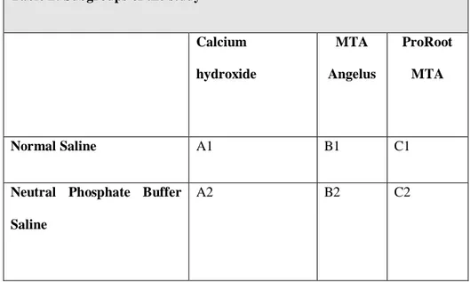
Table 2: Subgroups of the study
Calcium hydroxide MTA Angelus ProRoot MTA Normal Saline A1 B1 C1
Neutral Phosphate Buffer Saline
formation were observed the same in both Ca(OH)2 groups. Dissolution and corrosion were observed in groups A1, A2 (Ca(OH)2) (Figure 1).
Discussion
This study intended to evaluate the surface topography of three retrograde filling materials;
calcium hydroxide, MTA Angelus and ProRoot MTA in different solutions.
Healthy periradicular complex includes multiple tissues like cementum, periodontal ligament and bone. The ability to increase the regeneration of the functional periradicular complex is a feature on demand for root-end filling
materials [15]. MTA has ability of
promoting hard tissue
accumulation, especially cement formation [2]. The ability of hydroxyapatite formation on its interfaces when in contact with physiological or simulated body fluids such as phosphate buffer saline indicates that this material is bioactive. The fact that a material is bioactive makes it more
preferable in use. The surface topography of the materials which used in this study was examined thus their bioactivity was also identified [3].
Sarkar et al. [15] suggest that the physico-chemical reactions of the MTA changes according to root-end filling material’s ability of good sealing and bio-compatibility. Samples which stored in phosphate buffered saline (PBS) solution were photographed with scanning electron microscopy (SEM), examined energy dispersive X-ray analysis (EDXA), and X-ray diffraction (XRD) techniques after 2 weeks. SEM examination of the surface of MTA which were predisposed to the STF showed to be covered with sediments of similar morphology and chemical composition. [15] Asgary et al. [3] used extracted teeth. Samples prepared with MTA or new experimental cement (NEC) and subgroups were stored in normal saline solution or neutral phospate buffered solution. Samples were photographed with SEM before and after placement in solution. The samples which prepared with MTA and stored with normal saline did not show any change but other samples showed white crystal formation and sedimentation over the retrograde filling materials, the edges of the materials and root-end cavities’, and around the dentin surfaces as a white plaque. This situation is similar to our study. [3].
Saghiri et al. [16] studied how storage medium affects MTA and Biodentine cement in terms of their surface porosity. The specimens were utilized under SEM X1000 magnifications. They concluded the least surface crystallization surveyed in MTA samples were stored in STF. The
most surface crystallization
surveyed in Biodentine samples were stored in distilled water
(DW). Biodentine samples
demonstrated higher surface porosities when compared to the MTA samples which were stored in DW and STF with significantly
lower surface porosities [16].
Endosequence Root Repair
Material (ERRM) Putty and Paste were evaluated in terms of biocompatibility and compared to gray MTA by Ma et al. [17] Each material evaluated by SEM.. IRM and Cavit G showed inadequate crystallized superficial structure. ERRM Putty, ERRM Paste, and MTA displayed similar crystalline surface
structures. These crystals
contained calcium, carbon, and oxygen and low level of
phosphorus [17].
Vajja et al. [18] investigated in a vitro study that whether the thickness of three root-end filling materials have an effect on sealing ability. Cavities were filled with MTA, RMGIC and IRM. Teeth were examined under stereomicroscope at 30X magnification. The MTA had better sealing properties in proportion to other materials at
periapical area [18].
Gandolfi et al.[19] tested apatite-forming ability on ProRoot MTA cement after placement in PBS. Samples were photographed with an ESEM-EDX analysis and the analysis showed different surface morphologies related on the absorbing time. Result of this study proved that ProRoot MTA surface morphology is rapidly modified by the phosphate solution. Similar results were obtained in our study
[19].
Asgary et al. [20] compared the properties of NEC and mineral trioxide aggregate (MTA). All specimens were photographed and analyzed with scanning electron microscope and electron probe microanalysis (EPMA). White MTA and NEC specimens both showed the existence of crystalline
particles [20].
All these studies demonstrated that MTA specimens’ surface shows crystalline particles under SEM examination. These results are similar to properties of our
study. In the present study, higher
crystallization and porosities occurred in ProRoot MTA, MTA Angelus than Calcium Hydroxide, which is in agreement with previous studies.
This study was performed in vitro conditions; which may not imitate the exact situation of the oral cavity as blood or moisture contamination may affect the properties of sealant which are sensitive to moisture. Longitudinal in vivo studies are required to
check the surface topography and crystal precipitation for establishment of protocols for routine clinical usage.
Conclusion
The consequences of this study revealed that Ca(OH)2 is more
resorbable than MTA Angelus and ProRoot MTA as expected. The
crystals formation and
precipitation observed in NPBS solution was more than NS solution for all groups as hydroxiapatite crystals.
References
1. Modern endodontic
surgery concepts and
practice: a review. Kim S, Kratchman S. J. Endod
2006;32:601–23. PMID:
16793466
2. Histologic assessment of mineral trioxide aggregate as a root-end filling in monkeys. Torabinejad M, Pitt Ford TR, McKendry DJ, Abedi HR, Miller DA, Kariyawasam SP. J Endod 1997; 23: 225–8. PMID: 9594770
3. Effect of two storage
solutions on surface
topography of two root-end fillings. Asgary S, Eghbal MJ, Parirokh M, Ghoddusi J. Australian Endodontic Journal. 2009; 35: 147–152. PMID: 19961453 4. Comparison of mineral trioxide aggregate and
calcium hydroxide for
apexification of immature
permanent teeth: A
systematic review and meta-analysis. Lin JC, Lu JX, Zeng Q, Zhao W, Li WQ, Ling JQ. Journal of the Formosan Medical Association 2016 Jul; 115(7): 523-30. PMID: 26911724 5. Biointeractivity-related versus chemi/physisorption-related apatite precursor-forming ability of current root end filling materials. Gandolfi MG, Taddei P, Modena E, Siboni F, Prati C. 2013. J Biomed Mater Res Part B 2013;101B:1107– 1123. PMID: 23559495 6. Mineral trioxide aggregate:
A comprehensive literature review—Part III: Clinical
applications, drawbacks,
and mechanism of action. Parirokh M, Torabinejad M. J Endod 2010;36:400–413. PMID: 20171353
7. Properties of a new root-end filling material. Chong HK, Islam I, Yap AU, Tong YW, Koh ET. J Endod
2005;31:665–8. PMID: 16123702 8. Treatment options: biological basis of regenerative endodontic procedures. Hargreaves KM, Diogenes A, Teixeira FB. Pediatr Dent 2013;35:129-40. PMID: 23635981
9. Will mineral trioxide
aggregate replace calcium hydroxide in treating pulpal
and periodontal healing complications subsequent to dental trauma? A review. Bakland LK, Andreasen JO.
Dental Traumatology.
2012; 28: 25–32. PMID: 21895969
10. Apatite formation on
bioactive calciumsilicate
cements for dentistry
affects surface topography
and human marrow
stromal cells proliferation. Gandolfi MG, Ciapetti G, Taddei P, Perut F, Tinti A, Cardoso M, Van Meerbek B, Prati C. Dent Mater 2010;26: 974–992. PMID: 20655582
11. Biyoseramik esaslı kök
kanal patları: Derleme. Bilgiç A, Bodrumlu E. Atatürk Üniv. Diş Hek. Fak. Derg. 2016; 14:114-17.
12. Mineral trioxide aggregate: A comprehensive literature review—Part II: Leakage
and biocompatibility
investigations. Torabinejad M, Parirokh M. J Endod 2010;36:190–202. PMID: 20113774
13. Physical and chemical properties of a new
root-end filling material.
Torabinejad M, Hong CU, McDonald F, Pitt Ford TR. J Endod 1995; 21: 349–53. PMID: 7499973
14. Cementoblasts maintain
expression of osteocalcin in the presence of mineral
trioxide aggregate.
Thomson TS, Berry JE, Somerman MJ, Kirkwood
KL. J Endod 2003; 29: 407– 12. PMID: 12814226
15. Physicochemical basis of the biologic properties of mineral trioxide aggregate. Sarkar NK, Caicedo R, Ritwik P, Moiseyeva R, Kawashima I. J Endod 2005;31: 97–100. PMID: 15671817
16. Storage Medium Affects the Surface Porosity of Dental Cements. Saghiri MA, Shabani A, Asatourian A, Sheibani N. Journal of clinical and diagnostic
research: JCDR. 2017;
11(8), ZC116. PMID:
28969288
17. Biocompatibility of two novel root repair materials. Ma J, Shen Y, Stojicic S, Haapasalo M. J Endod. 2011; 37(6), 793-798. PMID: 21787491 18. Influence of Different Thickness of Mineral
Trioxide Aggregate, Resin Modified Glass Ionomer Cement and Intermediate Restorative Material on Sealing Ability of Root End Fillings: An in vitro Study.
Vajja S, Naik BD,
Vummidisetti SV,
Yarlagadda V. Journal of
Clinical & Diagnostic
Research: JCDR 2018;
12(1):ZC10-13. PMID:
29899639
19. Apatite-forming ability
(bioactivity) of ProRoot MTA. Gandolfi MG, Taddei P, Tinti A, Prati C. Int Endod J. 2010; 43(10), 917-929. PMID: 20646080
20. The properties of a new
endodontic material. Asgary S, Shahabi S, Jafarzadeh T, Amini S, Kheirieh S. J Endod 2008; 34(8), 990-993. PMID: 18634932