Eur J Orthop Surg Traumatol (2012) 22:25–28 DOI 10.1007/s00590-011-0777-y
123
O R I G I N A L A R T I C L EArthroscopic release of the subscapularis for shoulder
contracture of obstetric palsy
Mehmet Armangil · Burak Akan · Kerem Basarir · Sirri Sinan Bilgin · Serkan Gürcan · Mehmet Demirtas
Received: 22 January 2011 / Accepted: 21 February 2011 / Published online: 9 March 2011 © Springer-Verlag 2011
Abstract
Objective Retrospective study of one surgeon’s experience with arthroscopic release in obstetrical brachial plexus palsy.
Methods Over a four-year period, 6 patients who pre-sented with a shoulder contracture secondary to obstetric palsy before the age of 8 years were treated arthroscopi-cally. Small arthroscopy instruments, small shaver blades, including a 2.7-mm, 30° angled scope, and 90° radiofre-quency probe, were used for this surgery. Patient selection for this approach was based on the lack of bone deformity and targeted soft tissue release. Postoperative brace immo-bilization for 6 weeks was used in all patients. Functional status of the patients was evaluated pre- and postopera-tively with the assessment of external rotation.
Results The case series consisted of 2 girls and 4 boys with a mean age of 5.1 years (range, 3–8 years). No patient was lost to follow-up, and all patients completed a mini-mum 1 year of clinical and radiographic follow-up.
Increases in external rotation were observed in all patients. There was no intra- or postoperative complications. Conclusion Arthroscopic treatment of the shoulder con-tracture in obstetric palsy was found to be a safe and eVec-tive procedure in patients who are likely to undergo future tendon transfer or bone surgery.
Keywords Obstetric palsy · Shoulder arthroscopy · Pediatric shoulder arthroscopy · Subscapular release
Introduction
Obstetrical brachial plexus paralysis (OBPP) refers to com-plete or partial injury of the brachial plexus produced at the time of birth. OBPP involves most commonly the upper cervi-cal roots (C5, C6) resulting in muscle imbalance between internal and external rotators of the shoulder [1]. Although the majority of the patients (80–90%) recover spontaneously, some have a remaining imbalance causing severe functional impairment [1, 2]. In addition to diminished function, pro-longed muscle contractures result in bony deformities in both the glenoid and proximal humerus. Internal rotation contrac-tures due to external rotational weakness are the most common deformity in OBPP. Contracture release, tendon transfers, and rotational osteotomies are surgical methods used, depending on the level of deformity and the patient’s age. To achieve func-tional external rotation of the arm, contracted structures should be released Wrst. Currently, there is a lack of consensus on which procedures oVer the best outcome for mobility and func-tion, and what are the precise indications for each type of sur-gery. There is also a large variation of practice between centers [1]. This is further compounded by the heterogeneity in the assessment methods between the diVerent studies, making comparison diYcult.
M. Armangil (&) · K. Basarir · S. S. Bilgin · S. Gürcan · M. Demirtas
Department of Orthopedics and Traumatology, Faculty of Medicine, A.Ü.T.F. Ãbn-i Sina Hospital, Ankara University, 06100 SamanpazarÂ, Ankara, Turkey e-mail: [email protected]
B. Akan
Department of Orthopedics and Traumatology, Faculty of Medicine, Ufuk University, Ankara, Turkey
S. Gürcan
Department of Orthopedics and Traumatology, DiyarbakÂr State Hospital, DiyarbakÂr, Turkey
26 Eur J Orthop Surg Traumatol (2012) 22:25–28
123
Achieving shoulder balance in a timely manner may help in centering the humeral head in the glenoid achieving joint congruence. This will result in better clinical scores and joint geometry. Isolated release of the subscapularis is the preferred method in the younger age group (<4 years old) before latissimus dorsi or teres major tendon transfers are needed [3]. Subscapularis release was generally performed as an open procedure, though there have been reports using arthroscopic release [4]. To our knowledge, little informa-tion has been published on the outcome of arthroscopic subscapularis release [5]. This study observes the results and outcome in six cases of arthroscopic subscapularis release.
Patients and methods
The institutional review board (IRB) from our institution approved this retrospective study. The series included six children with OBPP treated for internal rotation deformity in our institution. All the patients were treated by a senior surgeon (D. M.). The medical records and radiographs of the patients were reviewed. From the total of 10 children treated for OBPP, four were eliminated due to lost to fol-low-up (n = 1), incomplete data (n = 1), and not meeting the minimum of one-year follow-up (n = 2). Remaining six children with full data underwent complete evaluation for this study. Demographic information (gender, age at pre-sentation) as well as clinical Wles, surgical records, and medical history were reviewed.
Patients were examined for ranges of motion both preop-eratively and postoppreop-eratively. Indications of surgical inter-vention with this technique were internal rotation contractures,and a deWcit in passive external rotation with no contractures of the elbow joint, and below age nine ide-ally earlier than 6 years before unreconstructible glenoid deformities occurred. Physical examination revealed that none of the patients were able to elevate the hand up to the mouth level. Stretching exercises and conservative therapy were recommended Wrst. Surgery was decided after 2 months of unsatisfactory conservative treatment. Two patients had a previous sural nerve grafting for upper OBPP.
Surgical technique
Surgical technique was performed in lateral decubitus under general anesthesia. Small arthroscopy instruments, small shaver blades, including a 2.7 mm, 30° angled scope, and 90° radiofrequency probe, were used for this surgery. A posterior portal was established Wrst. The posterior portal was made at the posterior glenohumeral joint line about
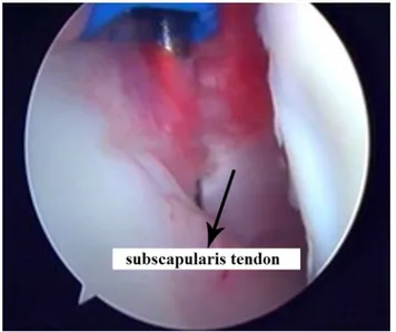
1 cm below the level of the posterior part of the acromion. Because of the limited range of motion and contracture, it was helpful to abduct and apply traction to the arm to enter the joint with a blunt trocar. An anterior portal was made from outside in, under direct visualization through the pos-terior portal. The anpos-terior portal was used after placing a 5.5-mm working cannula. To visualize the entire subscapu-laris tendon, it is necessary to release the anterior capsular ligaments at their attachment to the glenoid labrum. A radiofrequency probe set on 25 W was the most useful instrument to perform the release. The tendinous portion of the subscapularis was released. Low-proWle forceps and arthroscopic scissors were helpful, especially for releasing the capsular ligaments. After release of the anterior soft tis-sues, the axillary nerve was commonly seen.
After standard arthroscopic evaluation, subscapular tenotomy and anterior release were performed in all patients. Subscapular tenotomy was done from the tendon’s insertion (Fig.1). The increased external rotation arc was ampliWed by releasing the tissues from the rotator interval to the coracoid process.
After surgery, all patients were immobilized in a cast brace in the external rotation for 4 weeks. Then, physical therapy was initiated including stretching exercises and bracing in external rotation at nights for another 4 weeks. Patients were seen for follow-up at routine postoperative intervals by the treating surgeon until consolidation and were revisited for this study. Clinical examination was based on evaluation of shoulder function. The method of follow-up included an assessment made by an independent observer (M. A.). Clinical results including ranges of motion, shoulder function, and muscle strength were care-fully noted. The results were compared as of the situation at
Eur J Orthop Surg Traumatol (2012) 22:25–28 27
123
clinical presentation, and data from this observation weredisplayed in the “Results” section.
Results
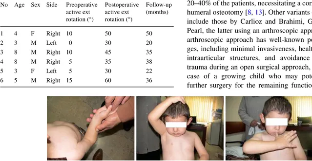
The study included 4 boys and 2 girls with an average age of 5.1 years (range, 3–8 years). The observed level of the OBPP was C5–C6 in 4 cases and C5–C6–C7 in 2. Hand function was good in all patients. The demographic and clinical characteristics of the patients as well as the preop-erative and follow-up results are summarized in Table1.
There were no intraoperative problems such as neuro-vascular injury associated with the arthroscopic approach. The mean operative time was 35 min (range, 25–60 min). No postoperative complications were observed in this series including wound infection or nerve palsy. Average follow-up was 33.5 months (20–50).
A signiWcant improvement in active abduction was observed with an average of +32.5° (mean 47.5–80°). Active external rotation was increased with an average of +34° (mean from 7.5–41.5°) (Fig.2).
Discussion
OBPP occurs in 0.4–2.5 per 1,000 live births, and the most commonly injured brachial plexus nerve roots in these inju-ries are the upper cervical roots (C5, C6) [6–9]. Despite
considerable improvement observed in most cases, in a signiWcant percentage of patients (5–19%), a residual dysfunction may result due to muscular imbalance related to the primary neurologic lesion [10]. Weakness of external rotators and abductors may lead to internal rotation and Xexion contracture, and Wnally to posterior subluxation of the humeral head [11]. Continuous muscular imbalance causes deformities of the humeral head and scapula in the growing bones of children, which may persist despite neu-rological improvement [11, 12]. Therefore, timely interven-tion for balancing the forces among the shoulder joint is of paramount importance. Since Fairbank’s deWnition in 1913, a number of procedures have been deWned and used for cor-recting diVerent levels of contracture and deformity, includ-ing isolated soft tissue release or combined muscle transfer and bone osteotomy [4, 6]. Despite this signiWcant experi-ence, there is currently no general agreement on the timing, exact indications or surgical procedures to be used in the treatment of these deformities [6, 8]. Basically, soft tissue procedures are the preferred modalities in the younger age group before development of bone deformities, while exter-nal rotation osteotomy of the humerus is preferred in chil-dren over 8 years with an incongruent glenohumeral joint [13, 14]. Soft tissue procedures may include isolated release of both the subscapular tendon and the anterior shoulder capsule or tendon transfer of the latissimus dorsi muscle [1, 4]. Long-term shoulder stiVness is the main
issue after Fairbank’s isolated subscapularis, capsular release, and open reduction [8, 14]. Considering this com-plication, Birch deWned shortening of the abnormal corac-oids process and subscapular release [13]. Although active shoulder function improved with this procedure, a very dis-abling external rotation contracture was observed in up to 20–40% of the patients, necessitating a corrective rotational humeral osteotomy [8, 13]. Other variants of this procedure include those by Carlioz and Brahimi, Gilbert et al., and Pearl, the latter using an arthroscopic approach [3, 4]. The arthroscopic approach has well-known potential advanta-ges, including minimal invasiveness, healthy evaluation of intraarticular structures, and avoidance of unnecessary trauma during an open surgical approach, especially in the case of a growing child who may potentially undergo further surgery for the remaining functional deWcits. The Table 1 Demographic and clinical characteristics of six patients with
obstetrical palsy
No Age Sex Side Preoperative active ext rotation (°) Postoperative active ext rotation (°) Follow-up (months) 1 4 F Right 10 50 50 2 3 M Left 0 30 20 3 8 M Right 10 45 35 4 8 M Right 5 35 38 5 3 F Left 5 30 22 6 5 M Right 15 60 36
28 Eur J Orthop Surg Traumatol (2012) 22:25–28
123
low infection rates and decreased blood loss are the other advantages of arthroscopy compared to open surgical tech-niques.
The strength of this study is that it brings information about a procedure that is not very common and that may be very helpful in the management of this not-so-rare condition. The results are very good in terms of functional outcome, and the invasiveness of the procedure is limited. Recommending this procedure in the early development of the disease may bring hope, comfort, better life quality, and better mobility to a group of speciWc patients at an early age.
The limitations of this study include the lack of a control group and the limited number of patients. Our conclusion is that arthroscopic subscapularis and anterior capsular release for internal rotation deformity in OBPP shows promising results in the younger age groups.
ConXict of interest No beneWts in any form have been or will be re-ceived from a commercial party related directly or indirectly to the sub-ject of this manuscript.
References
1. Hale HB, Bae DS, Waters PM (2010) Current concepts in the man-agement of brachial plexus birth palsy. J Hand Surg Am 35:322–331 2. Greenwald AG, Schute PC, Shiveley JL (1984) Brachial plexus birth palsy: a 10-year report on the incidence and prognosis. J Pediatr Ortho 4:689–692
3. Gilbert A, Brockman R, Carlioz H (1991) Surgical treatment of brachial plexus birth palsy. Clinical Orthop Relat Res 264:39–47 4. Pearl LM, Edgerton WB, KazimiroV AP, Burchette JR, Wong K
(2006) Arthroscopic release and latissimus dorsi transfer for
shoulder internal rotation contractures and glenohumeral defor-mity secondary to brachial plexus birth palsy. J Bone Joint Surg Am 88:564–574
5. Pearl LM, Bradford WE, Darissa SK, Darakjian AB, Kosco AE, KazimiroV PB, Burchette RJ (2003) Comparison of arthroscopic Wndings with magnetic resonance imaging and arthrography in children with glenohumeral deformities secondary to brachial plexus birth palsy. J Bone Joint Surg Am 85:890–898
6. Newman CJ, Morrison L, Lynch B, Hynes D (2006) Outcome of subscapularis muscle release for shoulder contracture secondary to brachial plexus palsy at birth. J Pediatr Orthop 26:647–651 7. Jackson ST, HoVer MM, Parrish N (1988) Brachial-plexus palsy
in the newborn. J Bone Joint Surg Am 70:1217–1220
8. van der Sluijs JA, van Ouwerkerk WJ, de Gast A, Nollet F, Winters H, Wuisman PIJ (2004) Treatment of internal rotation contracture of the shoulder in obstetric brachial plexus lesions by subscapular tendon lengthening and open reduction: early results and complications. Pediatr Orthop B 13:218–224
9. Bell MJ, Beauchamp CG, Kellam JK, McMurtry RY (1985) The results of plating humeral shaft fractures in patients with multiple injuries. The Sunnybrook experience. J Bone Joint Surg Br 67:293–296
10. Bae DS, Waters PM, Zurakowski D (2003) Reliability of three classiWcation systems measuring active motion in brachial plexus birth palsy. J Bone Joint Surg Am 85-A:1733–1738
11. Nath RK, Paizi M (2007) Scapular deformity in obstetric brachial plexus palsy: a new Wnding. Surg Radiol Anat 29:133–140 12. van der Sluijs JA, van Ouwerkerk WJ, de Gast A, Wuisman PI,
Nollet F, Manoliu RA (2001) Deformities of the shoulder in infants younger than 12 months with an obstetric lesion of the brachial plexus. J Bone Joint Surg Br 83:551–555
13. Birch R (1998) Birth lesions of the brachial plexus. In: Birch R, Bonney G, Perrycb Wynn (eds) Surgical disorders of the periphe-ral nerves. Curchill Livingstone, London, pp 209–233
14. Waters PM, Peljovich AE (1999) Shoulder reconstruction in patients with chronic brachial plexus birth palsy. A case control study. Clin Orthop Relat Res 364:144–152