507
Journal of Neurological Sciences [Turkish] 32:(3)# 45; 507-518, 2015 http://www.jns.dergisi.org/text.php3?id=901
Research Article
Effects of Low Dose Methotrexate in Cerebral Ischemia Reperfusion Injury in Rat
Bulent BAKAR1, Emine Arzu KOSE2, Elif SARI3, Bahram SARKARATI4, Pinar ATASOY5
1
Kirikkale University, School of Medicine, Neurosurgery, Kırıkkale, Türkiye 2Istanbul Medipol University, Anaestesiology and Reanimation, Istanbul, Türkiye 3Kirikkale University,
School of Medicine, Plastic and Reconstructive Surgery, Kırıkkale, Türkiye 4Hacettepe University, School of Medicine, Biochemistry, Ankara, Türkiye 5Kirikkale University, School
of Medicine, Pathology, Kırıkkale, Türkiye Summary
Background: During cerebral ischemia reperfusion injury, oxidative stress leads to
excitotoxicity, blood brain barrier dysfunction and inflammation. This study was designed to evaluate possible protective effects of low dose methotrexate on cerebral transient ischemia reperfusion injury in rat.
Methods: Except CONTROL group, temporary aneurysm clips were performed to both
common carotid arteries of rats for duration of 30 minutes. Four hours later, except CONTROL and SHAM groups, methotrexate (1.25 mg/kg/day) was administered intraperitoneally. Seventy two hours later, animals of CONTROL, MTX-A and SHAM-A group; and ten days later animals of MTX-C and SHAM-C group were sacrificed and hippocampal pyknotic neuronal cell count results and tissue lipid peroxidation (LPO) values were analyzed statistically.
Results: Pyknotic cell count values of CONTROL group were lower than A,
SHAM-C, MTX-A and MTX-C group values. Cell count values of SHAM-A and MTX-A group were higher than SHAM-C and MTX-C values, respectively. LPO values of CONTROL group were lower than SHAM-A and MTX-A values, but not different from MTX-C and SHAM-C values. LPO values of MTX-A group higher than MTX-C group values.
Conclusion: Cell count values and LPO values demonstrated that low dose methotrexate
could not prevent neuronal cells from destructive effects of transient ischemia reperfusion injury in rat.
Key words: Low dose methotrexate, ischemia reperfusion injury, pyknotic neuron, lipid
peroxidation
Düşük Doz Metotreksatın Ratlarda Serebral İskemi Reperfüzyon Üzerine Olan Etkileri Özet
Amaç: Serebral iskemi reperfüzyon yaralanmalarında oluşan oksidatif stress
eksitotoksisiteye, kan-beyin bariyerinde bozulmaya ve inflamasyon süreçlerinin oluşmasına neden olur. Bu çalışmada ratlarda oluşturulan hipoksi reperfüzyon yaralanmasında düşük doz metotreksatın olası koruyucu etkileri araştırılmıştır.
Yöntem ve Gereç: Serebral iskemi reperfüzyon yaralanması oluşturmak amacıyla
KONTROL grubu haricindeki tüm deney hayvanlarının (N=30) iki taraflı karotis arterlerine 30 dakika süreyle geçici anevrizma klibi konulmuştur. Yaralanmadan 4 saat sonra KONTROL ve SHAM grubu haricindeki hayvanlara intraperitoneal düşük doz metotreksat (1.25 mg/kg/gün) verilmiştir. Takiben 72 saatin sonunda KONTROL (N=6), MTX-A (N=8) ve SHAM-A (N=6) grubundaki hayvanlara ve 10 günün sonunda MTX-C (N=8) ve SHAM-C
508
(N=8) grubundaki hayvanlara ötenazi uygulanıp beyin dokuları çıkarılmış; hipokampustaki dejenere (piknotik) nöron hücre sayıları ve dokulardaki lipid peroksidasyon (LPO) düzeyleri istatistiksel analize tabi tulmuştur.
Bulgular: KONTROL grubunun piknotik nöron sayılarının SHAM-A, SHAM-C, MTX-A ve
MTX-C gruplarınınkinden düşük olduğu saptanmıştır. Öte yandan SHAM-A ve MTX-A grubuna ait sayım değerlerinin sırası ile SHAM-C ve MTX-C gruplarının değerlerine göre belirgin yüksek olduğu tespit edilmiştir. KONTROL grubunun LPO düzeylerinin SHAM-A and MTX-A gruplarından düşük ancak MTX-C ve SHAM-C gruplarının düzeyleri ile aynı olduğu bulunmuştur. Ayrıca, MTX-A grubunun LPO değerlerinin MTX-C grubuna göre daha yüksek olduğu da görülmüştür.
Sonuç: Araştırmanın sonunda, düşük doz metotreksat tedavisinin ratların nöronal hücelerini
serebral iskemi reperfüzyon yaralanmasının yıkıcı etkilerinden koruyamadığı gözlenmiştir.
Anahtar Kelimeler: Düşük doz metotreksat, iskemi reperfüzyon yaralanması, lipid
peroksidasyon, piknotik nöron
INTRODUCTION
Cerebrovascular diseases including ischemic stroke are associated with high morbidity and mortality worldwide. When the ischemia reperfusion injury occurs in the cerebral tissue, oxidative stress induced by ischemic cascades leads to excitotoxicity, blood brain barrier dysfunction and initiates post-ischemic inflammation(1). It also leads to enhanced generation of oxidants (such as superoxide and hydroxyl free radicals), and inflammatory mediators (such as interleukins (IL), tumor necrosis factor-alpha (TNF-α), interferon gamma (IF-γ) by activation of the endothelial cells, microglia, leukocytes and fibroblasts that can contribute to stroke-related brain injury). Oxidants and hydrogen peroxide are neurotoxic agents and initiate the free radical-mediated fatty acid chain reactions (i.e. lipid peroxidation) located in neuronal cell membrane, essential cellular proteins and DNA(1,20). These reactions can destroy the neuronal cell membrane permeability and cause to decrease in membrane-bound Na⁺-K⁺ ATPase enzyme activity and alter
protein synthesis. Thus, free radicals are important target for improving the outcome of the ischemic stroke today(24). On the other hand, recent reports demonstrate that adenosine receptors (A1, A2A, A2B, A3) are important targets in the treatment of stroke because extracellular adenosine
concentrations increase dramatically soon after cerebral ischemia. It has been shown that adenosine receptors locate both on central nervous system cells and on leukocytes and their neuroprotective role through A1 receptor subtype during ischemia is accepted, but it is well known that the use of selective A1 agonist drugs is hampered by undesirable side effects such as sedation, bradycardia, and hypotension.(13,19)
Recently, low dose methotrexate (MTX) which can inhibit lymphocyte proliferation in inflammation areas, and inhibit the destructive capacity of those leukocytes that do arrive at the inflamed site has been used to treatment the rheumatoid arthritis and other inflammatory diseases(4). Although, exact mechanism of its anti-inflammatory action has not been understood clearly yet, low dose MTX promotes extracellular adenosine accumulation which has cytoprotective effects by inhibiting the generation of the toxic oxygen metabolites at inflammation areas. It may also decrease the adhesion of neutrophils to the endothelium and decrease the neutrophil (but not macrophage) leukotriene synthesis(3,6,8,15). With all these findings mentioned above, this preliminary study was designed to evaluate the possible protective effects of low dose MTX on the cerebral transient ischemia reperfusion injury in rat.
509
MATERIAL AND METHODS
Materials:
This experimental study was performed in accordance with the guidelines for the use of laboratory animal subjects in research set by the Ethical Committee of Ankara Education and Research Hospital, Ankara, Turkey (Date: 04.04.2013; Number: 0013). In clinical experience, low dose methotrexate is administered at dose of 15-17.5 mg weekly to the patients with rheumatoid arthritis(4). In this study, methotrexate (Methotrexate DBL, Hospira Australia PYY Ltd., Mulgrave, Victoria, Australia) was administered to the rats at dose of 1.25 mg/ kg once a day intraperitoneally. The density of Methotrexate DBL is 25 mg/ ml and its intraperitoneal LD50 is 6 mg/ kg in rats.
Except the CONTROL group (no surgical procedure was performed and no chemical material was infused; n: 6) 30 Wistar albino rats of 250-350 g weight were randomly divided into two main groups used for the acute stage investigation (72 hours after the surgical procedures) and chronic stage investigation (10 days after the surgical procedures).
1. The acute stage group divided into two groups randomly was listed as below: - SHAM-A group (no chemical material was infused after the surgical procedures; n: 6).
- MTX-A group (low dose methotrexate was infused once-daily for 3 days after the surgical procedures; n: 8)
2. The chronic stage group divided into two groups randomly was listed as below: - SHAM-C group (no chemical material was infused after the surgical procedures; n: 8).
- MTX-C group (low dose methotrexate was infused once-daily for 7 days after the surgical procedures; n: 8)
Anesthesia was performed with intraperitoneal administration of 40 mg/ kg ketamine HCl (Ketalar®; Pfizer Inc, USA), and 5 mg/ kg xylazine HCl (Rompun® %2; Bayer HealthCare AG, Germany).
Methods:
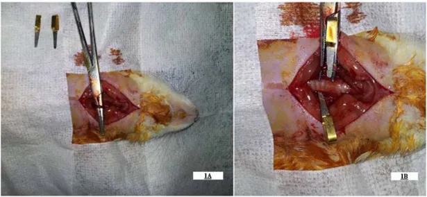
All animals were placed under sedation anesthesia with intraperitoneal ketamine HCl 40 mg/kg and xylazine HCl 6 mg/kg during spontaneous respiration at room temperature. Under an operating microscope, bifurcation of the right and left common carotid arteries were exposed through a midline neck incision, and after the vagus nerve dissection temporary aneurysm clips were attempted to both internal carotid arteries to constitute cerebral transient ischemia reperfusion injury for duration of 30 minutes (Figure 1); and then carotid arteries were unclamped(9,20). After this procedure, all rats were removed from sedation anesthesia spontaneously under the blanket. Four hours later from the surgical procedure, except CONTROL and SHAM groups, 1.25 mg/ kg once a day intraperitoneal methotrexate was administered slowly (within 10 seconds). Seventy two hours later, all animals except those of the MTX-C and SHAM-C groups; and ten days later the remaining of the animals were re-sedated with intraperitoneal ketamine HCl 40 mg/kg and xylazine HCl 6 mg/kg for sacrification. For sacrification, the whole body blood of each rat was collected from the vena cava inferior; and then rats were decapitated (Figure 2, Figure 3) and all hippocampal formations of right hemispheres (i.e. parietal region) were dissected and stored in 10% buffered formaldehyde solution at room temperature for future histopathological examination. For biochemical examination, remaining part of right hemispheres (i.e. frontal region) were immediately stored at -30 0C in dry air.
510
Figure 1: Figure demonstrates the dissected (1A) and clipped (1B) both common carotid arteries of the rat.
Figure 2: The macroscopic appearance of the whole cerebrum, and cerebellum of the rat following the acute
stage of the ischemia reperfusion injury.
Figure 3: The macroscopic appearance of the whole cerebrum, and cerebellum of the rat following the
511 Histopathological Analysis:
For histopathological examination, all tissue samples were fixated in 10% buffered formaldehyde and processed according to the routine light microscopic tissue processing technique. Serial sections of 5 micrometer thickness stained with haematoxylene-eosin (H&E) were examined and photographed by using a microscope (Leica Microsystems, Wetzlar GmbH). Each section was evaluated by experienced pathologist blinded to the groups, and test materials. The number of pyknotic neuronal cells (Pyknosis is called condensation and reduction in the size of a cell or cell nucleus, and irreversible condensation of chromatin in the nucleus of a cell undergoing necrosis or apoptosis) in each group was counted separately in areas per section of the hippocampal CA1, CA2, CA3 and DG region. Then the number of the degenerated neuron cells (i.e. pyknotic neurons) was calculated as an average per rat (Figure 4, Figure 5)(20). Biochemical Analysis:
The biochemical data consisted of the brain tissue malonyldialdehyde (MDA) concentration which is an important indicator of lipid peroxidation (LPO) level. The MDA levels were measured according to the method of Mihara et al. The principle of the method was based on the
spectrophotometric measurement of the color that occurred during the reaction of thiobarbituric acid with MDA. The concentration of thiobarbituric acid reactive substances (TBARS) was calculated by the absorbance coefficient of malondialdehyde–thiobarbituric acid complex(14). In this study, all specimens
were evaluated by an experienced biochemist blinded to the study groups, and experimental material. The lipid peroxidation (LPO) level values in nanomoles per gram of wet tissue were obtained by performing of the 532 nanometer spectrophotometry (Shimadzu® UV-120-02 spectrophotometer) to the specimens applied thiobarbituric acid.
Statistical Analysis:
The values of the pyknotic neuronal cell count values and lipid peroxidation level values in both stages were normally distributed and variations were homogenous among the groups. Therefore, One-Way Analysis of Variance (ANOVA) was performed to all values. To determine the statistical differences between the groups, post hoc evaluation (One-Way ANOVA-Tukey HSD Multiple Comparisons test) was performed. p values less than 0.05 were considered to be significant(17).
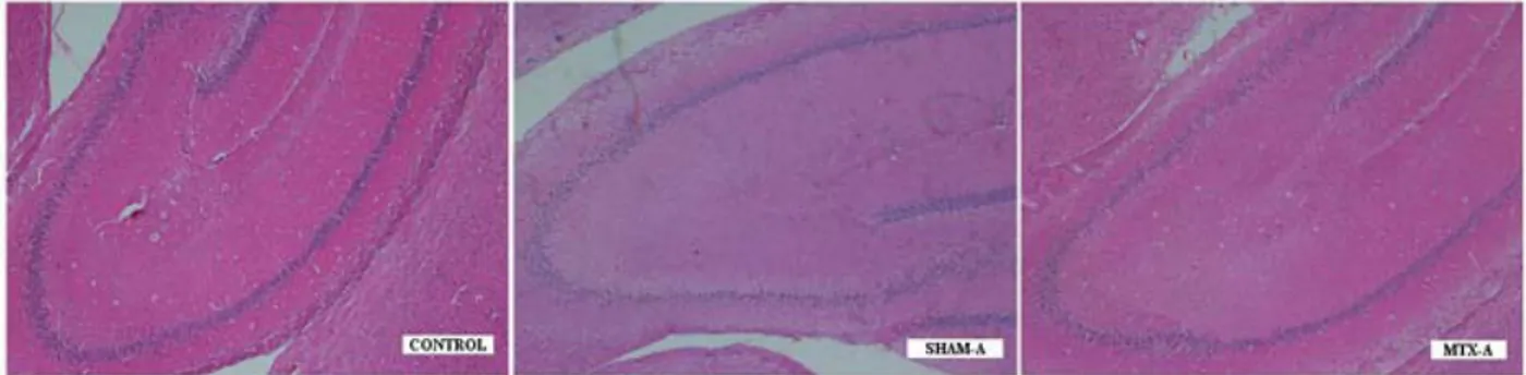
Figure 4: Histopathological examination of the CONTROL group demonstrated capillary stasis and minimal
edema with mild neuronal degeneration. But, SHAM-A and MTX-A group demonstrated severe edema located neuropil and perivascular area, significant capillary stasis, cellular swelling, and shrinkage of nuclei (HEx40).
512
RESULTS
Light Microscopy:
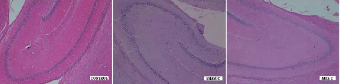
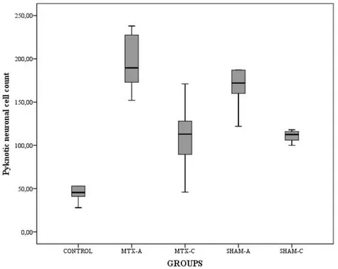
Histopathological examination of the CONTROL group demonstrated capillary stasis in some of the capillary vessels and minimal edema in some areas of neuropil with mild neuronal degeneration. But, SHAM-A and MTX-A group demonstrated severe edema both at neuropil and perivascular area with significant capillary stasis, disappearance of typical cellular arrangement, irregularities of intercellular organization, cellular swelling, and shrinkage of nuclei (pyknosis) (Figure 4). However, the histopathological findings were less severe in SHAM-C and MTX-C group. The degenerated (pyknotic) neuronal cell counts, degree of degeneration, and irregularities of intercellular organizations were significantly less than acute stage groups but more than CONTROL group (Figure 5). When compared the SHAM groups to the MTX groups, the distribution of the degenerated neuronal cells in hippocampal regions were more homogenous in the SHAM groups, however in MTX groups the degenerated neuronal cells were mostly accumulated in the CA2 region (Table 1).
Histopathological Analysis:
The pyknotic neuronal cell count values were statistically significant among the groups (F=24.506, p<0.001). The post hoc analysis results of the pyknotic neuronal cell count values revealed that there were significant differences between CONTROL/ MTX-A (p<0.001); CONTROL/ SHAM-A (p<0.001); CONTROL/ MTX-C (p=0.008); CONTROL/ C (p=0.003), SHAM-A/ SHAM-C (p=0.007), and MTX-SHAM-A/ MTX-C groups (p<0.001). But, there was no statistically significant difference between the SHAM-A/ MTX-A (p=0.754), and SHAM-C/ MTX-C (p=0.995) groups (Table 1, Table 2, Table 3, Figure 6). Biochemical Analysis:
The LPO values were statistically significant among the groups (F=6.079, p=0.001). The post hoc analysis results performed to the LPO values revealed that there was only significant difference between the MTX-A/ MTX-C groups (p=0.002) (Table 1, Table 2, Table 4, Figure 7)
Figure 5: Histopathological findings were less severe in the SHAM-C and MTX-C group. The degenerated
neuronal cell counts, degree of degeneration, and irregularities of intercellular organizations were significantly less than acute stage groups; but more than CONTROL group (HEx40).
513
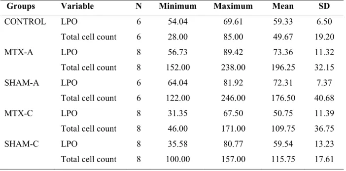
Table 1: Descriptive table of the pyknotic neuronal cell count values and lipid peroxidation
level values of all groups (LPO: lipid peroxidation; SD: standard deviation)
Groups Variable N Minimum Maximum Mean SD
CONTROL LPO 6 54.04 69.61 59.33 6.50
Total cell count 6 28.00 85.00 49.67 19.20
MTX-A LPO 8 56.73 89.42 73.36 11.32
Total cell count 8 152.00 238.00 196.25 32.15
SHAM-A LPO 6 64.04 81.92 72.31 7.37
Total cell count 6 122.00 246.00 176.50 40.68
MTX-C LPO 8 31.35 67.50 50.75 11.39
Total cell count 8 46.00 171.00 109.75 36.75
SHAM-C LPO 8 35.58 80.77 59.54 13.23
Total cell count 8 100.00 157.00 115.75 17.61
Table 2: One-Way Analysis of Variance (ANOVA) test results demonstrate that pyknotic
neuronal cell count values and lipid peroxidation level values were statistically different among the groups. p<0.05
Variable F p
Picnotic cell count 24.506 <0.001
514
Table 3: The post hoc analyses results revealed that pyknotic neuronal cell count values of
the CONTROL group were lower than values of other groups in both stages. Additionally, when cell count values of the acute stage were compared to chronic stage values, MTX-A, and SHAM-A group values were higher than MTX-C, and SHAM-C values. One-Way ANOVA-Tukey HSD Multiple Comparisons test. p<0.05 (df: degrees of freedom)
Stage Variable (I-J) Mean Difference (I-J)
Standart error
p
Acute CONTROL/ MTX-A 146.58* 16.51 <0.001
CONTROL/ SHAM-A -126.83* 17.65 <0.001
MTX-A/ SHAM-A -19.75 16.51 0.754
Chronic CONTROL/ MTX-C 60.08* 16.51 0.008
CONTROL/ SHAM-C 66.08* 16.51 0.003
MTX-C/ SHAM-C 6.00 15.29 0.995
Acute/ Chronic SHAM-A/ SHAM-C 60.75* 16.51 0.007
MTX-A/ MTX-C 86.50* 15.29 <0.001
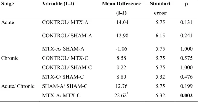
Table 4: The lipid peroxidation level values of CONTROL group were lower than MTX-A,
and SHAM-A values in acute stage but there was no statistical difference among the groups in chronic stage. Additionally, lipid peroxidation level values of the MTX-A group were higher than MTX-C values, but SHAM groups values were not different each other. One-Way ANOVA-Tukey HSD Multiple Comparisons test. p<0.05. (df: degrees of freedom)
Stage Variable (I-J) Mean Difference (I-J)
Standart error
p
Acute CONTROL/ MTX-A -14.04 5.75 0.131
CONTROL/ SHAM-A -12.98 6.15 0.241
MTX-A/ SHAM-A -1.06 5.75 1.000
Chronic CONTROL/ MTX-C 8.58 5.75 0.575
CONTROL/ SHAM-C 0.22 5.75 1.000
MTX-C/ SHAM-C 8.80 5.32 0.476
Acute/ Chronic SHAM-A/ SHAM-C 12.76 5.75 0.199
515
DISCUSSION
In literature, experimental animal stroke models demonstrated that TNF-α, IL-1 and IL-6 are potent inflammatory cytokines which are able to modulate the ischemic
damage size. Several studies concluded that TNF-α, and IL-6 levels are elevated in cerebrospinal fluid and blood of stroke patients(18,23,26). Additionally, postmortem studies in human stroke revealed that microglia is the major source of the
TNF-Figure 6: The mean values of the pyknotic neuronal cell count values of the both stages groups compared to the
CONTROL group. Each error bar shows the minimum and maximum of the cell count values.
Figure 7: The mean values of the tissue lipid peroxidation levels of the both stages groups compared to the
516 α(10,21). IL-1, and TNF-α induces the expression of adhesion molecule which promotes neutrophil and other subtype leukocytes accumulation. TNF-α also stimulates acute-phase protein production, blood–brain barrier breakdown and induction of other inflammatory mediators. Low dose MTX inhibits transmethylation reactions and promotes extracellular adenosine accumulation at the sites of inflammation. It is known that adenosine is a key mediator in the anti-inflammatory actions of the MTX. Low concentrations of the adenosine inhibit Fc-gamma-receptor mediated neutrophil phagocytosis via its A2 receptors. In addition, adenosine also inhibits the TNF-induced generation of elastase by neutrophils; generation of superoxide anions by monocytes; and proliferation of the lymphocytes in inflamed areas.(2,5,16). IL-4, IL-13, IFN-γ, TNF-α and granulocyte-macrophage colony stimulating factor are among the cytokines inhibited by MTX(6). It may also decrease the neutrophil (but not macrophage) leukotriene synthesis(3).
Many reports in literature concluded that the ischemia reperfusion injury induced by occlusion of both common carotid arteries could not be developed in rat brain, because the rat cerebral blood circulation is supported by rich anastomoses between the anterior and posterior circulation vessel(7,11,25). On the other hand many other studies accepted in scientific literature demonstrated that this surgical method is efficient to develop the ischemia reperfusion injury in rat(16,20). Because many stroke patients administered to hospital four hours later from occurrence of the stroke, we decided to infuse the experimental material to the rats four hours later from the removing of the clip. Actually, in present study pyknotic neuronal cell count values of the SHAM and MTX groups were significantly different from the CONTROL group values, and this means that our surgical method is efficient to produce transient ischemia reperfusion injury in rat easily
and adequately without additional morbidity.
In present study, pyknotic neuronal cell count values of both stages groups demonstrated that low dose MTX could not prevent the neuronal cells from the destructive effects of transient ischemia reperfusion injury in rat. It may mean that low dose MTX could not prevent the neuronal cells which try to be alive in areas exposed to transient ischemia from the destructive effects of the ischemic injury in rat. Additionally, statistical analyses results revealed that low dose MTX could not reduce the lipid peroxidation cascades in both stages of the ischemia, although the LPO level values of the MTX-C group lower than all other group values numerically. Finally, it can be said that low dose MTX could not effect to any stage of the cerebral ischemia reperfusion injury in rat, although the LPO and pyknotic neuronal cell count values of the acute stage groups were higher than chronic stage groups values statistically.
Study limitations
Present study has some limitations. First, because of some financial and technical limitations this study could not be supported by using of the some specific radiological imaging methods (such as MR-spectrometry, SPECT, radionuclides, angiography) to show the defective cerebral metabolism, cerebral infarct size etc. Second, because of some technical and financial difficulties this study could not be supported with immunohistochemical, and electron microscopic findings which can show ultrastructural details of the inflammatory response, neuronal necrosis and edema in the acute and/ or chronic stages of the cerebral transient ischemia reperfusion injury in rat. Third, this study should be supported by using more specific biochemical analyses for other detailed inflammatory pathways (such as apoptotic pathways, glutathione level, nitrite/ nitrate level, and xhantine oxidase activity level measurements). Because of some technical
517 and financial difficulties the other possible beneficial effects of the low dose MTX could not be evaluated properly. Fourth, because many studies showed the neurotoxic effects of the intrathecal MTX on the spinal cord tissue by developing of the axonal swelling and loss, demyelination and astrocytosis, present study did not contain the study groups to evaluate the effectiveness of the MTX infused via intrathecal route(5,12,22). But, in future studies, low dose methotrexate may be administered via the intrathecal route to evaluate its possible effects to the cerebral ischemia reperfusion injury.
CONCLUSION
At the end of this study, pyknotic neuronal cell count values and LPO values demonstrated that low dose methotrexate could not prevent neuronal cells from destructive effects of transient ischemia reperfusion injury in rat. Finally, it could be said that low dose MTX has no beneficial effect in transient cerebral ischemia reperfusion injury in rat.
Acknowledgement
The authors would like to express their thanks and gratitude to Mr. Erkan Kaya for his skilled assistance during the study.
Conflict of Interest Statement
The authors declare that they have no conflict of interest.
Correspondence to:
Bulent Bakar
E-mail: [email protected]
Received by: 12 March 2015 Revised by: 10 August 2015 Accepted: 15 August 2015
The Online Journal of Neurological Sciences (Turkish) 1984-2015
This e-journal is run by Ege University Faculty of Medicine,
Dept. of Neurological Surgery, Bornova, Izmir-35100TR
as part of the Ege Neurological Surgery World Wide Web service.
Comments and feedback: E-mail: [email protected] URL: http://www.jns.dergisi.org
Journal of Neurological Sciences (Turkish) Abbr: J. Neurol. Sci.[Turk]
ISSNe 1302-1664
REFERENCES
1. Brüning CA, Prigol M, Luchese C, Jesse CR,
Duarte MM, Roman SS, Nogueira CW: Protective effect of diphenyl diselenide on ischemia and reperfusion-induced cerebral injury: involvement of oxidative stress and pro-inflammatory cytokines. Neurochem Res 2012; 37: 2249-2258.
2. Chan ESL, Bruce N Cronstein BN. Molecular
action of methotrexate in inflammatory diseases. Arthritis Res 2002; 4: 266-273.
3. Cronstein BN, Naime D, Ostad E: The
antiinflammatory mechanism of methotrexate. Increased adenosine release at inflamed sites diminishes leukocyte accumulation in an in vivo model of inflammation. J Clin Invest 1993; 92: 2675-2682.
4. Cronstein BN. Low-dose methotrexate: a
mainstay in the treatment of rheumatoid arthritis. Pharmacol Rev 2005; 57: 163-172.
5. Gregorios JB, Gregorios AB, Mora J, Marcillo
A, Fojaco RM, Green B: Morphologic alterations in rat brain following systemic and intraventricular methotrexate injection: light and electron microscopic studies. Neuropathol Exp Neurol 1989; 48: 33-47.
6. Haskó G, Cronstein BN. Adenosine: an
endogenous regulator of innate immunity. Trends Immunol 2004; 25: 33-39.
7. Herz RC, Hillen B, Versteeg DH, De Wildt DJ.
Collateral hemodynamics after middle cerebral artery occlusion in Wistar and Fischer-344 rats. Brain Res. 1998; 793: 289-296.
8. Johnston A, Gudjonsson JE, Sigmundsdottir H,
Ludviksson BR, Valdimarsson H: The anti-inflammatory action of methotrexate is not mediated by lymphocyte apoptosis, but by the suppression of activation and adhesion molecules. Clin Immunol 2005; 114: 154-163.
9. Kose EA, Bakar B, Kasimcan O, Atilla P,
Kilinc K, Muftuoglu S, Apan A: Effects of intracisternal and intravenous dexmedetomidine on ischemia-induced brain
518
injury in rat: a comparative study. Turk Neurosurg 2013; 23: 208-217.
10. Lambertsen KL, Biber K, Finsen B: Inflammatory cytokines in experimental and human stroke. J Cereb Blood Flow Metab 2012; 32: 1677-1698.
11. Li L, Ke Z, Tong KY, Ying M. Evaluation of cerebral blood flow changes in focal cerebral ischemia rats by using transcranial Doppler ultrasonography. Ultrasound Med Biol. 2010; 36: 595-603.
12. Mahoney DH Jr, Shuster JJ, Nitschke R, Lauer SJ, Steuber CP, Winick N, Camitta M: Acute neurotoxicity in children with B-precursor acute lymphoid leukemia: an association with intermediate-dose intravenous methotrexate and intrathecal triple therapy--a Pediatric Oncology Group study. J Clin Oncol 1998; 16: 1712–1722.
13. Melani A, Pugliese AM, Pedata F. Adenosine receptors in cerebral ischemia. Int Rev Neurobiol. 2014;119:309-48. doi: 10.1016/B978-0-12-801022-8.00013-1.
14. Mihara M, Uchiyama M: Determination of malonaldehyde precursor in tissues by thiobarbituric acid test. Anal Biochem 1977; 86: 271-278.
15. Montesinos MC, Takedachi M, Thompson LF, Wilder TF, Fernández P, Cronstein BN: The antiinflammatory mechanism of methotrexate depends on extracellular conversion of adenine nucleotides to adenosine by nucleotidase: findings in a study of ecto-5'-nucleotidase gene-deficient mice. Arthritis Rheum 2007; 56: 1440-1445.
16. Nagakannan P, Shivasharan BD, Thippeswamy BS, Veerapur VP: Effect of tramadol on behavioral alterations and lipid peroxidation after transient forebrain ischemia in rats. Toxicol Mech Methods 2012; 22: 674-678. 17. Nie NH, Hull CH, Jenkins JG. SPSS: Statistical
Package for Social Science. New York, Mc Graw Hill Inc., 1975.
18. Pasarica D, Gheorghiu M, Topârceanu F, Bleotu C, Ichim L, Trandafir T. Neurotrophin-3, TNF-alpha and IL-6 relations in serum and cerebrospinal fluid of ischemic stroke patients. Roum Arch Microbiol Immunol. 2005; 64: 27-33.
19. Pedata F, Pugliese AM, Coppi E, Dettori I, Maraula G, Cellai L, Melani A. Adenosine A2A receptors modulate acute injury and neuroinflammation in brain ischemia. Mediators Inflamm. 2014;2014:805198. doi: 10.1155/2014/805198. Epub 2014 Aug 5. 20. Quartu M, Serra MP, Boi M, Pillolla G, Melis
T, Poddighe L, Del Fiacco M, Falconieri D, Carta G, Murru E, Cordeddu L, Piras A, Collu M, Banni S: Effect of acute administration of Pistacia lentiscus L. essential oil on rat cerebral cortex following transient bilateral common carotid artery occlusion. Lipids Health Dis 2012; 11: 8.
21. Tuttolomondo A, Pecoraro R, Pinto A. Studies of selective TNF inhibitors in the treatment of brain injury from stroke and trauma: a review
of the evidence to date. Drug Des Devel Ther. 2014 Nov 7;8:2221-2239. eCollection 2014. 22. Vezmar S, Becker A, Bode U, Jaehde U:
Biochemical and clinical aspects of methotrexate neurotoxicity. Chemotherapy 2003; 49: 92-104.
23. Vila N, Castillo J, Dávalos A, Chamorro A. Proinflammatory cytokines and early neurological worsening in ischemic stroke. Stroke. 2000; 31: 2325-2329.
24. White BC, Grossman LI, Krause GS: Brain injury by global ischemia and reperfusion: A theoretical perspective on membrane damage and repair. Neurology 1993; 43: 1656-1665. 25. Woitzik J, Schneider UC, Thomé C, Schroeck
H, Schilling L. Comparison of different intravascular thread occlusion models for experimental stroke in rats. J Neurosci Methods. 2006; 151: 224-231.
26. Zaremba J, Losy J. Cytokines in clinical and experimental ischemic stroke. Neurol Neurochir Pol. 2004; 38(1 Suppl 1): S57-62.
View publication stats View publication stats