ORIGINAL ARTICLE
Change in species distribution and antifungal susceptibility
of candidemias in an intensive care unit of a university hospital
(10-year experience)
Bilgul Mete1 &Esra Yerlikaya Zerdali2&Gokhan Aygun3&Nese Saltoglu1&Ilker Inanc Balkan1&Ridvan Karaali1& Sibel Yildiz Kaya4&Berna Karaismailoglu1&Abdurrahman Kaya5&Seval Urkmez6&Gunay Can7&Fehmi Tabak1& Recep Ozturk8
Received: 28 May 2020 / Accepted: 20 July 2020
# Springer-Verlag GmbH Germany, part of Springer Nature 2020 Abstract
Candidemia is a nosocomial infection mostly found in critically ill patients. Our objectives were to evaluate the change in distribution and resistance profile of Candida spp. isolated from candidemic patients in our intensive care unit over two 5-year periods spanning 15 years and to evaluate the risk factors. Records from the microbiology laboratory were obtained, from January 2004 to December 2008 and from January 2013 to December 2017, retrospectively. Antifungal susceptibility was performed by E-test and evaluated according to EUCAST breakpoints. A total of 210 candidemia cases occurred; 238 Candida spp. were isolated in 197 patients (58.8% male; mean age, 59.2 ± 19.6 years). The most predominant risk factor was central venous catheter use. Species distribution rates were 32%, 28%, 17%, and 11% for C. albicans (n = 76), C. parapsilosis (n = 67), C. glabrata (n = 40), and C. tropicalis (n = 27), respectively. Resistance rate to anidulafungin was high in C. parapsilosis over both periods and increased to 73% in the second period. Fluconazole showed a remarkable decrease for susceptibility in C. parapsilosis (94 to 49%). The prevalence of MDR C. parapsilosis (6%/33%) and C. glabrata (0%/44%) increased in the second period. We observed a predominance of non-albicans Candida spp., with C. parapsilosis being the most frequent and C. glabrata infections presenting with the highest mortality. High level of echinocandin resistance in C. parapsilosis and increasing prevalences of MDR C. parapsilosis and C. glabrata seem emerging challenges in our institution.
Keywords Candida . Species . Candidemia . Susceptibility . E-test
Introduction
Candidemia is one of the three most frequently encountered nosocomial bloodstream infections [1,2]. The incidence of candidemia was reported as 1.2 and 25 cases per 100,000
persons, 1.22 episodes per 1000 discharges in studies from Europe, United States, and Asia, respectively. [3–5].
Candidemia is a nosocomial infection found mostly in crit-ically ill, immunosuppressed, and surgical patients; it leads to prolonged hospitalization and presents with high morbidity
* Bilgul Mete [email protected]
1 Department of Infectious Diseases and Clinical Microbiology, Cerrahpasa School of Medicine, Istanbul University-Cerrahpasa, Istanbul, Turkey
2
Department of Infectious Diseases and Clinical Microbiology, Istanbul Haseki Research and Training Hospital, Istanbul, Turkey 3 Department of Medical Microbiology, Cerrahpasa School of
Medicine, Istanbul University-Cerrahpasa, Istanbul, Turkey
4
Department of Infectious Diseases and Clinical Microbiology, Sungurlu State Hospital, Corum, Turkey
5 Department of Infectious Diseases and Clinical Microbiology, Istanbul Research and Training Hospital, Istanbul, Turkey 6
Department of Anesthesiology and Reanimation, Cerrahpasa School of Medicine, Istanbul University-Cerrahpasa, Istanbul, Turkey 7
Department of Public Health, Cerrahpasa School of Medicine, Istanbul University-Cerrahpasa, Istanbul, Turkey
8 Department of Infectious Diseases and Clinical Microbiology, Medical Faculty, Istanbul Medipol University, Istanbul, Turkey
https://doi.org/10.1007/s10096-020-03994-6
and mortality rates [6, 7]. Attributable mortality rate may reach 50% or higher in septic shock [8]. Use of broad-spectrum antibiotics and immunosuppressive drugs, prolonged hospitalization, intensive care unit (ICU) stay, his-tory of intra-abdominal surgery, parenteral nutrition, hemodi-alysis, and use of central venous catheters (CVC) are among the important predisposing risk factors for candidemia [9,10]. Prolonged hospital stay and hospital costs lead to signifi-cant economic burden. In a systematic review, mean cost per hospitalization associated with candidemia was demonstrated to range from $10,216 to $37,715 [11].
The incidence of candidemia has steadily increased over the recent years and the epidemiology of candidemia has changed over the decades, shifting from C. albicans to non-albican species. Although species distribution differs by geo-graphic areas due to different underlying conditions, wide-spread therapeutic and prophylactic use of antifungal agents has led to a worldwide increase in prevalence of infections with non-albicans Candida species such as C. glabrata, C. parapsilosis, and C. tropicalis [12–15]. Furthermore, ex-tensive use of fluconazole may have led to selection of isolates that are resistant or less sensitive to fluconazole [16–18]. Increasing rates of acquired fluconazole resistance in non-albicans Candida spp. have been reported in both SENTRY study and population-based surveillance programs [15,
19–21].
Data regarding the incidence of candidemia is sparse in Turkey: incidence was reported as 1–1.5 per 1000 admissions in some studies [22–24]. There is only one multicenter study reporting in vitro resistance in candidemia isolates in Turkey [25]. Other reports evaluating candidemia cases are mostly from single centers and no prior study from Turkey evaluated candidemia cases by means of clinical aspects, species distri-bution, susceptibilities, and microbiological changes over time.
In this study, we aimed to evaluate the change in distribu-tion and resistance profiles of Candida spp. isolated from blood cultures of candidemic patients followed up in the ICU of our hospital over two 5-year periods spanning 15 years and determine the underlying risk factors and outcome.
Material and methods
Documented candidemia in patients followed in the ICU of our university hospital over two 5-year periods (January 2004 to December 2008 and January 2013 to December 2017) were analyzed retrospectively.
Our university hospital has 1200 beds and is located at the center of Istanbul. Patients from all parts of our country may be referred to our center. Our 13-bed medical surgery inten-sive care unit admits 600–800 patients annually. In cases of candidemia, the first preferred regimen is fluconazole and it is
re-evaluated and changed if necessary, according to the sus-ceptibility results.
Patients and episodes
Records from the microbiology laboratory were evaluated to identify patients with positive peripheral blood cultures for Candida spp. from January 2004 to December 2008 and from January 2013 to December 2017, retrospectively, giving a gap of 5 years to be able to evaluate the evolution of resistance with time. Isolation of Candida spp. from at least one blood culture of a patient was defined as candidemia. Isolation of Candida spp. from the same patient was considered as a new candidemia episode if the isolation times were ≥ 1 month apart.
Clinical data were collected from the patients’ available medical files and underlying risk factors were extracted for analysis. Presence of an underlying disease, intra-abdominal infection, sepsis, malignancies, CVC use, and recent surgery were the only accessible risk factors that could be analyzed.
A 30-day overall mortality (mortality attributed to all causes within 30 days after candidemia) was determined in eligible patients with available adequate data and compared at the level of species and antifungal drugs used for treatment.
Species identification
Blood cultures were monitored by an automated blood culture system (BacT/Alert; bioMerieux, France). Isolated Candida spp. were stored in cryoBank (Mast group, United Kingdom) at− 70 °C. Stored Candida spp. were subcultured onto Sabouraud dextrose agar (Oxoid, England) and incubat-ed for at least 72 h at 30 °C. Candida spp. grown in cultures were evaluated after inoculation into CHROMagar (CHROMagar, Paris, France) and species identification was performed using the commercial identification system API 32C (bioMerieux, France).
Susceptibility testing
Antifungal susceptibility for amphotericin B, anidulafungin, fluconazole, voriconazole, and posaconazole was performed by E-test method (AB BIODISK, Sweden/bioMerieux, France) according to the manufacturer’s instructions. Yeast cell suspensions were adjusted to 0.5 McFarland. Agar plates containing RPMI 1640 (with L-glutamine) medium supple-mented with 2% glucose buffered with MOPS in a pH of 7.0 (Wisent Bioproducts, Canada) were used to perform E-test. The plates were incubated at 35 °C for 48 h and evaluated for minimal inhibitory concentration (MIC) results. MIC levels were determined as the lowest concentration at which the border of the elliptical zone of growth inhibition intersected the E-test strips. According to the manufacturer’s
instructions, an 80% inhibition of growth was used as the endpoint when reading the MIC levels of azoles, whereas for amphotericin B, a complete inhibition of growth was required to determine the MIC endpoint. Interpretations of MIC levels were evaluated according to the European Committee on Antimicrobial Susceptibility Testing (EUCAST) Antifungal Clinical Breakpoints. Quality control was performed by test-ing the strains recommended by EUCAST: Candida krusei ATCC 6258 and Candida parapsilosis ATCC 22019 (http:// www.eucast.org/fileadmin/src/media/PDFs/EUCAST_files/ AFST/Clinical_breakpoints/Antifungal_breakpoints_v_9.0_
180212.pdf). Candida spp. that were multidrug resistant
(MDR, defined as resistance to > 1 drug class) and extensively drug resistant (XDR, defined as resistance to three drug classes) were identified and compared in terms of rate between the two periods [26].
Statistical analysis
Data were analyzed using SPSS, version 25.0 (SPSS) for Windows. Chi-square test and analysis of variance (ANOVA) were used for evaluation of data. A p value < 0.05 was considered to be statistically significant.
Results
Over the two 5-year periods, a total of 210 candidemia cases were encountered and 238 Candida spp. were isolated in 197 patients. One hundred and sixteen (58.8%) of the patients were male and the mean age was 59.2 ± 19.63 years.
Risk factors
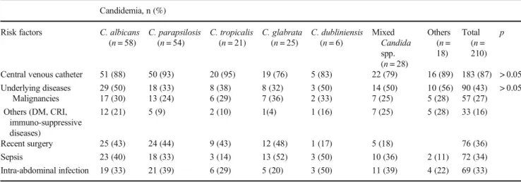
Patients’ medical records were examined to evaluate the avail-able risk factors. The most predominant risk factor was pres-ence of CVC (87%), followed by underlying diseases (43%) and history of recent surgery (36%). Statistical analysis did not reveal significant relationship between any species and available underlying risk factors. No significant difference was observed when the underlying factors were compared between C. albicans and non-albicans Candida spp. or be-tween C. parapsilosis and other Candida spp. Patient charac-teristics and underlying risk factors are demonstrated in Table1.
Mortality
A 30-day overall mortality was determined in patients with available adequate data and compared at the level of species and antifungal drug used for treatment. In total, 100 patients were eligible for this analysis. Overall mortality was 64%, with 56%, 58%, 70%, and 94% mortality for C. albicans,
C. parapsilosis, C. tropicalis, and C. glabrata, respectively. When compared at the species level, the rate of mortality was significantly higher in patients with C. glabrata infections (p = 0.045). Thirty-day overall mortality rates and relationship between antifungal susceptibility and antifungal treatment are demonstrated in Table2.
Epidemiology
Non-albicans Candida spp. were predominant with a rate of 68% within the entire study period. Overall species distribu-tion was 32%, 28%, 17%, and 11% for C. albicans, C. parapsilosis, C. glabrata, and C. tropicalis, respectively.
Over the first 5-year period, 100 candidemia episodes were identified in 100 patients and 124 Candida spp. were isolated. The most frequent species were C. albicans (29%), followed by C. parapsilosis (24%), C. glabrata (18%), C. tropicalis (11%), and C. dubliniensis (6%). Non-albicans Candida spp. constituted 71% of the isolates. Twenty-three episodes (18.5%) involved mixed candidemia with C. glabrata in 15 (65.2%), C. albicans in 14 (60.8%), and C. parapsilosis in 9 (39.1%). The distribution of the Candida spp. in the first 5-year period is demonstrated in Table3.
Over the second 5-year period, 110 candidemia episodes were identified in 97 patients and 114 Candida spp. were isolated. Two episodes of candidemia were encountered in nine patients and four episodes of candidemia were detected in one patient. The most frequent species were C. albicans (35.1%), followed by C. parapsilosis (28.9%), C. glabrata (15.8%), and C. tropicalis (11.4%). Non-albicans Candida spp. constituted approximately 64.9% of the isolates. Five episodes (4.5%) were accepted as mixed candidemia with C. albicans and C. glabrata involved in 3 (30%) of the epi-sodes. The distribution of the Candida spp. in the second period is demonstrated in Table3.
Antifungal susceptibility
Over the first 5-year period, susceptibility to amphotericin B was 100% among all Candida spp. All of C. albicans and almost all of the non-albicans Candida spp. were susceptible to anidulafungin. On the other hand, only 56% of C. parapsilosis were intermediately susceptible to anidulafungin. Fluconazole susceptibility rate was 94% for C. albicans and C. parapsilosis and 100% for C. tropicalis. Susceptibility rates of Candida spp. to major antifungals in the first 5-year period are demonstrated in Table4.
In the second 5-year period, amphotericin B susceptibility remained high at 95% for C. albicans, while it decreased to 79%, 72%, and 85% for C. parapsilosis, C. glabrata, and C. tropicalis, respectively. Susceptibility rates of all Candida spp. to anidulafungin were approximately 85%, except for C. parapsilosis, which showed 3% susceptibility and 24%
intermediate susceptibility. Fluconazole susceptibility remained high for C. albicans and C. tropicalis (93% and 92%, respectively), but showed a remarkable decrease to 49% for C. parapsilosis (MIC50= 4 μg/mL and MIC90= 256μg/mL). Susceptibility rates of Candida spp. to major antifungals in the second 5-year period are demonstrated in Table4.
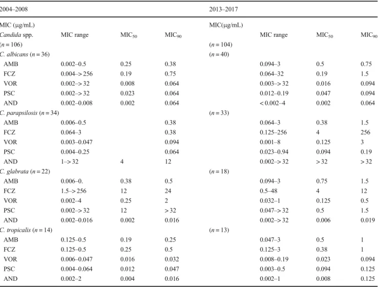
Ranges of MIC and MIC50 and MIC90 levels of the Candida spp. are demonstrated in Table5.
Resistance to more than one drug was detected in C. parapsilosis (15/67 22%), C. glabrata (8/40; 20%), and C. albicans (1/76; 1.3%) over 10 years and 79% were classi-fied as MDR. Over the first 5-year period, only two C. parapsilosis isolates (2/34; 6%) were determined to be resistant to azole and echinocandin class of drugs. On the other hand, over the second 5 years, 11 of 33 C. parapsilosis isolates (33%) were resistant to azole and echinocandins, one was resistant to azole and amphotericin B, and one was resis-tant to amphotericin B and echinocandins. Five isolates (15%) were resistant to all three drug classes and were classified as XDR. Eight isolates of C. glabrata (8/18; 44%) were defined as MDR: five were resistant to azole and amphotericin B, and three were resistant to amphotericin B and echinocandins.
Discussion
Candidemia remains an important cause of morbidity and mortality, especially in hospitalized and critically ill patients [6, 7]. The distribution of Candida species has changed in recent years with a trend of increasing rate of non-albicans Candida spp. [12–15,27,28] while Asian studies revealed that C. albicans is still predominant [5,29]. Although current study also revealed a predominance of non-albicans Candida spp. (68%), C. albicans is leading one, with a rate of 32%, followed by C. parapsilosis (28%), and C. glabrata and C. tropicalis, similar to the other reports from Turkey [22,24]. When compared over the two 5-year periods, the distribution of the species was almost stable: non-albicans Candida spp. constituted 71% of the isolates in the first period, while it de-creased to 64.9% in the second period. Although statistically insignificant, the frequency of C. albicans and C. parapsilosis increased from 29% to 35% and 24% to 29%, respectively. It may be due to the fact that more patients with intra-abdominal surgery were recruited in the second period.
Table 1 Characteristics and risk factors in candidemia cases Candidemia, n (%)
Risk factors C. albicans (n = 58) C. parapsilosis (n = 54) C. tropicalis (n = 21) C. glabrata (n = 25) C. dubliniensis (n = 6) Mixed Candida spp. (n = 28) Others (n = 18) Total (n = 210) p
Central venous catheter 51 (88) 50 (93) 20 (95) 19 (76) 5 (83) 22 (79) 16 (89) 183 (87) > 0.05 Underlying diseases 29 (50) 18 (33) 8 (38) 8 (32) 3 (50) 14 (50) 10 (56) 90 (43) > 0.05 Malignancies 17 (30) 13 (24) 6 (29) 7 (36) 2 (33) 7 (25) 5 (28) 57 (27) Others (DM, CRI, immuno-suppressive diseases) 12 (21) 5 (9) 2 (10) 1(4) 1 (16) 7 (25) 5 (28) 33 (16) Recent surgery 25 (43) 24 (44) 9 (43) 12 (48) 1 (17) 5 (18) 76 (36) Sepsis 23 (40) 18 (33) 3 (14) 13 (52) 3 (50) 10 (36) 2 (11) 72 (34) Intra-abdominal infection 19 (33) 21 (39) 6 (29) 5 (20) 3 (50) 11 (39) 4 (22) 69 (33) DM diabetes mellitus, CRI chronic renal insufficiency
Table 3 Distribution of Candida spp. in the intensive care unit 2004–2008 2013–2017 Candida spp. n= 124 (%) n = 114 (%) Total n = 238 C. albicans 36 (29) 40 (35) 76 (32) C. parapsilosis 34 (24) 33 (29) 67 (28) C. glabrata 22 (18) 18 (16) 40 (17) C. tropicalis 14 (11) 13 (11) 27 (11) C. dubliniensis 7 (6) 1 8 (3) C. lusitaniae 3 (2) - 3 (1) C. krusei 2 (2) 2 (2) 4 (2) Others 6 (5) 7 (6) 13 (5)
Table 2 Thirty-day overall mortality
Overall mortality n (%) p C. albicans (n = 43) 24 (56) NS C. parapsilosis (n = 31) 18 (58) NS C. glabrata (n = 16) 15 (94) 0.045 C. tropicalis (n = 10) 7 (70) NS Total (n = 100) 64(64)
Among non-albicans species, C. parapsilosis was the most prominent in Latin America, India, South Africa, and Asia [5,
13,15, 30,31]. It is also frequently encountered in some Mediterranean countries including Turkey [32] with isolation rates ranging from 6 to 66% [33,34]. The prevalence of different Candida spp. may vary due to patientrelated or -unrelated factors. C. parapsilosis is more common in neonatal or surgical ICUs; CVC, total parenteral nutrition, recent sur-gery, use of echinocandins, and poor infection control are among important risk factors [6,35–37]. Recent fluconazole therapy, older age, gastrointestinal surgery, and intravenous drug use are the prevalent risk factors for C. glabrata infec-tions [10,35]. The most predominant risk factor was use of
CVC in our study, although the risk factors were comparable among Candida spp. in our study. This may be due to the small number of cases and similar characteristics of the pa-tients. Prior gastrointestinal surgery, antifungal use, and CVC were reported as significant risk factors for non-albicans candidemia [38, 39]. Although statistically insignificant, ICU stay, CVC use, recent surgery, and poor infection control may have led to the predominance of C. parapsilosis among non-albicans species.
The incidence rate of candidemia cases due to mixed spe-cies was reported in the range of 2–6% [7,40]. In our study, this rate was 13.3%, higher than the other studies. Although this higher rate could not be attributed to any risk factor by Table 4 Susceptibility rates of Candida spp. to major antifungals
2004–2008 2013–2017 Candida spp. (n = 113) S n (%) I n (%) R n (%) (n = 104) S n (%) I n (%) R n (%) C. albicans (n = 36) (n = 40) AMB 36 (100) 38 (95) 2 (5) FCZ 34 (94) 1 (3) 1 (3) 37 (93) 3 (7) VOR 32 (88) 2 (6) 2 (6) 35 (88) 3 (7) 2 (5) PSC 32 (88) 4 (12) 34 (85) 6 (15) AND 36 (100) 35 (88) 5 (12) C. parapsilosis (n = 34) (n = 33) AMB 34 (100) 26 (79) 7 (21) FCZ 32 (94) 2(6) 16 (49) 1 (2) 16 (49) VOR 33 (97) 1 (3) 17 (52) 1 (3) 15 (45) PSC 32 (94) 2(6) 16 (48) 17 (52) AND 19 (56) 15 (44) 1 (3) 8 (24) 24 (73) C. glabrata (n = 22) (n = 18) AMB 22 (100) 13 (72) 5 (28) FCZ 21(95) 1 (5) 17 (94) 1 (6) VOR IE IE PSC IE IE AND 22 (100) 15 (83) 3 (17) C. tropicalis (n = 14) (n = 13) AMB 14 (100) 11 (85) 2 (15) FCZ 14 (100) 12 (92) 1 (8) VOR 14 (100) 12 (92) 1 (8) PSC 14 (100) 6 (46) 7 (54) AND 13 (93) 1 (7) 11 (85) 2 (15) C. dubliniensis (n = 7) AMB IE FCZ IE VOR 7 (100) PSC 7 (100) AND IE
AMB amphotericin B, FCZ fluconazole, VOR voriconazole, PSC posaconazole, AND anidulafungin, IE insufficient evidence, S susceptible, R resistant, I intermediate
statistical analysis, recent intra-abdominal surgery might be the contributing factor.
Overall mortality of candidemia may reach 50% or even exceed 60% in treated patients [8,41]. In a review of random-ized trials, increased mortality was seen with C. tropicalis infections, while C. parapsilosis infections were associated with lower mortality [42]. In our study, overall mortality was 64%. Mortality rate was higher for C. glabrata and lower for C. parapsilosis and C. albicans infections.
Due to their broad-spectrum activity against Candida spe-cies, the echinocandins are preferred extensively for the treat-ment of candidemia. The highest echinocandin MICs are found for C. parapsilosis, C. glabrata, C. tropicalis, and C. guilliermondii [15,43]. In the current study, almost all Candida spp. (except C. parapsilosis) were susceptible to anidulafungin in the first period but the resistance rate was high in C. parapsilosis (44%) and even increased to 73% in the second period.
In the SENTRY program, resistance to echinocandins was uncommon among all Candida spp. [15]. On the other hand, 51 isolates were reported to have MIC values of 4 μg/mL to anidulafungin from Europe, Latin and North America [44]. Similarly, 22 years of candidemia study from Norway revealed that none of C. parapsilosis isolates were susceptible with intermediate susceptibility of 89% (28.3% with MIC > 4μg/mL), similar to our results [45]. These discrepancies may be due to the difference in the analysis of susceptibilities between Clinical Laboratory Standards Institute (CLSI) referred in SENTRY and EUCAST in Norwegian and our study [26, 46]. A study from Israel evaluated 899 candidemia cases using the E-test for antifungal susceptibility and interpreted the results a c c o r d i n g t o C L S I b r e a k p o i n t s . N o n e o f t h e C. parapsilosis isolates were resistant to caspofungin. Since we have used EUCAST breakpoints, this might have led to an overestimation of resistance in our study [47]. Table 5 MIC range, MIC50and MIC90levels of the Candida spp.
2004–2008 2013–2017
MIC (μg/mL) MIC(μg/mL)
Candida spp. MIC range MIC50 MIC90 MIC range MIC50 MIC90
(n = 106) (n = 104) C. albicans (n = 36) (n = 40) AMB 0.002–0.5 0.25 0.38 0.094–3 0.5 0.75 FCZ 0.004–> 256 0.19 0.75 0.064–32 0.19 1.5 VOR 0.002–> 32 0.008 0.064 0.003–> 32 0.016 0.094 PSC 0.002–> 32 0.023 0.064 0.012–0.19 0.047 0.094 AND 0.002–0.008 0.002 0.064 < 0.002–4 0.002 0.064 C. parapsilosis (n = 34) (n = 33) AMB 0.006–0.5 0.38 0.064–3 0.38 1.5 FCZ 0.064–3 0.38 0.125–256 4 256 VOR 0.003–0.047 0.094 0.001–8 0.125 3 PSC 0.004–0.25 0.064 0.023–0.94 0.094 0.19 AND 1–> 32 4 12 0.002–> 32 > 32 > 32 C. glabrata (n = 22) (n = 18) AMB 0.006–0. 0.38 0.5 0.094–3 0.75 1.5 FCZ 1.5–> 256 12 24 0.5–48 4 12 VOR 0.002–4 0.25 2 0.032–1 0.125 0.5 PSC 0.002–> 32 12 > 32 0.047–> 32 0.5 1.5 AND 0.002–0.016 0.002 0.016 0.002–> 32 0.006 0.019 C. tropicalis (n = 14) (n = 13) AMB 0.125–0.5 0.19 0.25 0.047–3 0.5 1 FCZ 0.125–0.5 0.25 0.5 0.125–3 0.38 1 VOR 0.006–0.047 0.016 0.032 0.008–0.19 0.023 0.094 PSC 0.004–0.064 0.012 0.047 0.003–0.5 0.094 0.125 AND 0.002–2 0.004 0.016 0.002–1 0.008 0.125
AMB amphotericin B, FCZ fluconazole, VOR voriconazole, PSC posaconazole, AND anidulafungin, IE insufficient evidence, S susceptible, R resistant, I intermediate
C. parapsilosis species complex and C. guilliermondii have higher MIC values to echinocandins, but since glucan synthesis is still inhibited at therapeutic levels, treatment with echinocandins is generally effective [48–51]. C. parapsilosis sensu stricto is reported to be more resistant to anidulafungin, and Borghi et al. reported 34 C. parapsilosis sensu stricto strains having MIC values of 4μg/mL from Italy [50,52,
53]. Since a subclassification has not been performed, C. parapsilosis sensu stricto might have predominated and this might have also led to higher MIC values to anidulafungin in our strains. Although a clonal analysis has not been per-formed, another hypothesis for this high resistance rate might be a single resistant C. parapsilosis clone circulating in our unit.
Susceptibility rate of Candida spp., except for C. glabrata to fluconazole, voriconazole, and posaconazole, was high in the first period. Because of C. albicans ranked the first and C. parapsilosis the second in our unit, fluconazole was pre-scribed more frequently empirically. In case of determination of azole resistance, echinocandins or amphotericin B (espe-cially in C. parapsilosis infections) were preferred for antifun-gal treatment. Susceptibility rate to fluconazole and v o r i c o n a z o l e r e m a i n e d h i g h f o r C . a l b i c a n s a n d C. tropicalis, but only half of C. parapsilosis were susceptible in the second period. Since fluconazole is recommended as the first-line therapy for C. parapsilosis infections and resistance to echinocandins was noted in our center, fluconazole was prescribed very intensely [8]. As a consequence, susceptibility rate to fluconazole might have significantly decreased in C. parapsilosis. Decreased susceptibility of C. parapsilosis to other azoles in our study may be related to cross-resistance, which has been described also in previous studies [15].
In the second period, although susceptibility to amphotericin B remained high in C. albicans, it decreased in the non-albicans group (72%–85%). Since C. parapsilosis and C. glabrata predominated in non-albicans spp., amphotericin B was frequently used empirically besides flu-conazole. This might have led to the decrease in rate of sus-ceptibility. Multidrug resistance is uncommon and usually involves acquired resistance in species with intrinsic resis-tance. It is mostly encountered in C. glabrata and C. auris. Antifungal use, subtherapeutic drug levels, and poor infection control are among the risk factors [27]. Resistance to more than one drug class was detected predominantly in C. parapsilosis and C. glabrata in our study. We think that this high rate of multidrug resistance might be due to poor infection control.
Lastly, we have compared our results with the results of the first multicenter study reporting resistance rates according to CLSI breakpoints in 1991 candidemia cases in our country [25]. MIC90levels are similar in terms of resistance in most Candida spp., except for C. albicans and C. parapsilosis: MIC9 0 levels for fluconazole and anidulafungin in
C. parapsilosis were higher in our study. These discrepancies may be due to the E-test method used in our study and due to a probably endemic C. parapsilosis isolate circulating in our unit.
There are some limitations to our study. First, this is a retrospective and mainly laboratory-based study. Since the medical records were incomplete in certain parameters such as previous hospitalization, total parenteral nutrition, previous antibiotic, antifungal, and corticosteroid use for an important number of patients, these parameters could not be evaluated. Second, we have used the E-test, which is an alternative method for antifungal susceptibility. EUCAST and CLSI recommend broth microdilution as the reference method, but as this method is labor-intensive and expensive, the E-test method is preferred for being more practical and easier to perform [26, 46,
54,55]. The agreement rates of the E-test with the refer-ence method are usually favorable [55]. However, microdilution broth test was suggested to be more conve-nient for non-albicans Candida spp., in particular for the isolates with high MIC values against azoles [56]. Morace et al. demonstrated that the E-test provides valuable re-sults with the exception of C. glabrata for azoles and C. parapsilosis for echinocandins. [57]. Thus, the use of E-test in our study might explain the high resistance rates of C. parapsilosis to anidulafungin. On the other hand, L o v e r o e t a l . f o u n d 9 5 . 6–97.8% agreement for C. parapsilosis when compared the EUCAST and E-test method for echinocandins and favored the use of commer-cial methods [58].
In conclusion, although there is a predominance of non-albicans Candida spp. in our institution, C. non-albicans remained the most commonly encountered Candida species. Among non-albicans Candida spp., C. parapsilosis ranked first followed by C. glabrata. Predominant risk factors were pres-ence of an underlying disease, use of CVC, and history of recent surgery. Mortality rate was higher in patients infected with C. glabrata. High level of echinocandin resistance and MDR/XDR among C. parapsilosis and C. glabrata isolates is an important issue in our unit. Distribution and antifungal susceptibilities of Candida spp. differ throughout years. Every center should follow their change in species and sus-ceptibility profile closely and direct empirical antifungal ther-apies based on the local resistance profile.
Compliance with ethical standards
Conflict of interest The authors declare that they have no conflict of interest.
Ethical approval Not applicable. Informed consent Not applicable.
References
1. Bassetti M, Peghin M, Timsit JF (2016) The current treatment land-scape: candidiasis. J Antimicrob Chemother 71(suppl 2):ii13–ii22 2. Kullberg BJ, Arendrup MC (2015) Invasive candidiasis. N Engl J
Med 373(15):1445–1456
3. Cleveland AA, Farley MM, Harrison LH, Stein B, Hollick R, Lockhart SR et al (2012) Changes in incidence and antifungal drug resistance in candidemia: results from population-based laboratory surveillance in Atlanta and Baltimore, 2008–2011. Clin Infect Dis 55:1352–1361
4. Klingspor L, Tortorano AM, Peman J, Willinger B, Hamal P, Sendid B et al (2015) Invasive Candida infections in surgical pa-tients in intensive care units: a prospective, multicentre survey ini-tiated by the European Confederation of Medical Mycology (ECMM)(2006–2008). Clin Microbiol Infect 21:87
5. Tan BH, Chakrabarti A, Li RY, Patel AK, Watcharananan SP, Liu Z, Asia Fungal Working Group (AFWG) et al (2015) Incidence and species distribution of candidaemia in Asia: a laboratory-based sur-veillance study. Clin Microbiol Infect 21(10):946–953
6. Eggiman P, Garbino J, Pittet D (2003) Epidemiology of Candida species infections in critically ill non-immunosuppressed patients. Lancet Infect Dis 3:685–702
7. Sellami A, Sellami H, Neji S, Makni F, Abbes S, Cheikhrouhou F et al (2011) Antifungal susceptibility of bloodstream Candida iso-lates in Sfax Hospital: Tunisia. Mycopathologia 171(6):417–422 8. Pappas PG, Kauffman CA, Andes DR, Clancy CJ, Marr KA,
Ostrosky-Zeichner L et al (2016) Clinical practice guideline for the management of Candidiasis: 2016 update by the Infectious Diseases Society of America. Clin Infect Dis 62(4):e1–e50 9. Schelenz S (2008) Management of candidiasis in the intensive care
unit. J Antimicrob Chemother 61(Suppl 1):i31–i34
10. Horn DL, Neofytos D, Anaissie EJ, Fishman JA, Steinbach WJ, Olyaei AJ et al (2009) Epidemiology and outcomes of candidemia in 2019 patients: data from the prospective antifungal therapy alli-ance registry. Clin Infect Dis 48:1695–1703
11. Wan Ismail WNA, Jasmi N, Khan TM, Hong YH, Neoh CF (2019) The economic burden of candidemia and invasive candidiasis: a systematic review. Value Health Reg Issues 21:53–58
12. Pfaller MA, Diekema DJ (2010) Epidemiology of invasive mycoses in North America. Crit Rev Microbiol 36:1–53
13. Nucci M, Queiroz-Telles F, Tobon AM, Restrepo A, Colombo AL (2010) Epidemiology of opportunistic fungal infections in Latin America. Clin Infect Dis 51:561–570
14. Wang H, Xiao M, Chen SC, Kong F, Sun ZY, Liao K et al (2012) In vitro susceptibilities of yeast species to fluconazole and voriconazole as determinedby the 2010 National China Hospital Invasive Fungal Surveillance Net (CHIF-NET) study. J Clin Microbiol 50:3952–3959
15. Pfaller MA, Diekema DJ, Turnidge JD, Castanheira M, Jones RN (2019) Twenty years of the SENTRY antifungal surveillance pro-gram: results for Candida species from 1997-2016. Open Forum Infect Dis 6(Suppl 1):S79–S94
16. Hachem R, Hanna H, Kontoyiannis D, Jiang Y, Raad I (2007) The changing epidemiology of invasive candidiasis. Cancer 112:2334– 2337
17. Nguyen MH, Peacock JE Jr, Morris AJ, Tanner DC, Nguyen ML, Snydman DR et al (1996) The changing face of candidemia : emer-gence of non C. albicans species and antifungal resistance. Am J Med 100:617–623
18. Kontoyiannis DP, Lewis RE (2002) Antifungal drug resistance of pathogenic fungi. Lancet 359:1135–1144
19. Chapman B, Slavin M, Marriott D, Halliday C, Kidd S, Arthur I, et al, Australian and New Zealand Mycoses Interest Group (2017=
changing epidemiology of candidaemia in Australia. J Antimicrob Chemother. 2017;72:1103–8.
20. Trouve C, Blot S, Hayette MP, Jonckheere S, Patteet S, Rodriguez-Villalobos H et al (2017) Epidemiology and reporting of candidaemia in Belgium: a multi-Centre study. Eur J Clin Microbiol Infect Dis 36:649–655
21. Pinhati HM, Casulari LA, Souza AC, Siqueira RA, Damasceno CM, Colombo AL (2016) Outbreak of candidemia caused by flu-conazole resistant Candida parapsilosis strains in an intensive care unit. BMC Infect Dis 16:433
22. Alp S, Arikan-Akdagli S, Gulmez D, Ascioglu S, Uzun O, Akova M (2015) Epidemiology of candidaemia in a tertiary care university hospital: 10-year experience with 381 candidaemia episodes be-tween 2001 and 2010. Mycoses 58(8):498–505
23. Yeşilkaya A, Azap Ö, Aydın M, Akçil Ok M (2017) Epidemiology, species distribution, clinical characteristics and mortality of candidemia in a tertiary care university hospital in Turkey, 2007-2014. Mycoses 60(7):433–449
24. Kazak E, Akın H, Ener B, Sığırlı D, Özkan Ö, Gürcüoğlu E et al (2014) An investigation of Candida species isolated from blood cultures during 17 years in a university hospital. Mycoses 57(10): 623–629
25. Arikan-Akdagli S, Gülmez D, Doğan Ö, Çerikçioğlu N, Doluca Dereli M, Birinci A et al (2019) First multicentre report of in vitro resistance rates in candidaemia isolates in Turkey. J Glob Antimicrobiol Res 18:230–234
26. Arendrup MC, Patterson TF (2017) Multidrug-resistant Candida: epidemiology, molecular mechanisms, and treatment. J Infect Dis 216(3):S445–S451
27. Pfaller MA, Diekema DJ, Gibbs DL, Newell VA, Ellis D, Tullio V, the Global Surveillance Group et al (2010) Results from the ARTEMIS DISK global antifungal surveillance study, 1997 to 2007: a 10.5-year analysis of susceptibilities of Candida species to fluconazole and voriconazole as determined by CLSI standardized disk diffusion. J Clin Microbiol 48:1366–1377
28. Vaezi A, Fakhim H, Khodavaisy S, Alizadeh A, Nazeri M, Soleimani A et al (2017) Epidemiological and mycological charac-teristics of candidemia in Iran: a systematic review and meta-anal-ysis. J Mycol Med 27(2):146–152
29. Zhou ZL, Lin CC, Chu WL, Yang YL, Lo HJ, TSARY Hospitals (2016) The distribution and drug susceptibilities of clinical Candida species in TSARY 2014. Diagn Microbiol Infect Dis 86(4):399– 404
30. Chakrabarti A, Sood P, Rudramurthy SM, Chen S, Kaur H, Capoor M et al (2015) Incidence, characteristics and outcome of ICU-acquired candidemia in India. Intensive Care Med 41:285–295 31. Govender NP, Patel J, Magobo RE, Naicker S, Wadula J, Whitelaw
A, TRAC-South Africa group et al (2016) Emergence of azole-resistant Candida parapsilosis causing bloodstream infection: re-sults from laboratory-based sentinel surveillance in South Africa. J Antimicrob Chemother 71:1994–2004
32. Montagna MT, Lovero G, Borghi E, Amato G, Andreoni S, Campion L et al (2014) Candidemia in intensive care unit: a na-tionwide prospective observational survey (GISIA-3 study) and review of the European literature from 2000 through 2013. Eur Rev Med Pharmacol Sci 18(5):661–674
33. Tukenmez Tigen E, Bilgin H, Perk Gurun H, Dogru A, Ozben B, Cerikcioglu N et al (2017) Risk factors, characteristics, and out-comes of candidemia in an adult intensive care unit in Turkey. Am J Infect Control 45(6):e61–e63
34. Horasan ES, Ersöz G, Göksu M, Otag F, Kurt AO, Karaçorlu S et al (2010) Increase in Candida parapsilosis fungemia in critical care units: a 6-years study. Mycopathologia. 170(4):263–268
35. Pfaller MA, Diekema DJ (2007) Epidemiology of invasive candi-diasis: a persistent public health problem. Clin Microbiol Rev 20: 133–163
36. Lockhart SR, Messer SA, Pfaller MA, Diekema DJ (2008) Geographic distribution and antifungal susceptibility of the newly described species Candida orthopsilosis and Candida metapsilosis in comparison to the closely related species Candida parapsilosis. J Clin Microbiol 46:2659–2664
37. Forrest GN, Weekes E, Johnson JK (2008) Increasing incidence of Candida parapsilosis candidemia with caspofungin usage. J Inf Secur 56:126–129
38. Playford EG, Marriott D, Nguyen Q, Chen S, Ellis D, Slavin M et al (2008) Candidemia in nonneutropenic critically ill patients: risk factors for non-albicans Candida spp. Crit Care Med 36:2034 39. Chow JK, Golan Y, Ruthazer R, Karchmer AW, Carmeli Y,
Lichtenberg D et al (2008) Factors associated with candidemia caused by non-albicans Candida species versus Candida albicans in the intensive care unit. Clin Infect Dis 46:1206
40. Sipsas NV, Lewis RE, Tarrand J, Hachem R, Rolston KV, Raad II et al (2009) Candidemia in patients with hematologic malignancies in the era of new antifungal agents (2001-2007). Cancer 115:4745– 4752
41. Kollef M, Micek S, Hampton N, Doherty JA, Kumar A (2012) Septic shock attributed to Candida infection: importance of empiric therapy and source control. Clin Infect Dis 54 /12:1739
42. Andes DR, Safdar N, Baddley JW, Playford G, Rebolş AC, Rex JH, Mycoses Study Group et al (2012) Impact of treatment strategy on outcomes in patients with candidemia and other forms of invasive candidiasis: a patient-level quantitative review of randomized trials. Clin Infect Dis 54(8):1110
43. Spellberg BJ, Filler SG, Edwards JE Jr (2006) Current treatment strategies for disseminated candidiasis. Clin Infect Dis 42:244 44. Pfaller MA, Boyken L, Hollis RJ, Kroeger J, Messer SA, Tendolkar
S et al (2008) In vitro susceptibilities of invasive isolates of Candida spp. to anidulafungin, caspofungin and micafungin: six years of global surveillance. J Clin Microbiol 46:150–156
45. Hesstvedt L, Gaustad P, Andersen CT, Haarr E, Hannula R, Haukland HH et al (2015) Twenty-two years of candidaemia sur-veillance: results from a Norwegian national study. Clin Microbiol Infect 21(10):938–945
46. Clinical and Laboratory Standards Institute. Reference method for broth dilution antifungal susceptibility testing of yeasts; fourth in-formational supplement. CLSI document M27-S4. 2012; Clinical and Laboratory Standards Institute
47. Israel S, Amit S, Israel A, Livneh A, Nir-Paz R, Korem M (2019) The epidemiology and susceptibility of candidemia in Jerusalem, Israel. Front Cell Infect Microbiol 9:352
48. Tavanti A, Davidson AD, Gow NA, Maiden MC, Odds FC (2005) Candida orthoplosis and Candida metapsilosis spp. nov. to replace Candida parapsilosis groups II and III. J Clin Microbiol 43:284–292
49. Gonçalves SS, Amorim CS, Nucci M, Padovan AC, Briones MR, Melo AS et al (2010) Prevalence rates and antifungal susceptibility profiles of the Candida parapsilosis species complex: results from a nationwide surveillance of candidemia in Brazil. Clin Microbiol Infect 16(7):885–887
50. Gomez-Lopez A, Alastruey-Izquierdo A, Rodriguez D, Almirante B, Pahissa A, Rodriguez-Tudela JL, Barcelona Candidemia Project Study Group et al (2008) Barcelona Candidemia project study group prevalence and susceptibility profile of Candida metapsilosis and Candida orthopsilosis: results from population-based surveil-lance of candidemia in Spain. Antimicrob Agents Chemother 52: 1506–1509
51. Perlin DS (2015) Echinocandin resistance in Candida. Clin Infect Dis 61(6):S612–S617
52. Szabó Z, Szilágyi J, Tavanti A, Kardos G, Rozgonyi F, Bayegan S et al (2009) In vitro efficacy of 5 antifungal agents against C. parapsilosis, Candida orthopsilosis and Candida metapsilosis as determined by time-kill methodology. Diagn Microbiol Infect 64: 283–288
53. Borghi E, Sciota R, Iatta R, Biassoni C, Montagna MT, Morace G (2011) Characterization of Candida parapsilosis complex strains isolated from invasive fungal infections. Eur J Clin Microbiol Infect Dis 30(11):1437–1441
54. Alexander BD, Byrne TC, Smith KL, Hanson KE, Anstrom KJ, Perfect JR et al (2007) Comparative evaluation of Etest and sensititre yeastone panels against the Clinical and Laboratory Standards institute M27-A2 reference broth microdilution method for testing Candida susceptibility to seven antifungal agents. J Clin Microbiol 45(3):698–706
55. Arikan S (2007) Current status of antifungal susceptibility testing methods. Med Mycol 45:569–587
56. Metin DY, Hilmioglu Polat S, Samlioglu P, Doganay Oflazoglu B, Inci RI, Tumbay E (2011) Evaluation of antifungal susceptibility testing with microdilution and E test methods of Candida blood isolates. Mycopathologia 172(3):187–199
57. Morace G, Borghi E, Iatta R, Amato G, Anderoni S, Brigante G et al (2011) Antifungal susceptibility of invasive yeast isolates in Italy: the GISIA3 study in critically ill patients. BMC Infect Dis 11: 130–138
58. Lovero G, Borghi E, Balbino S, Cirasola D, De Giglio O, Perdoni F et al (2016) Molecular identification and echinocandin susceptibil-ity of candida parapsilosis complex bloodstream isolates in Italy, 2007-2014. PLoS One 11(2):e0150218
Publisher’s note Springer Nature remains neutral with regard to jurisdic-tional claims in published maps and institujurisdic-tional affiliations.